External Beam RT for Prostate Cancer - ASTRO
External Beam RT for Prostate Cancer - ASTRO
External Beam RT for Prostate Cancer - ASTRO

You also want an ePaper? Increase the reach of your titles
YUMPU automatically turns print PDFs into web optimized ePapers that Google loves.
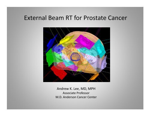
<strong>External</strong> <strong>Beam</strong> <strong>RT</strong> <strong>for</strong> <strong>Prostate</strong> <strong>Cancer</strong><br />
Andrew K. Lee, MD, MPH<br />
Associate Professor<br />
M.D. Anderson <strong>Cancer</strong> Center
Disclosure<br />
• I have no relevant conflicts of interest to<br />
disclose.<br />
• This presentation will NOT discuss<br />
iinvestigational i i l or off-label ff l b l use of fddrugs<br />
or<br />
therapies.
Learning Objectives<br />
• Understand the basics of simulating and<br />
planning a patient <strong>for</strong> prostate cancer<br />
radiation therapy<br />
• UUnderstand d d the h methods h d f<strong>for</strong> appropriate i<br />
delivery of dose-escalated and image-guided<br />
therapy h iin<br />
prostate cancer
Localized prostate cancer<br />
Low risk<br />
T1-2 T1 2<br />
Gleason 6<br />
PSA 20<br />
EB<strong>RT</strong> + HT<br />
EB<strong>RT</strong> + Brachy +HT<br />
Surgery (select)
2010 AJCC Staging/Prognostic GROUPS<br />
7th 7 Edition<br />
• Group I (Low risk)<br />
T1 T1a-2a, 2 Gleason Gl 66, PSA
MDACC prostate EB<strong>RT</strong> recommendations<br />
• Low risk 78 Gy (2 Gy)PTV Gy) PTV<br />
(>80 Gy CTV)<br />
• Intermediate risk <strong>Prostate</strong> & “proximal” SV<br />
6mo HT <strong>for</strong> select pts<br />
(2 mos TAB then leuprolide alone)<br />
• High risk & T3 <strong>Prostate</strong> & most of SV<br />
(Select pts LN)<br />
2 years HT
Dose-escalation Dose escalation w/ less toxicity<br />
• Delivery y techniques q<br />
– IM<strong>RT</strong>/ VMAT<br />
– Protons<br />
• Reduce PTV<br />
– Target localization (e.g. IG<strong>RT</strong>)<br />
– Target g immobilzation (e.g. ( g rectal balloon) )<br />
– Reduce CTV<br />
• Selective dose-escalation
At simulation…<br />
minimize systematic errors<br />
• If using fiducials: ≥3 fiducials ≥5 days prior to sim<br />
– If you cannot wait 5 days, then consider verification CT (CT or CBCT)<br />
the first week of treatment<br />
• Com<strong>for</strong>tably semi-full semi full bladder (do NOT overfill)<br />
• Not overly distended rectum (+/- enema)<br />
• Supine<br />
• Leg immobilization<br />
• Make sure patient is relaxed<br />
• Scan from L5 through lesser trochanters<br />
• 2-3 mm slices (make sure TPS can expand PTV correctly w/<br />
chosen slice thickness)
Systematic errors are clinically relevant
PSA control by mean rectal<br />
Cross Sectional Area at simulation
• Impact of rectal<br />
distention may be<br />
more significant than<br />
risk group
Take home message<br />
Do not miss posterior aspect of prostate!<br />
>2/3 cancers arise in peripheral zone<br />
Si Simulate l t with ith empty t rectum t ( (e.g. enema) )<br />
Thi d h f i i i l<br />
This decreases chances of missing posteriorly<br />
regardless of PTV
• Rectum<br />
• Bladder l dd<br />
• Femoral heads<br />
• CTV<br />
Defining Structures<br />
– Low risk = <strong>Prostate</strong> only<br />
– IInt risk ik= P<strong>Prostate</strong> + prox SV<br />
– High risk = <strong>Prostate</strong> (+EPE) + SV (+/- nodes)<br />
• Use zoom<br />
• Use window and level<br />
• Use other planes of view (especially sag <strong>for</strong> apex)
Don’t <strong>for</strong>get to window-level<br />
HHelps l w/ / ddefining fi i ti tissue planes l<br />
Pelvic W/L Head W/L
Urogenital diaghragm<br />
insertion into symphysis<br />
NOTE: Do NOT under-contour apex.<br />
Almost all PZ, no capsule, common site <strong>for</strong> <strong>RT</strong><br />
failures failures.<br />
In very advanced cases, disease can track along<br />
membranous urethra.
Defining Organs at Risk (OAR)<br />
Avoidance structures and denominator <strong>for</strong> DVH<br />
• RECTUM:<br />
From inferior ischium to anterior flexion of sigmoid<br />
Alt Alternatively ti l can use li linear llength th of f 10 10-11cm 11<br />
• BLADDER:<br />
Entire bladder (interpolate contour)<br />
• FEMORAL HEADS:<br />
Entire femoral head to lesser trochanter
Rectum and Bladder<br />
• Want to contour the rectum- rectum not the peri- peri<br />
rectal muscles.<br />
• If you using ultrasound-based guidance, pay<br />
special i l attention i to bl bladder-prostate<br />
dd<br />
interface.<br />
– Also will need to contour non-CTV portion of SV’s<br />
as reference structure
Ultrasound Alignment (sagittal)
IG<strong>RT</strong> PTV: VOLUMETRICALLY expand CTV 5-7 mm except<br />
posteriorly 4-5mm.<br />
Review PTV prior to planning. Validate PTV margin <strong>for</strong> your clinic.<br />
Current MDACC technique is 78Gy (2Gy) prescribed to PTV
MDACC DVH Plan Evaluation: Clinical Constraints<br />
(Clinical constraints ≠ Planning parameters)<br />
PTV: >96%V @ 78Gy<br />
<strong>Prostate</strong>: 100%V @ @ 78Gy<br />
SV: >98%V @ 78Gy<br />
Rectum:
8 angles <strong>for</strong> virtually any anatomy w/ 6 MV<br />
U Use
MDACC IM<strong>RT</strong> planning method<br />
• Use series of avoidance structures or “rings” in addition<br />
to iindividual di id l organs<br />
• Con<strong>for</strong>mal plans with “compact” dose distributions<br />
• Fewer trials are needed<br />
• 8 beam angles or VMAT<br />
• Minimize total monitor units<br />
• Fewer beam segments
OAR and Objectives<br />
<strong>External</strong><br />
Ring 2<br />
Ring 1<br />
<strong>Prostate</strong> + SV<br />
Max Dose 3800 cGy<br />
Max Dose 5500 cGy<br />
Max DVH 7500 2%<br />
Max Dose 8100 cGy<br />
Uni<strong>for</strong>m Dose7800 cGy
ROI Objectives (IM<strong>RT</strong>)<br />
ROI Type Target cGy % Volume Weight<br />
<strong>Prostate</strong> + SV Uni<strong>for</strong>m Dose 7760 100<br />
<strong>Prostate</strong> + SV Max Dose 8000 20<br />
Rectum Max DVH 6000 10% 1<br />
Rectum Max DVH 4000 30% 1<br />
Rectum Max DVH 2000 45% 1<br />
Bladder Max DVH 6000 8% 1<br />
Bladder Max DVH 4000 12% 1<br />
Bladder Max DVH 1500 30% 1<br />
Ring 1 Max DVH 7500 2% 10<br />
Ring 2 Max Dose 5500 5<br />
Ring 2 Max DVH 3000 25% 1<br />
<strong>External</strong> Max Dose 3800 50
DVH Plan Evaluation: Clinical Constraints<br />
(Clinical constraints ≠ Planning parameters)<br />
( gp )<br />
<strong>Prostate</strong>:<br />
>100%V@78Gy<br />
>100%V@78Gy<br />
100%V@78Gy<br />
SV:<br />
>98%V@78Gy<br />
Rectum:<br />
Axial dose distribution<br />
(qualitative evaluation)
Sagittal g<br />
dose distribution
Minimum dose vs. Mean dose<br />
• >96% PTV receives ≥ 78 Gy<br />
• 100% CTV receives ≥78 Gy<br />
• Mean dose to prostate ≈ 80-81 Gy<br />
• Need to have threshold <strong>for</strong> heterogeneity<br />
Typically prescribe to >96% of mean dose
VMAT <strong>for</strong> prostate<br />
• Similar to IM<strong>RT</strong> <strong>for</strong> inverse planning<br />
• 2 arcs (clockwise then counter)<br />
• Gantry angles 210-150 °, 150-210 °<br />
– May y extend to 182-178 °, , 178-182 ° <strong>for</strong> LN cases<br />
• 6MV<br />
• Avoid 0° 0 collimators to reduce “tongue tongue & groove effect effect” of<br />
MLC<br />
– Usually 10-45° but rotated in opposite directions <strong>for</strong> 2 nd arc (e.g. 15 vs. 335 °)<br />
• Avoid couch kicks unless absolutely needed<br />
• Typically start w/ 10x10 cm field<br />
• WWatch t h ttotal t l MU’ MU’s (96% mean
Use complementary angles <strong>for</strong><br />
collimation <strong>for</strong> arc 1 vs. arc 2
VMAT
For both IM<strong>RT</strong> and VMAT<br />
• Monitor patients w/ large pannus<br />
• Have patient i centered d on table bl f<strong>for</strong> sim i and d<br />
treatment<br />
• Consider couch <strong>for</strong> planning p g and if not using g<br />
IG<strong>RT</strong> (carbon fiber) couch, bring rails in to<br />
avoid treating g through g<br />
them
Rails “in” in and patient centered
Rails “IN” s “OUT” co ld change deli ered<br />
Rails “IN” vs. “OUT” could change delivered<br />
dose by 2.6% dose <strong>for</strong> IM<strong>RT</strong> and 2.1% VMAT
K. Pulliam et al. Phy Med Biol 56 (2011)
Sharp dose-fall off with IM<strong>RT</strong> and especially VMAT requires<br />
careful f l and d accurate t DAILY ttarget t llocalization li ti
Reducing PTV through IG<strong>RT</strong><br />
(Image Guided Radiation Therapy)<br />
• RRequires i ddaily il iimaging i of fth the ttarget t<br />
• De Decrease rease “ssystematic stemati” set setup p error<br />
– From simulation to treatment<br />
• Correct <strong>for</strong> INTER-fractional movement<br />
– Pelvis<br />
– Rectal and bladder filling<br />
• May not account <strong>for</strong> INTRA-fractional movement
• Portal imaging<br />
• Ultrasound<br />
IG<strong>RT</strong><br />
• Fiducial markers (intraprostatic)<br />
• Volumetric on-board imaging<br />
– In-room CT<br />
– Cone-beam CT
Ultrasound is reasonably accurate.<br />
• PTV margins must still be<br />
employed!<br />
– 5 mm may be b enoughh<br />
• Careful contouring of<br />
bladder/prostate interface<br />
• Thinner patients easier<br />
• Therapists training and<br />
feedback important<br />
• Patient training and<br />
cooperation ti are important i t t (e.g. (<br />
bladder filling)
• Pros<br />
Ultrasound-based Ultrasound based alignment<br />
– Non-invasive<br />
– Reasonably good<br />
alignment<br />
– Visualize SV/ bladder<br />
– Visualize prostate<br />
surface contour<br />
• Cons<br />
– User-subjectivity<br />
– Patient anatomy may<br />
affect image quality<br />
– Impact of probe pressure<br />
on prostate position<br />
– Different imaging g g<br />
modality
Fiducial markers
Fiducial markers should <strong>for</strong>m a triangle in each dimension<br />
around the isocenter if possible<br />
Coronal Sagittal-2 options<br />
Left Right<br />
Fiducial markers should be >2mm away y from pprostate capsule, p<br />
urethra, SV.
Fiducials: MV vs vs. KV imaging<br />
• Most systems using<br />
• KV imaging g gless<br />
dose<br />
• 2D-2D matching<br />
MV calculate center of<br />
mass shift<br />
– Increased dose to<br />
patient<br />
– NNeed d to t incorporate<br />
i t<br />
daily MV dose<br />
– Easy<br />
– Allows use of 2 fiducial<br />
markers<br />
– Error 1-2mm<br />
• Can use smaller fiducials or<br />
non non-metallic metallic<br />
– Decrease CT artifacts at<br />
simulation
EPID based fiducial alignment<br />
AP and d RRt LLateral l
AP kV radiograph (>300 lb pt)<br />
Thanks to R. Kudchadker<br />
DRR kV<br />
Fiducials
Problem w/ gold
Alternative markers<br />
Ca ++ vs. Carbon-coated ZrO 2
Pros:<br />
Fiducial-based alignment<br />
– Less subjectivity<br />
– Good alignment<br />
– Allows target tracking<br />
– Better <strong>for</strong> large patients<br />
– Image fusion (e.g. MRI-<br />
CT)<br />
– Visualize rectal gas<br />
Cons:<br />
– Invasive<br />
– Daily<br />
– Fiducials may migrate<br />
– No oimage age of o SV, S , rectum/bladder<br />
ec u /b adde<br />
– No image of prostate surface<br />
contour<br />
– Shifts may not be representative of<br />
volume<br />
– Fiducial markers may not stay in<br />
prostate<br />
MDACC study comparing fiducials vs CT on rails:<br />
MDACC study comparing fiducials vs CT-on-rails:<br />
1=U/S guidance, 2 (base-apex)=90%, 3=best<br />
(IJROBP 2009, 74)
• Better dose delivery => Better bullet<br />
• Better targeting => Better aim<br />
• Leads to smaller treatment margins<br />
– Lower toxicity <strong>for</strong> a given dose<br />
– Minimize toxicity at higher doses
Still need adequate PTV with<br />
fid fiducials…especially i l i ll f<strong>for</strong> arc <strong>RT</strong><br />
• Study d of f238 men treated d 5/2000-11/2004<br />
/ /<br />
• 70-78 Gy<br />
• 25 treated w/ fiducials &arc<strong>RT</strong> & arc <strong>RT</strong><br />
• 5-y 5 y FFBF: 58% w/ / fiducials duc a s vs. s 9 91% % w/bone /bo eaalign g (p=0.02)! (p 0 0 )<br />
– Multivariable analysis: Worse <strong>for</strong> fiducials p=0.047<br />
– 4 & 3mm PTV expansion may have been too tight<br />
Engels. IJROBP 2009
Intra-fraction Intra fraction variation due to gas<br />
IJROBP 2007
Gas may migrate<br />
superiorly!<br />
Note change in<br />
rectum, bladder, and<br />
prostate (translation, (translation<br />
rotation)<br />
IJROBP 2007
Fiducials vs. MRI<br />
Maximum prostate p de<strong>for</strong>mations after translational matching gof<br />
fiducials: 6 mm x-direction, 13 mm in y, 7 mm in z<br />
[Nichol et al. IJROBP 67, 2007]
<strong>Prostate</strong> alignment does not guarantee SV alignment<br />
[Frank et al. IJROBP 71, 2008]
Larger margin needed to cover SV’s<br />
5mm margin will<br />
miss SV almost 30%<br />
of the time
Reduce PTV<br />
• Decrease inter and intra-fractional variation (not<br />
just j motion) )<br />
• Pro-actively minimizes anatomic distortion<br />
– Control the prostate-rectal interface!!<br />
• Typical IG<strong>RT</strong> does not guarantee SV coverage or<br />
account <strong>for</strong> changes in contour de<strong>for</strong>mation<br />
• PTV’s ≤3mm not optimal w/ standard IG<strong>RT</strong> alone
What we don’t like to see
ERB gives margin<br />
Do not need to rely on bladder filling
May need to re-define “adequate” PTV coverage<br />
(Don’t (Don t pump in more monitor units than you need)<br />
Remember Remember…building b ilding up p dose in air is diffic difficult!<br />
lt!
Water-filled Water filled ERB
Is there a way to increase therapeutic<br />
ratio w/ out increasing <strong>RT</strong> dose?
RCT: 70Gy vs 70Gy+HT<br />
D’A D’Amico i et al. l JAMA 292 292; 2004
Combined <strong>RT</strong> + HT<br />
• Intermediate risk<br />
– High dose <strong>RT</strong> alone (> 75Gy)<br />
– <strong>RT</strong> + “short-term” HT (4-6 months)<br />
• High risk<br />
– <strong>RT</strong> + “long-term” HT (>28 months)<br />
– <strong>RT</strong>+ “short-term” HT in select patients<br />
• Locally-advanced (T3+)<br />
– <strong>RT</strong> + “long-term” HT
Practical considerations of HT + <strong>RT</strong><br />
• Begin HT at least 2 mos prior to <strong>RT</strong><br />
– Leuprolide (LHRH agonist) or goserelin (GnRH agonist)<br />
– Bicalutamide (androgen receptor blocker)<br />
– If high AUA-SI, then start bicalutamide >2 weeks prior & consider<br />
adding alpha blocker (e.g. Flomax, Uroxatral, Rapaflo)<br />
• Total androgen blockade prior to <strong>RT</strong><br />
– <strong>Prostate</strong> volume may reduce >30% in first 2-3 months<br />
– Total androgen blockade results in faster volume reduction than<br />
LHRH agonist i t monotherapy th<br />
– Want stable target volume through radiation course, decrease<br />
dose to rectum<br />
• Consider pre-HT planning target volume <strong>for</strong> patients with locallyadvanced<br />
(T3) disease<br />
– <strong>Prostate</strong> volume reduction may be concentric but tumor<br />
regression may not be<br />
– Neoadjuvant HT studies prior to RP
Axial Sagittal Coronal<br />
Pre-HT<br />
volume<br />
Post-HT<br />
volume
MDACC EB<strong>RT</strong> recommendations<br />
• Low risk <strong>Prostate</strong> only<br />
78 Gy PTV (>80 Gy CTV)<br />
• Intermediate risk <strong>Prostate</strong> & “Proximal” Proximal SV<br />
6 mo HT <strong>for</strong> select pts<br />
(≥ 2 mos TAB then leuprolide)<br />
• High risk & T3 <strong>Prostate</strong> & most of SV<br />
(Select pts LN)<br />
2 years HT
Decreasing “systematic” setup error from<br />
simulation to treatment is over half the battle<br />
• At simulation: Goal is reproducible anatomy<br />
– Com<strong>for</strong>table position (a relaxed patient is a stable patient)<br />
– Empty rectum (allows use of tighter posterior margin)<br />
– Do not overfill bladder<br />
– Consider possible CT “table sag”<br />
• During treatment: Be proactive<br />
– Educate patients about bladder and rectal filling (give<br />
feedback)<br />
– IG<strong>RT</strong> primarily <strong>for</strong> translational rather than rotational shifts<br />
– Examine large variations
Proton therapy <strong>for</strong> <strong>Prostate</strong> Ca<br />
• Supine<br />
• ER Balloon<br />
MDACC ttechnique h i<br />
• Bony and d fid fiducial i l alignment li<br />
• 2-fields every day (opposed lats)<br />
• CTV = <strong>Prostate</strong> + proximal SV<br />
• 2CGEx39= 2 CGE x 39 78 CGE to “PTV” PTV<br />
• Mean dose to CTV ~81 CGE
Range depends on radiologic path length
Dancing prostate & hips using vacuum bag<br />
25 treatment CTs<br />
Acquired during a course<br />
of 42 fxs treatment<br />
Dong (MDA), 2002
• Immobilization and reproducible setup is<br />
important <strong>for</strong> protons just like IM<strong>RT</strong><br />
• Reproduce radiologic path length<br />
• “Pro-active” target localization
Rectal balloon<br />
Sagittal and Coronal<br />
Fiducial marker<br />
<strong>Prostate</strong><br />
Fiducial marker
Fiducial marker Fiducial marker
Newhauser et al: Dose Perturbations from Au Cylinders<br />
z / mm m<br />
10<br />
20<br />
30<br />
40<br />
-20 -10 0<br />
x / mm<br />
10 20
All 3 large fiducials to 3000 HU No fiducials (over-ridden to tissue density)
Ca ++ Ca vs vs. Carbon Carbon-coated coated ZrO 2
Carbon-coated Carbon coated Zr0 on kV<br />
2
Fusion at simulation between<br />
scan 1 and d 2<br />
Scan 2 Scan 1<br />
No need <strong>for</strong> verification plan
Planning parameters<br />
• Right & left lateral beams (daily)<br />
– IImproved dcon<strong>for</strong>mality f lit<br />
– Potentially more <strong>for</strong>giving and robust<br />
• Geometrically and biologically (RBE)<br />
– Trade off is patient throughout<br />
• 78 CGE (2 CGE/fxn) to 100% CTV+margin<br />
– Usually prescribe to 98-96% isodose line<br />
• CTV = <strong>Prostate</strong> + Proximal SV
• Setup uncertainty ≤5mm<br />
• Distal margin = (0.035 x distal CTV<br />
radiological depth) + (3mm)* (3mm)<br />
• Proximal margin ~ 1cm<br />
• Smear ~0.9 0.9 cm<br />
(*<strong>Beam</strong> ( <strong>Beam</strong> range uncertainty)
• LM = setup uncertainty +<br />
penumbra p<br />
Lateral Margin<br />
• Setup uncertainty = 0.5cm<br />
• 225-250 225 250 MeV beam penumbra<br />
(95-50%) = 1.0-1.2cm<br />
• LM = 1.2-1.7 cm
Two opposed lateral beams
Rectal DVH V70=10%
Patient alignment<br />
at PTC-H<br />
• Daily yorthogonal g kV images g to<br />
align bony anatomy with<br />
reference DRR’s using 2-D<br />
matching<br />
X-ray tubes<br />
Image receptors<br />
Positioning Image Analysis System, ‘PIAS’<br />
Hitachi
Take home points<br />
• Higher radiation doses yield higher PSA control rates<br />
• Do not use too tight of a margin<br />
• Proactively position the patient and target<br />
– Mi Minimize i i iinter- t and diintra-fraction t f ti variation i ti<br />
• Opposed lateral beams are relatively <strong>for</strong>giving<br />
• Do not treat more of seminal vesicles than needed