Recent advances in ovulation synchronization and superovulation in ...
Recent advances in ovulation synchronization and superovulation in ...
Recent advances in ovulation synchronization and superovulation in ...

Create successful ePaper yourself
Turn your PDF publications into a flip-book with our unique Google optimized e-Paper software.
Abstracts. II International Symposium on Animal Biology of Reproduction, Nov. 19-22, 2008, São Paulo, SP, Brazil.<br />
Progesterone profile secretion by Corpus luteum formed after follicular aspiration <strong>in</strong> mares<br />
F.D. Mozzaquatro 1 , J.P. Verstegen 2 , R.H. Douglas 3 , M.H.T. Troedsson 2 , F.D. De la Corte 1 ,<br />
M.I.B. Rub<strong>in</strong> 1 , C.A.M. Silva 1<br />
1 Laboratory of Animal Embryology, Department Large Animal Cl<strong>in</strong>ics, Federal University of Santa Maria, Santa Maria/RS, Brazil;<br />
2 College of Veter<strong>in</strong>ary Medic<strong>in</strong>e, University of Florida, Ga<strong>in</strong>esville, FL 32618, USA; 3 B.E.T. Laboratories University of Kentucky<br />
Coldstream Research Campus 1501, Lex<strong>in</strong>gton, KY, USA.<br />
Introduction<br />
Follicular aspiration has been used to study follicular dynamics <strong>and</strong> <strong>ovulation</strong> <strong>synchronization</strong> <strong>in</strong> animals. This study<br />
aimed to evaluate the development of the Corpus luteum (CL) follow<strong>in</strong>g follicular aspiration <strong>in</strong> mares. Luteal development<br />
was determ<strong>in</strong>ed by measur<strong>in</strong>g serum concentrations of progesterone (P 4), <strong>and</strong> by a subjective characterization of the CL.<br />
Materials <strong>and</strong> Methods<br />
Crossbreed mares (n = 26) from 5 to 22 years of age, weigh<strong>in</strong>g 350 to 500kg <strong>and</strong> kept on pasture, were used for follicular<br />
aspiration. Ovarian activity was accessed by the use of transrectal exam<strong>in</strong>ation. Mares with follicles ≥25 mm (regardless<br />
the oestrus cycle status) were assigned to groups accord<strong>in</strong>g to follicular diameter: 25-29 mm; 30-35 mm <strong>and</strong> >35 mm.<br />
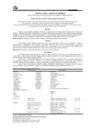
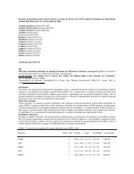
Transvag<strong>in</strong>al follicular aspiration (1) was performed <strong>in</strong> all follicles ≥10 mm. Echographic images of each follicle <strong>and</strong><br />
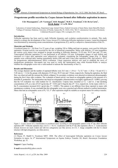
structures at 4 days after aspiration were subjectively evaluated, consider<strong>in</strong>g the echographic image (Fig. 1). Blood<br />
samples were collected daily start<strong>in</strong>g immediately before (D0) <strong>and</strong> until 8 days after aspiration (D8), <strong>and</strong> stored at -20°C<br />
for progesterone radioimmuneassay (RIA) evaluation. L<strong>in</strong>ear regression analysis was used to establish the curve of<br />
progesterone production. Qui-square test was used to verify the lute<strong>in</strong>ization rates while Kruskal-Wallis to analyze<br />
whether the echographic score was a valid method to <strong>in</strong>dicate the functionality of the CL.<br />
Results <strong>and</strong> Discussion<br />
The average diameter <strong>and</strong> the number of aspirated follicles were 26.3 mm ± 1.38 (n = 7); 32.7 mm ± 1.28 (n = 8) <strong>and</strong> 43.2 ±<br />
5.40 mm (n = 11) for the groups with diameters 25-29 mm; 30-35 mm <strong>and</strong> >35mm, respectively. Dur<strong>in</strong>g the aspiration, the fluid<br />
flow was observed until the moment the follicle collapsed. No secondary <strong>ovulation</strong>s were detected on transrectal ultrasonography<br />
dur<strong>in</strong>g the first 8 days after <strong>ovulation</strong>. The evaluation of luteal activity was based on echographic patterns <strong>in</strong> the presence of a<br />
desirable P 4 production. The lute<strong>in</strong>ization rate based on echographic characterization was highly significant (P < 0.0001), prov<strong>in</strong>g<br />
that the US evaluation was valid method for CL classification. Corpora lutea classified as CL 1 <strong>and</strong> CL 2 had decl<strong>in</strong><strong>in</strong>g<br />
concentrations of P 4 at a rate of 0.25 ng/mL/day (P = 0.03) <strong>and</strong> 0.14ng/mL/day (P = 0.2), respectively. In the mares with CL 3,<br />
the P 4 concentration <strong>in</strong>creased (0.61 ng/mL/day; P < 0.0001). This pattern is not different from those reported <strong>in</strong> mares with<br />
spontaneous <strong>ovulation</strong>. It was concluded that the echographic score was a practical <strong>and</strong> efficient method to confirm lute<strong>in</strong>ization.<br />
Mares that had an echographic score of P 4 “CL 3” after aspiration might be suitable as recipient mares for embryo transfer.<br />
Progesterone concentration<br />
(ng/mL)<br />
8<br />
6<br />
4<br />
2<br />
0<br />
CL 1<br />
0 1 2 3 4 5 6 7 8<br />
days after follicular aspiration<br />
Progesterone concentration<br />
(ng/mL)<br />
10<br />
8<br />
6<br />
4<br />
2<br />
0<br />
Anim. Reprod., v.6, n.1, p.229, Jan./Mar. 2009 229<br />
CL 2<br />
0 1 2 3 4 5 6 7 8<br />
days after follicular aspiration<br />
Progesterone concentration<br />
(ng/mL)<br />
6<br />
4<br />
2<br />
0<br />
CL 3<br />
0 1 2 3 4 5 6 7 8<br />
days after follicular aspiration<br />
Figure 1. Corpus luteum (CL) classification <strong>in</strong> crossbreed mares accord<strong>in</strong>g to ultrasonographic pattern at the 4 th day afterr follicular<br />
aspiration (D0 = Follicular aspiration day). (A) CL 1– No CL visualization until the 4 th day after aspiration; (B) CL 2– Luteal structure<br />
formed, however not much evident <strong>and</strong> with low echogenicity (see blue arrows). (C) CL 3– Image compatible with the CL (luteal<br />
structure with high echogenicity, see white arrows).<br />
References<br />
(1) Hayna JT, Madill S, Troedsson MHT. 2004. The effect of transvag<strong>in</strong>al follicular aspiration on Corpus luteum<br />
formation <strong>in</strong> mares. In: 6 th International Symposium on Equ<strong>in</strong>e Embryo Transfer. RJ, Brazil. Havemeyer Foundation<br />
Workshop. pp. 35.<br />
Support: Capes Fund<strong>in</strong>g.<br />
A<br />
A<br />
E-mail:mararub<strong>in</strong>90@yahoo.com.br<br />
B C