Recent advances in ovulation synchronization and superovulation in ...
Recent advances in ovulation synchronization and superovulation in ...
Recent advances in ovulation synchronization and superovulation in ...

You also want an ePaper? Increase the reach of your titles
YUMPU automatically turns print PDFs into web optimized ePapers that Google loves.
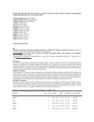
Abstracts. II International Symposium on Animal Biology of Reproduction, Nov. 19-22, 2008, São Paulo, SP, Brazil.<br />
Oct-4 <strong>and</strong> viment<strong>in</strong> expression <strong>in</strong> Agouti paca embryos<br />
A.L.R. Franciolli 1 , C.E. Ambrósio 1 , D.S. Mart<strong>in</strong>s 1 , C.V. Wenceslau 1 , F.R. Alves 1 , R.E.G. Rici 1 ,<br />
M.R.F. Machado 3 , A.C. Mor<strong>in</strong>i 1 , A.F. Carvalho 2 , M.A. Migl<strong>in</strong>o 1<br />
1 Dept. of Surgery, Faculty of Veter<strong>in</strong>ary Medic<strong>in</strong>e, University of São Paulo, São Paulo, Brazil; 2 Dept. of Morphology, UNIfeob,<br />
São João da Boa Vista, São Paulo, Brazil; 3 Paulista State University, Jaboticabal, São Paulo, Brazil.<br />
Introduction<br />
Research <strong>in</strong>volv<strong>in</strong>g stem cells showed that the prote<strong>in</strong> OCT-4 is the most used as a marker of pluripotential mobile<br />
(1) <strong>and</strong> first described <strong>in</strong> embryonic cells of mice (2). The viment<strong>in</strong> is a marker of mesenchyme of mesoderm<br />
embryonic cells <strong>and</strong> also used <strong>in</strong> embryos to colocalized as fibroblasts <strong>in</strong>termediary filament (3). The objective was<br />
to identify pluripotential of embryonic cells of Agouti paca through immunomarked with OCT-4 <strong>and</strong> viment<strong>in</strong>.<br />
Materials <strong>and</strong> Methods<br />
They were used 2 embryos of Agouti paca with 1.8 cm of Crow-rump (CR) <strong>and</strong> another with 1.2 cm of CR, set <strong>in</strong><br />
paraformaldeyde 4%, dehydrated <strong>and</strong> embedded <strong>in</strong> paraff<strong>in</strong>. Sequential slides of 5µm were carried out, followed by<br />
the process<strong>in</strong>g for the immunohistochemistry.<br />
Results <strong>and</strong> Discussion<br />
The Oct-4 as a marker of pluripotential showed up <strong>in</strong> the heart positively to the embryo of 1.2 cm <strong>in</strong> CR <strong>in</strong> the<br />
region of myocardium, as well for the kidney <strong>in</strong> both embryos <strong>in</strong> the tubules region <strong>and</strong>, liver, as <strong>in</strong> a important body<br />
haematopoiesis <strong>in</strong> the hepatocytes cords. These f<strong>in</strong>d<strong>in</strong>gs corroborate to the results of literature, which cites that Oct-<br />
4 expressed <strong>in</strong> cattle is located <strong>in</strong> blastomere, <strong>in</strong> the <strong>in</strong>ternal cell mass of the blastocysts, <strong>in</strong> epiblast <strong>and</strong> embryonic<br />
stem cells (4), as well as l<strong>in</strong>es of cells from human tissues of adult liver, heart <strong>and</strong> bone marrow (5). Also note that<br />
the same was not expressive <strong>in</strong> the lung, <strong>in</strong>test<strong>in</strong>e <strong>and</strong> somites for the two embryos. The viment<strong>in</strong> as a marker of<br />
mesenchyme cells <strong>and</strong> the embryonic mesoderm made up positive <strong>in</strong> both embryos, as <strong>in</strong> the lungs <strong>and</strong> bronchi <strong>in</strong><br />
the region of capillaries that nourish the region of epithelial cells. In the heart, this marker was positive <strong>in</strong> microvessels,<br />
mesenchyme <strong>and</strong> heart. In the <strong>in</strong>test<strong>in</strong>e mesenchyme around the <strong>in</strong>test<strong>in</strong>al tubules <strong>and</strong> blood capillaries<br />
proved to be positive for viment<strong>in</strong>, but also <strong>in</strong> somites, which were found around the cartilage <strong>and</strong> mesenchyme. In<br />
the liver, regions found positive for viment<strong>in</strong> were mesenchyme cells <strong>and</strong> the endothelium of s<strong>in</strong>usoids, while <strong>in</strong> the<br />
kidney, positive <strong>in</strong> mesenchyme <strong>and</strong> was seen <strong>in</strong> the region around the kidney tubules <strong>and</strong> the capillaries. As <strong>in</strong> our<br />
results, the term positive for viment<strong>in</strong> mesenchyme of embryos <strong>in</strong> pigs (6) <strong>and</strong> human (7) is expressed <strong>in</strong> cells of<br />
epiblast <strong>and</strong> hypoblast, which brought the mesoderm <strong>and</strong> endoderm, respectively.<br />
References<br />
(1) Vejsted M, Offenberg H, Maddox-Hyttel P. 2006. Reprod Fertil Dev, 18:1-2; (2) Okamoto K, et al. 1990. Cell,<br />
60:461-472; (3) Franke WW, et al. 1978. Proc Nat Acad Sci USA, 75:5034-5038; (4) Kurosaka S, Eckardt S,<br />
McLaughl<strong>in</strong> JK. 2004. Biol Reprod, 71:1578-1582; (5) Beltrami AP, et al. 2007. J Am Soc Hematol, 110:3438-<br />
3446; (6) Prelle K, Holtz W, Osborn M. 2001. Anat Histol Embryol, 30:339-344; (7) Moore KL, Persaud TVN.<br />
2004. Embriologia clínica. 7. ed. São Paulo: Elsevier Ltda. 609 pp. [<strong>in</strong> Portuguese].<br />
F<strong>in</strong>ancial support: FAPESP<br />
E-mail: <strong>and</strong>refranciolli@yahoo.com.br<br />
Anim. Reprod., v.6, n.1, p.243, Jan./Mar. 2009 243