dimensional numerical simulation of blood flow in mouse ... - CFD
dimensional numerical simulation of blood flow in mouse ... - CFD
dimensional numerical simulation of blood flow in mouse ... - CFD

Create successful ePaper yourself
Turn your PDF publications into a flip-book with our unique Google optimized e-Paper software.
N<strong>in</strong>th International Conference on <strong>CFD</strong> <strong>in</strong> the M<strong>in</strong>erals and Process Industries<br />
CSIRO, Melbourne, Australia<br />
10-12 December 2012<br />
THREE- DIMENSIONAL NUMERICAL SIMULATION OF BLOOD FLOW<br />
IN MOUSE AORTIC ARCH AROUND ATHEROSCLEROTIC PLAQUES<br />
Paul<strong>in</strong>e ASSEMAT 1* , Jillian HOUGH 1 , Karen K. SIU 2,3 , James A. ARMITAGE 4 , Karla G. CONTRERAS 1 ,<br />
Andrea APRICO 5 , Karen ANDREWS 5 , Anthony DART 5 ,Jaye CHIN-DUSTING 5 and Kerry HOURIGAN 1<br />
1 Department <strong>of</strong> Mechanical and Aerospace Eng<strong>in</strong>eer<strong>in</strong>g & Division <strong>of</strong> Biological Eng<strong>in</strong>eer<strong>in</strong>g, Monash University,<br />
Victoria 3800, AUSTRALIA<br />
ABSTRACT<br />
2 Monash Biomedical Imag<strong>in</strong>g, Monash University, Victoria 3800, AUSTRALIA<br />
3 Australian Synchrotron, 800 Blackburn Rd, Clayton, Victoria 3168, AUSTRALIA<br />
4 Department <strong>of</strong> Anatomy and Developmental Biology, Monash University, Victoria 3800, AUSTRALIA<br />
5 Baker IDI Heart and Diabetes Institute, 75 Commercial Rd, Melbourne, Victoria 3004, AUSTRALIA<br />
*Correspond<strong>in</strong>g author, E-mail address: Paul<strong>in</strong>e.assemat@monash.edu<br />
Atherosclerosis occurs due to the build-up and <strong>in</strong>filtration<br />
<strong>of</strong> lipid streaks <strong>in</strong> artery walls lead<strong>in</strong>g to plaques.<br />
Understand<strong>in</strong>g the development <strong>of</strong> atherosclerosis and<br />
plaque vulnerability is <strong>of</strong> critical importance s<strong>in</strong>ce plaque<br />
rupture can result <strong>in</strong> heart attack or stroke. Plaques can be<br />
divided <strong>in</strong>to two dist<strong>in</strong>ct types: those that rupture<br />
(vulnerable) and those that are less likely to rupture<br />
(stable). In the last decade, researchers have been<br />
<strong>in</strong>terested <strong>in</strong> study<strong>in</strong>g the <strong>in</strong>fluence <strong>of</strong> the mechanical<br />
effects (<strong>blood</strong> shear stress, pressure forces and structural<br />
stress) on the plaque formation and rupture processes.<br />
In the literature, physiological experimental studies are<br />
limited by the complexity <strong>of</strong> <strong>in</strong> vivo experiments to study<br />
such effects whereas the <strong>numerical</strong> approach <strong>of</strong>ten uses<br />
simplified models compared with realistic conditions so<br />
that no general agreement <strong>of</strong> the mechanisms responsible<br />
for plaque formation has yet been found. The purpose <strong>of</strong><br />
the present work is to <strong>in</strong>clude more realistic conditions for<br />
the calculations <strong>of</strong> the mechanical effects by implement<strong>in</strong>g<br />
real geometries <strong>in</strong> our <strong>numerical</strong> model. To this aim,<br />
computational fluid dynamics s<strong>of</strong>tware is used to simulate<br />
<strong>blood</strong> velocity and pressure fields <strong>in</strong> mice aortic arch.<br />
NOMENCLATURE<br />
Re Reynolds number<br />
WSS wall shear stress<br />
WSSG wall shear stress gradient<br />
OSI oscillatory shear <strong>in</strong>dex<br />
Greek Symbols<br />
ρ density<br />
μ dynamic viscosity<br />
µ-CT micro-computed tomography<br />
INTRODUCTION<br />
Atherosclerosis is an <strong>in</strong>flammatory disease characterized<br />
by lipid accumulation underneath the endothelium at the<br />
boundary <strong>of</strong> <strong>blood</strong> vessel walls. In addition to lipid<br />
deposition, atherosclerotic progression <strong>in</strong>volves a complex<br />
process <strong>of</strong> monocyte <strong>in</strong>filtration, lipid oxidation, foam cell<br />
formation, smooth muscle cell migration and extracellular<br />
Copyright © 2012 CSIRO Australia 1<br />
matrix production (Chatzizisis et al., 2007, Pello et al.,<br />
2011). The lipid core is separated from the circulat<strong>in</strong>g<br />
<strong>blood</strong> by a fibrous cap composed <strong>of</strong> smooth muscle cells<br />
and extracellular matrix (Chatzizisis et al., 2007). As<br />
plaques develop they can cause lum<strong>in</strong>al narrow<strong>in</strong>g<br />
(reduction <strong>of</strong> the volume <strong>of</strong> the fluid part), or may undergo<br />
expansive remodell<strong>in</strong>g to ma<strong>in</strong>ta<strong>in</strong> lumen diameter (Stone<br />
et al., 2007). Vulnerable plaques are prone to rupture, with<br />
disruption <strong>of</strong> the fibrous cap expos<strong>in</strong>g the thrombogenic<br />
plaque core to the circulat<strong>in</strong>g <strong>blood</strong> (Re<strong>in</strong><strong>in</strong>ger et al.,<br />
2010). Interactions between platelets and the lipid core<br />
can <strong>in</strong>duce thrombus formation on the plaque surface, with<br />
possible consequences <strong>in</strong>clud<strong>in</strong>g vessel occlusion,<br />
myocardial <strong>in</strong>farction or stroke. While it is accepted that<br />
plaque vulnerability is <strong>in</strong>fluenced by fibrous cap thickness<br />
and the size <strong>of</strong> the lipid core (Avger<strong>in</strong>os et al., 2011), it<br />
also depends on lum<strong>in</strong>al remodell<strong>in</strong>g and hemodynamic<br />
parameters for which no general agreement has been<br />
found.<br />
Atherosclerotic plaques are found at particular sites <strong>in</strong> the<br />
arterial tree, with plaques commonly found on the <strong>in</strong>ner<br />
curvatures <strong>of</strong> arteries and near bifurcations. This regional<br />
localization <strong>of</strong> atherosclerosis depends largely on<br />
hemodynamic factors such as wall shear stress WSS<br />
(Chatzizisis et al., 2007). The distribution <strong>of</strong><br />
hemodynamic field depends on the geometry patterns <strong>of</strong><br />
the arteries, with low WSS (uni-directional WSS with a<br />
low time-average) commonly observed on <strong>in</strong>ner curvatures<br />
<strong>of</strong> arteries and oscillatory WSS (bi-directional WSS with a<br />
low time-average) <strong>in</strong> regions <strong>of</strong> bifurcations (Cheng et al.,<br />
2006). This low WSS hypothesis for plaque formation and<br />
progression has been proposed by numerous groups<br />
(Gibson et al., 1993, Stone et al., 2007) and has been<br />
related to the alteration <strong>of</strong> cholesterol transport (Caro et<br />
al., 1971), up-regulation <strong>of</strong> the adhesion <strong>of</strong> molecules that<br />
attract leukocytes and <strong>of</strong> growth factors that promote<br />
smooth muscle cells to proliferate and migrate (Chatzizisis<br />
et al., 2007) or to the permeability <strong>of</strong> the endothelial layer<br />
(Stangeby et al., 2002). Other hemodynamic factors are<br />
also thought to play a role <strong>in</strong> plaque development<br />
<strong>in</strong>clud<strong>in</strong>g the oscillatory shear <strong>in</strong>dex (OSI), which<br />
quantifies the temporal variation <strong>in</strong> WSS, and the wall<br />
shear stress gradient (WSSG) correspond<strong>in</strong>g to spatial
WSS variation. Us<strong>in</strong>g a laser Doppler velocimetry method,<br />
Huo et al. (2008) showed that while the plaques grow <strong>in</strong><br />
regions <strong>of</strong> low WSS, atherosclerosis development is<br />
enhanced by oscillations <strong>in</strong> WSS. In an <strong>in</strong> vivo study <strong>in</strong><br />
<strong>mouse</strong> carotid arteries, stable plaques were shown to<br />
develop <strong>in</strong> regions <strong>of</strong> oscillatory WSS and vulnerable<br />
plaques <strong>in</strong> regions <strong>of</strong> low WSS (Cheng et al., 2006).<br />
In addition, DePaola et al. (1992) showed that large<br />
WSSG can <strong>in</strong>duce morphological and functional changes<br />
<strong>in</strong> the endothelium near <strong>flow</strong> reattachment sites. In<br />
addition to these parameters <strong>in</strong>dicat<strong>in</strong>g the <strong>in</strong>fluence <strong>of</strong> the<br />
viscous forces, pressure forces seem also to affect plaque<br />
formation. In their recent paper, Michell et al. (2011)<br />
showed that an <strong>in</strong>crease <strong>in</strong> the vessel <strong>in</strong>ternal pressure<br />
<strong>in</strong>creases the leukocyte adhesion to the endothelial layer<br />
and then enhances one <strong>of</strong> the biological processes<br />
responsible for plaque formation.<br />
Thus, while it is largely agreed that atherosclerotic<br />
development occurs <strong>in</strong> regions <strong>of</strong> disturbed <strong>flow</strong>, the exact<br />
contributions <strong>of</strong> various hemodynamic parameters such as<br />
WSS, OSI, WSSG and pressure are still under debate.<br />
The present paper will focus on plaque formation by<br />
<strong>in</strong>vestigat<strong>in</strong>g the hemodynamics <strong>of</strong> <strong>blood</strong> <strong>flow</strong> through the<br />
<strong>mouse</strong> aortic arch with and without plaques. The aims <strong>of</strong><br />
the present paper are: to validate a new <strong>numerical</strong><br />
approach which not only allows us to take <strong>in</strong>to account the<br />
plaques but which will also be directly adaptable as fluid<br />
structure <strong>in</strong>teraction code; and to provide some <strong>in</strong>sights<br />
<strong>in</strong>to the relation between hemodynamic parameters and<br />
plaque development. Three <strong>mouse</strong> models are be<strong>in</strong>g<br />
studied: results from wild type C57/B6 mice (no plaque)<br />
and ApoE deficient mice (stable plaques) will be presented<br />
here. Experiments on a third model, the SM22α-hDTR<br />
ApoE deficient mice (vulnerable plaques), are currently<br />
under study and will not be presented <strong>in</strong> this paper. After<br />
the tissues have been fixed, the samples are imaged by<br />
micro-computed tomography us<strong>in</strong>g synchrotron X-ray<br />
beams; the result<strong>in</strong>g images are reconstructed to get the<br />
3D geometries used for the <strong>numerical</strong> <strong>simulation</strong>s. The<br />
orig<strong>in</strong>ality <strong>of</strong> our approach consists <strong>in</strong> remov<strong>in</strong>g<br />
<strong>numerical</strong>ly the plaques to study the plaque formation and<br />
development processes on effectively the same aorta. The<br />
precise density contrast obta<strong>in</strong>ed by Synchrotron imag<strong>in</strong>g<br />
enables us to differentiate the plaque from the artery wall.<br />
A high def<strong>in</strong>ition is clearly observed for each tissue which<br />
can be reconstructed separately us<strong>in</strong>g segmentation tools.<br />
In addition, tak<strong>in</strong>g <strong>in</strong>to account the complete real<br />
geometry will allow us to study the l<strong>in</strong>k between the<br />
topography <strong>of</strong> the plaques and their rupture sites. This<br />
work is the first step towards a more complex undertak<strong>in</strong>g<br />
where it is planned that fluid structure <strong>in</strong>teractions will be<br />
taken <strong>in</strong>to account. After the description <strong>of</strong> the<br />
experimental and <strong>numerical</strong> approach, validation and<br />
results will be presented.<br />
MODEL DESCRIPTION<br />
Animal preparation<br />
Animal models provide a key <strong>in</strong>sight to understand<br />
atherosclerosis formation. In this study, the<br />
Apolipoprote<strong>in</strong> E knockout (ApoE -/-) <strong>mouse</strong> model fed a<br />
high fat diet (21% fat; 0.15% cholesterol) is used as a<br />
model for spontaneous atherosclerotic development (Bond<br />
Copyright © 2012 CSIRO Australia 2<br />
and Jackson, 2011). In the near future, the SM22α-hDTR<br />
ApoE-/- will be used as a model which develops<br />
vulnerable plaques. Normal mice do not develop<br />
atherosclerosis and are used as controls <strong>in</strong> this<br />
<strong>in</strong>vestigation. Mice were given a term<strong>in</strong>al dose <strong>of</strong><br />
anaesthetic and then transcardially perfused with<br />
hepar<strong>in</strong>ised sal<strong>in</strong>e to clear <strong>blood</strong> from the vasculature.<br />
Tissue was then perfusion fixed with Karnovsky’s fluid<br />
(2% glutaraldehyde + 4% paraformaldehyde <strong>in</strong> 0.1M<br />
phosphate buffer, pH 7.4). This procedure preserves vessel<br />
morphology <strong>in</strong> the native orientation. Tissues were then<br />
dissected, dehydrated through graded butanol and<br />
embedded <strong>in</strong> paraff<strong>in</strong>.<br />
Imag<strong>in</strong>g<br />
Imag<strong>in</strong>g <strong>of</strong> the <strong>mouse</strong> aortas has been obta<strong>in</strong>ed by microcomputed<br />
tomography (µ-CT) us<strong>in</strong>g synchrotron X-ray<br />
beams (PSI, Swiss Light Synchrotron, Switzerland) and<br />
then reconstructed to derive the 3D geometries used for<br />
the <strong>numerical</strong> <strong>simulation</strong>s (figure 1). The µ-CT data are<br />
acquired us<strong>in</strong>g monochromatic X-rays <strong>of</strong> 15keV, and a<br />
CCD detector with X-ray converter and <strong>in</strong>terchangeable<br />
objective lenses to achieve pixel sizes <strong>in</strong> the range 2-6 µm<br />
(as dictated by the sample size).The samples are imaged<br />
us<strong>in</strong>g a propagation distance (~2-10cm) to enhance the<br />
contrast <strong>of</strong> the lipid core/fibrous cap. The reconstruction<br />
employs the Pagan<strong>in</strong> algorithm (Pagan<strong>in</strong> et al., 2002)<br />
us<strong>in</strong>g X-TRACT s<strong>of</strong>tware developed at the CSIRO<br />
(Gureyev et al., 2011 http://www.tsimag<strong>in</strong>g.net/Services/AppInfo/X-TRACT.aspx).<br />
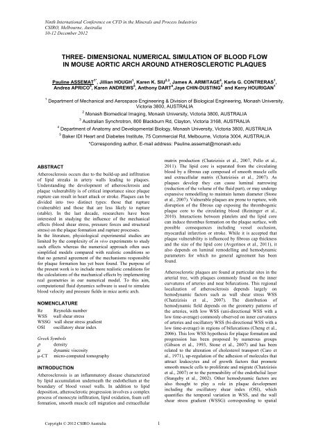
Figure 1: Reconstructed <strong>mouse</strong> aortic arch.<br />
Numerical methods<br />
Geometry<br />
The s<strong>of</strong>tware X-TRACT provides two <strong>dimensional</strong> section<br />
images <strong>of</strong> the geometry which are then reconstructed <strong>in</strong><br />
the 3D segmentation s<strong>of</strong>tware AVIZO. The high spatial<br />
resolution <strong>of</strong> the images obta<strong>in</strong>ed by µ-CT and the f<strong>in</strong>e<br />
density contrast allowed us to differentiate the plaques<br />
from the artery walls. An example <strong>of</strong> an obta<strong>in</strong>ed geometry<br />
can be seen <strong>in</strong> figure 2. To verify that the plaques were<br />
well def<strong>in</strong>ed <strong>in</strong> AVIZO, histochemical analyses were<br />
performed withMasson's Trichrome. Figures 3a and 3b
show, respectively, a section <strong>of</strong> reconstructed µ-CT image<br />
and the associated histology data. On figure 3b, the blue<br />
colour corresponds to collagen, the p<strong>in</strong>k colour<br />
corresponds to the cytoplasm that characterises the<br />
presence <strong>of</strong> cells. On the right, foam cells which are<br />
characteristic <strong>of</strong> the early stage <strong>of</strong> plaque formation are<br />
visible whereas on the left, the collagen present <strong>in</strong> the<br />
fibrous cap is observed.<br />
Figure 2: Half aortic arch, coloured blue corresponds to<br />
the artery wall, and atherosclerotic plaque coloured green.<br />
The black l<strong>in</strong>e corresponds to the section plane<br />
represented on figure 3.<br />
(a)<br />
(b)<br />
Figure 3: Sections <strong>of</strong> the three-<strong>dimensional</strong> geometry<br />
located by the l<strong>in</strong>e figure 2. (a) Image obta<strong>in</strong>ed by µ-CT,<br />
the blue colour corresponds to the artery wall, the green to<br />
the plaque. (b) Histological analyse <strong>of</strong> the aortic arch.<br />
Sections were cut at 5 µm through the entire aorta and<br />
sta<strong>in</strong>ed with Masson's Trichrome <strong>in</strong> order to visualise the<br />
vascular structures and plaque.<br />
Copyright © 2012 CSIRO Australia 3<br />
Mesh<br />
The geometry is imported <strong>in</strong> ICEM <strong>CFD</strong> to generate the<br />
mesh. In the follow<strong>in</strong>g, only the fluid compartment<br />
(lumen) will be considered. The ends <strong>of</strong> the vessels are<br />
extended to prevent boundary condition <strong>in</strong>teractions <strong>in</strong> the<br />
aortic arch zone. Two methods have been tested:<br />
tetrahedral and hexahedral mesh<strong>in</strong>g. The first type <strong>of</strong><br />
element is easier to implement but requires a lot <strong>of</strong><br />
memory for the calculations. Block<strong>in</strong>g and O-grid<br />
methods are used to generate the mesh with hexahedral<br />
elements. These methods allow us to control with<br />
precision the mesh density and topology, <strong>in</strong> particular, the<br />
size and number <strong>of</strong> the boundary layers along the<br />
geometry can be controlled. In addition, this approach has<br />
been shown to tolerate large deformation <strong>in</strong> a straight<br />
vessel model <strong>in</strong> a study under progress. This not the case<br />
for the tetrahedral approach. An example <strong>of</strong> a hexahedral<br />
mesh is given <strong>in</strong> figure 4. A comparison between these<br />
two approaches will be presented <strong>in</strong> the results section,<br />
however the implementation <strong>of</strong> the hexahedral mesh is<br />
under progress and the rema<strong>in</strong><strong>in</strong>g results are obta<strong>in</strong>ed by<br />
tetrahedral methods.<br />
Figure 4: Example <strong>of</strong> hexahedral mesh designed for a<br />
<strong>mouse</strong> aortic arch without plaque. The p<strong>in</strong>k colour<br />
corresponds to the geometry obta<strong>in</strong>ed by µ-CT whereas<br />
the blue corresponds to extensions.<br />
Numerical model<br />
The Navier-Stokes equations are solved us<strong>in</strong>g the element<br />
based f<strong>in</strong>ite volume methods s<strong>of</strong>tware ANSYS CFX. A<br />
pseudo time-stepp<strong>in</strong>g technique method is used to<br />
calculate the steady <strong>flow</strong>. The <strong>blood</strong> is considered<br />
Newtonian as the shear rates calculated are greater than<br />
150s -1 , a condition that makes the Newtonian<br />
approximation relatively accurate (Wells et al., 1961). The<br />
density ρ and the dynamic viscosity µ are, respectively,<br />
ρ=1060kg/m 3 and µ= 0.0035Pa.s (Trachet et al., 2009).<br />
Mass <strong>flow</strong> rate boundary conditions have been applied at
the <strong>in</strong>let and the outlet. No-slip boundary conditions have<br />
been applied along the rigid walls.<br />
RESULTS<br />
The <strong>flow</strong> rates used as boundary conditions were obta<strong>in</strong>ed<br />
from Huo et al. (2008) <strong>in</strong> which the <strong>flow</strong> is measured<br />
experimentally <strong>in</strong> the same wild type mice used for the<br />
present study. In the follow<strong>in</strong>g, steady <strong>flow</strong> is considered<br />
to validate our approach. Figure 5 gives the nomenclature<br />
for each artery branch, table 1 <strong>in</strong>dicates the <strong>flow</strong> rates, the<br />
diameters and Reynolds from Huo et al. (2008) as well as<br />
the measured diameters and calculated Reynolds numbers<br />
for the present study.<br />
AA<br />
BCA<br />
LCC<br />
Figure 5: Lumen distribution <strong>of</strong> a <strong>mouse</strong> aortic arch with<br />
plaques. The <strong>in</strong>let and outlets are labelled accord<strong>in</strong>g to the<br />
notations used <strong>in</strong> table 1.<br />
The Reynolds number is calculated from the equivalent<br />
diameter obta<strong>in</strong>ed from the measurement <strong>of</strong> the surface<br />
area. In table 1, it is observed that the diameters are<br />
smaller than the ones measured by Huo et al. (2008), a<br />
difference which probably comes from the difference <strong>in</strong><br />
the tissue fix<strong>in</strong>g method but also due to the different<br />
perfusion pressure. In current experiments, agarose is used<br />
as an embedd<strong>in</strong>g medium to prevent shr<strong>in</strong>kage effects<br />
caused by paraff<strong>in</strong>.<br />
As mentioned previously, the first step <strong>of</strong> the validation is<br />
to show <strong>in</strong>variance <strong>of</strong> computed parameter magnitudes<br />
which are derived from different types <strong>of</strong> meshes. Figure 5<br />
shows the iso-contours <strong>of</strong> the WSS for tetrahedral (Figure<br />
6a) and hexahedral (Figure 6b) meshes <strong>in</strong> rescaled<br />
geometries. This scal<strong>in</strong>g implies that the equivalent<br />
diameter <strong>of</strong> the ascend<strong>in</strong>g aorta is equal to the one<br />
measured <strong>in</strong> Huo et al. (2008). The maximum <strong>of</strong> WSS <strong>in</strong><br />
both configurations are respectively 18.38 and 18.85 Pa<br />
for the two different meshes. The difference between these<br />
two values is ma<strong>in</strong>ly due to the weak control <strong>of</strong> the<br />
boundary layers <strong>in</strong> the tetrahedral mesh for which the<br />
number <strong>of</strong> elements is 2 times bigger than for the<br />
hexahedral. A mesh study has been done on meshes to<br />
optimize the <strong>numerical</strong> parameters but will not be<br />
presented here. The performance <strong>of</strong> our hexahedral<br />
approach for a complex real geometry is shown here.<br />
Copyright © 2012 CSIRO Australia 4<br />
LS<br />
DA<br />
Experimental<br />
Re<br />
Flow Rate, Q<br />
(mL/m<strong>in</strong>) Huo<br />
et al. (2008)<br />
Diameter, D<br />
(mm) Huo et<br />
al. (2008)<br />
Re, Huo et al.<br />
(2008)<br />
Experimental<br />
Diameter (mm)<br />
Experimental<br />
Re<br />
Scaled<br />
Diameters(mm)<br />
93 AA BCA 22 LCC 26 17 LS DA 60<br />
12.02 1.87 1.35 1.06 7.74<br />
1.3 0.85 0.64 0.64 -<br />
61 14 14 11 -<br />
0.84 0.56 0.33 0.38 0.82<br />
93 22 26 17 60<br />
1.3 0.68 0.31 0.51 1.08<br />
Table 1: Comparison <strong>of</strong> diameter and Reynold’s number<br />
from experimental f<strong>in</strong>d<strong>in</strong>gs and the values stated <strong>in</strong> Huo et<br />
al. (2008). The experimental dimensions <strong>of</strong> the aorta and<br />
its ma<strong>in</strong> branches were measured from the reconstructed<br />
three-<strong>dimensional</strong> geometry. The <strong>flow</strong> rates used as<br />
boundary conditions were obta<strong>in</strong>ed from Huo et al (2008).<br />
For the fifth row, Re=ρQD/(Aµ) where Q is the <strong>flow</strong> rate,<br />
A is the surface area and D is the equivalent diameter. The<br />
discrepancy between the dimensions comes from the<br />
shr<strong>in</strong>kage caused by embedd<strong>in</strong>g <strong>in</strong> paraff<strong>in</strong>. The last row<br />
corresponds to scaled diameters with Huo et al. (2008).<br />
Figure 6: Iso-contours <strong>of</strong> WSS, (a) tetrahedral mesh, (b)<br />
hexahedral mesh.
Consistent with the literature (Huo et al., 2008, Trachet et<br />
al., 2009), we f<strong>in</strong>d that the local m<strong>in</strong>imum <strong>of</strong> wall shear<br />
stress occurs before the bifurcation (figure 7a) and <strong>in</strong> the<br />
<strong>in</strong>ner curvature <strong>of</strong> the aortic arch (figure 8a). Qualitatively<br />
the iso-contours <strong>of</strong> WSS have a similar distribution except<br />
for the second bifurcation where our geometry presents<br />
specificities that make the WSS locally high (small<br />
equivalent diameter <strong>of</strong> this branch). In addition,<br />
differences arise from the fact that <strong>in</strong> both articles, the<br />
authors use skeletonisation methods which smooth the<br />
geometry and give axisymmetric configurations that do<br />
not take <strong>in</strong>to account stenotic plaques. To f<strong>in</strong>ish, both<br />
cited papers report results for time-averaged WSS. A<br />
parallel study (not presented here), tak<strong>in</strong>g <strong>in</strong>to account<br />
realistic pulsatile <strong>flow</strong> conditions, showed that the<br />
distribution <strong>of</strong> the WSS on the systole peak is similar to<br />
the time averaged WSS itself similar to the results<br />
presented <strong>in</strong> figures 6-8. A quantitative comparison with<br />
the previous reference studies will be communicated <strong>in</strong> the<br />
future.<br />
Figure 7: (a) Isocontours <strong>of</strong> WSS <strong>in</strong> a geometry without<br />
plaque, zoom on the bifurcations, (b) zoom <strong>of</strong> figure 2.<br />
Figures 7a and 7b, 8a and 8b illustrate that plaques<br />
develop around the local m<strong>in</strong>ima <strong>of</strong> wall shear stress and<br />
their expansion seems to be directed through the zone<br />
where the wall shear stress <strong>in</strong>creases or decreases the least<br />
(small gradient <strong>of</strong> WSS). This observation <strong>in</strong>dicates that<br />
the amplitude <strong>of</strong> WSS is not only important for the<br />
formation process but also for the plaque development and<br />
lumen remodell<strong>in</strong>g. In addition, it suggests that the<br />
assumption <strong>of</strong> plaques development downstream (Olgac et<br />
al. 2009) may be limited, more specifically <strong>in</strong> complex<br />
geometries such as aortic arches. Figure 9 shows the WSS<br />
<strong>in</strong> a geometry where plaques have been considered. A<br />
local <strong>in</strong>crease <strong>of</strong> WSS is observed (<strong>in</strong>dicated by arrows)<br />
on all plaques due to the constrictive remodell<strong>in</strong>g <strong>of</strong> the<br />
vessel. A number <strong>of</strong> studies (Groen et al. 2008, Tang et al.<br />
2009), report a l<strong>in</strong>k between high WSS and plaque<br />
rupture, however structural stresses are thought to play a<br />
more important role (Sadat et al., 2010) <strong>in</strong> this process.<br />
Further <strong>in</strong>vestigations with the implementation <strong>of</strong> fluid-<br />
Copyright © 2012 CSIRO Australia 5<br />
structure <strong>in</strong>teraction is required to resolve this question<br />
under debate.<br />
Figure 8: (a) Isocontours <strong>of</strong> WSS <strong>in</strong> a geometry without<br />
plaque, zoom on the <strong>in</strong>ner curvature, (b) zoom <strong>of</strong> figure 2.<br />
Figure 9: Isocontours <strong>of</strong> wall shear stress around plaques.
CONCLUSION<br />
This study is a first step <strong>in</strong> understand<strong>in</strong>g the effect <strong>of</strong><br />
hemodynamic parameters on plaque formation and<br />
development under realistic conditions.<br />
In this article, we have successfully applied an approach<br />
that permits the calculation <strong>of</strong> the <strong>blood</strong> <strong>flow</strong> <strong>in</strong> complex<br />
real artery geometries <strong>in</strong> the presence <strong>of</strong> plaques. The<br />
method <strong>of</strong> remov<strong>in</strong>g the plaque <strong>numerical</strong>ly to study the<br />
formation and the development <strong>of</strong> the plaque <strong>in</strong> the same<br />
artery predicts correctly the formation sites around local<br />
m<strong>in</strong>imum wall shear stress. In addition, we report that<br />
plaque development appears to be <strong>in</strong> the direction <strong>of</strong> the<br />
smallest gradient <strong>of</strong> wall shear stress, not only downstream<br />
the formation sites as suggested <strong>in</strong> the literature.<br />
ACKNOWLEDGEMENTS<br />
This work was supported by the Multi-modal Australian<br />
ScienceS Imag<strong>in</strong>g and Visualisation Environment<br />
(MASSIVE) (www.massive.org.au). The authors<br />
acknowledge f<strong>in</strong>ancial support from the Australian<br />
Research Council, under grant no. DP110100434.<br />
REFERENCES<br />
AVGERINOS, E.D., GIANNAKOPOULOS, T.G.,<br />
KADOGLOU, N.P., LIAPIS, C.D., MOULAKAKIS, K.G.<br />
and PAPAPETROU, A., (2011), “Biomarkers for<br />
diagnosis <strong>of</strong> the vulnerable atherosclerotic plaque.”,<br />
Interventional Cardiology, 3(2),223.<br />
BOND, A.R and C. L. JACKSON, (2011), “The fat-fed<br />
Apolipoprote<strong>in</strong> E Knockout <strong>mouse</strong> brachiocephalic artery<br />
<strong>in</strong> the study <strong>of</strong> atherosclerotic plaque rupture.”, J. Biomed.<br />
Biotechnol, 2011, 379069.<br />
CARO, C.G., FITZ-GERALD, J.M. and SCHROTER,<br />
R.C., (1971), “Atheroma and Arterial wall shear.<br />
Observation, correlation and proposal <strong>of</strong> a shear<br />
dependent mass transfer mechanism for atherogenesis.”, P.<br />
R. Soc. London, 177(1046), 109-133.<br />
CHATZIZISIS, Y.S., COSKUN, A.U., JONAS, M.,<br />
EDELMAN, E.R., FELDMAN, C.L. and STONE, P.H.,<br />
(2007), “Role <strong>of</strong> endothelial shear stress <strong>in</strong> the natural<br />
history <strong>of</strong> coronary atherosclerosis and vascular<br />
remodell<strong>in</strong>g: molecular, cellular, and vascular behaviour.”<br />
J. Am. Coll. Cardiol., 49(25), 2379-2393.<br />
CHENG, C., TEMPEL, D., VAN HAPEREN, R., VAN<br />
DER BANN, A., GROSVELD, F., DAEMEN M.J.,<br />
KRAMS, R. and DE CROM R., (2006), “Atherosclerotic<br />
lesion size and vulnerability are determ<strong>in</strong>ed by patterns <strong>of</strong><br />
fluid shear stress.”, J. Am. Heart Assoc., 113, 2744-2753.<br />
DEPAOLA, N., GIMBORNE, MA., DAVIS, J.F., and<br />
DEWEY, F., (1991), “Vascular endothelium responds to<br />
fluid shear stress gradients”. Atherosclerosis and<br />
Thrombosis, 12, 1254-1257.<br />
GIBSON, C.M., DIAZ, L., KANDARPA, K., SACKS,<br />
F.M., PASTERNAK, R.C., SANDOR, T., FELDMAN, C.<br />
and STONE, P.H., (1993), “Relation <strong>of</strong> vessel wall shear<br />
stress to atherosclerosis progression <strong>in</strong> human coronary<br />
arteries.”, J. Am. Heart Assoc., 13, 210-315.<br />
GROEN, H.C., GIJSEN, F.J.H., VAN DER LUGT, A.,<br />
HATSUKAMI, T.S., VAN DER STEEN, A.F.W., YUAN<br />
, C. and WENTZEL, J.J., (2007), “ Plaque Rupture <strong>in</strong> the<br />
Carotid Artery Is Localized at the High Shear Stress<br />
Region.”, Stroke, 38: 2379-2381.<br />
GUREYEV, T.E, NESTERETS, Y., TERNOVSKI, D.,<br />
THOMPSON, D., WILKINS, S. W., STEVENSON, A.<br />
Copyright © 2012 CSIRO Australia 6<br />
W., SAKELLARIOU, A. and TAYLOR J.A, (2011),<br />
“Toolbox for advanced X-ray image process<strong>in</strong>g,” Proc.<br />
SPIE 8141, 81410B, 81410B-14.<br />
HUO, Y., GUO, X. and KASSAB, G.S., (2008), “The<br />
<strong>flow</strong> field along the entire length <strong>of</strong> the <strong>mouse</strong> aorta and<br />
primary branches.”, Ann. Biomed. Eng., 36(5), 685-699.<br />
MICHELL, D. L., ANDREWS, K. L., WOOLLARD, K.<br />
J. and CHIN-DUSTING, J. P., (2011), “Imag<strong>in</strong>g leukocyte<br />
adhesion to the vascular endothelium at high <strong>in</strong>tralum<strong>in</strong>al<br />
pressure.”, J. Vis. Exp., 54, e3221.<br />
OLGAC, U., POULIKAKOS, D., SAUR, S.C.,<br />
ALKADHI, H. and KURTCUOGLU, V., (2009), “<br />
Patient-specific three-<strong>dimensional</strong> <strong>simulation</strong> <strong>of</strong> LDL<br />
accumulation <strong>in</strong> a human left coronary artery <strong>in</strong> its healthy<br />
and atherosclerotic states.”, Am. J. Physiol. Heart Circ.<br />
Physiol., 296,969-982.<br />
PAGANIN. D.P., MAYO, S.C., GUREYEV, T.E.,<br />
MILLER, P.R. and WILKINS S.W., (2002),<br />
“Simultaneous phase and amplitude extraction from a<br />
s<strong>in</strong>gle defocused image <strong>of</strong> a homogeneous object.”, J.<br />
Microscopy, 206(1), 33-40.<br />
PELLO, O.M., SILVESTRE, C., PIZZOL, M.D. and<br />
ANDRES, V., (2011), “A glimpse on the phenomenon <strong>of</strong><br />
macrophage polarization dur<strong>in</strong>g atherosclerosis.”,<br />
Immunobiology, 216,1172-1176.<br />
REININGER, A.J., BERNLOCHNER, I., PENZ, S.M.,<br />
RAVANAT, C., SMETHURST, P., FARNDALE R, W.,<br />
GACHET, C., BRANDL, R. and SIESS, W., (2010), “A<br />
2-Step Mechanism <strong>of</strong> Arterial Thrombus Formation<br />
Induced by Human Atherosclerotic Plaques.”, J. Am. Coll.<br />
Cardiol., 55(11), 1147-1158.<br />
SADAT , U., TENG. Z. and GILLARD, J.H., (2010),<br />
“Biomechanical structural stresses <strong>of</strong> atherosclerotic<br />
plaques.”, Expert Rev. <strong>of</strong> Cardiovasc. Therapy, 8(10):<br />
1469-1481.<br />
STANGEBY, D.K. and ETHIER, C.R., (2002),<br />
“Computational analysis <strong>of</strong> coupled <strong>blood</strong>-wall arterial<br />
LDL transport.”, T. ASME, 124, 1-8.<br />
STONE, P.H., COSKUN, A.U., KINLAY, S., POPMA,<br />
J.J., SONKA, M., WAHLE, A., YEGHIAZARIANS, Y.,<br />
MAYNARD, C., KUNTZ, R.E. and FELDMAN, C.L.,<br />
(2007), “Regions <strong>of</strong> low endothelial shear stress are the<br />
sites where coronary plaque progresses and vascular<br />
remodell<strong>in</strong>g occurs <strong>in</strong> humans: and <strong>in</strong> vivo serial study.”,<br />
Eur. Heart J., 28,705-710.<br />
TANG, D., TENG, Z., CANTON, G., YANG, C.,<br />
FERGUSON, M., HUANG, X., ZHENG, J., WOODARD,<br />
P.K. and YUAN, C., (2009), “Sites <strong>of</strong> rupture <strong>in</strong> human<br />
atherosclerotic carotid plaques are associated with high<br />
structural stresses: an <strong>in</strong> vivo mri-based 3d fluid-structure<br />
<strong>in</strong>teraction study.”, Stroke, 40: 3258-3263.<br />
TRACHET, B., SWILLENS, A., VAN LOO, D.,<br />
CASTELEYN, C., DE PAEPE, A., LOEYS, B. and<br />
SEGERS P., (2009), “The <strong>in</strong>fluence <strong>of</strong> aortic dimensions<br />
on calculated wall shear stress <strong>in</strong> the <strong>mouse</strong> aortic arch.”,<br />
Comput. Method Biomec., 12, 491-499.<br />
WELLS. R.E., MERRIL, J.W. and MERRILL, E.W.,<br />
(1961), “Shear rate dependence <strong>of</strong> the viscosity <strong>of</strong> whole<br />
<strong>blood</strong> and plasma.”, Science, 133(3455), 763-764.