Phycoerythrins of the oxyphotobacterium Prochlorococcus marinus ...
Phycoerythrins of the oxyphotobacterium Prochlorococcus marinus ...
Phycoerythrins of the oxyphotobacterium Prochlorococcus marinus ...

Create successful ePaper yourself
Turn your PDF publications into a flip-book with our unique Google optimized e-Paper software.
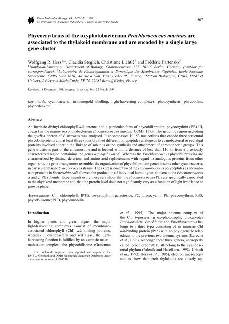
Plant Molecular Biology 40: 507–521, 1999.<br />
© 1999 Kluwer Academic Publishers. Printed in <strong>the</strong> Ne<strong>the</strong>rlands.<br />
<strong>Phycoerythrins</strong> <strong>of</strong> <strong>the</strong> <strong>oxyphotobacterium</strong> <strong>Prochlorococcus</strong> <strong>marinus</strong> are<br />
associated to <strong>the</strong> thylakoid membrane and are encoded by a single large<br />
gene cluster<br />
Wolfgang R. Hess 1,∗ , Claudia Steglich, Christiane Lichtlé 2 and Frédéric Partensky 3<br />
1 Humboldt-University, Department <strong>of</strong> Biology, Chausseestrasse 117, 10115 Berlin, Germany ( ∗ author for<br />
correspondence); 2 Laboratoire de Photorégulation et Dynamique des Membranes Végétales, Ecole Normale<br />
Supérieure, CNRS URA 1810, 46 rue d’Ulm, Paris Cedex 05, France; 3 Station Biologique, CNRS, INSU et<br />
Université Pierre et Marie Curie, BP 74, 29682 Rosc<strong>of</strong>f Cedex, France<br />
Received 18 December 1998; accepted in revised form 22 March 1999<br />
Key words: cyanobacteria, immunogold labelling, light-harvesting complexes, photosyn<strong>the</strong>sis, phycobilins,<br />
phytoplankton<br />
Abstract<br />
An intrinsic divinyl-chlorophyll a/b antenna and a particular form <strong>of</strong> phycobiliprotein, phycoerythrin (PE) III,<br />
coexist in <strong>the</strong> marine <strong>oxyphotobacterium</strong> <strong>Prochlorococcus</strong> <strong>marinus</strong> CCMP 1375. The genomic region including<br />
<strong>the</strong> cpeB/A operon <strong>of</strong> P. <strong>marinus</strong> was analysed. It encompasses 10 153 nucleotides that encode three structural<br />
phycobiliproteins and at least three (possibly five) different polypeptides analogous to cyanobacterial or red algal<br />
proteins involved ei<strong>the</strong>r in <strong>the</strong> linkage <strong>of</strong> subunits or <strong>the</strong> syn<strong>the</strong>sis and attachment <strong>of</strong> chromophoric groups. This<br />
gene cluster is part <strong>of</strong> <strong>the</strong> chromosome and is located within a distance <strong>of</strong> less than 110 kb from a previously<br />
characterized region containing <strong>the</strong> genes aspA-psbA-aroC. Whereas <strong>the</strong> <strong>Prochlorococcus</strong> phycobiliproteins are<br />
characterized by distinct deletions and amino acid replacements with regard to analogous proteins from o<strong>the</strong>r<br />
organisms, <strong>the</strong> gene arrangement resembles <strong>the</strong> organization <strong>of</strong> phycobiliprotein genes in some o<strong>the</strong>r cyanobacteria,<br />
in particular marine Synechococcus strains. The expression <strong>of</strong> two <strong>of</strong> <strong>the</strong> <strong>Prochlorococcus</strong> polypeptides as recombinant<br />
proteins in Escherichia coli allowed <strong>the</strong> production <strong>of</strong> individual homologous antisera to <strong>the</strong> <strong>Prochlorococcus</strong><br />
α and β PE subunits. Experiments using <strong>the</strong>se sera show that <strong>the</strong> <strong>Prochlorococcus</strong> PEs are specifically associated<br />
to <strong>the</strong> thylakoid membrane and that <strong>the</strong> protein level does not significantly vary as a function <strong>of</strong> light irradiance or<br />
growth phase.<br />
Abbreviations: Chl, chlorophyll; IPTG, iso-propyl-thiogalactoside; PC, phycocyanin; PE, phycoerythrin; PBS,<br />
phycobilisome; PUB, phycourobilin<br />
Introduction<br />
In higher plants and green algae, <strong>the</strong> major<br />
light-harvesting complexes consist <strong>of</strong> membraneassociated<br />
chlorophyll (Chl) a/b-binding proteins,<br />
whereas in cyanobacteria and red algae, <strong>the</strong> lightharvesting<br />
function is fulfilled by an extrinsic macromolecular<br />
complex, <strong>the</strong> phycobilisome (Grossman<br />
The nucleotide sequence data reported will appear in <strong>the</strong><br />
EMBL, GenBank and DDBJ Nucleotide Sequence Databases under<br />
<strong>the</strong> accession number AJ001230.<br />
507<br />
et al., 1995). The major antenna complex <strong>of</strong><br />
<strong>the</strong> Chl b-possessing oxyphototrophic prokaryotes<br />
Prochlorothrix, Prochloron and <strong>Prochlorococcus</strong> belongs<br />
to a third type consisting <strong>of</strong> an intrinsic Chl<br />
a/b-binding protein (Pcb) with no phylogenetic relatedness<br />
to <strong>the</strong> previous two antenna systems (Laroche<br />
et al., 1996). Although <strong>the</strong>se three genera, unproperly<br />
called ‘prochlorophytes’, all belong to <strong>the</strong> cyanobacterial<br />
phylum (Palenik and Haselkorn, 1992; Urbach<br />
et al., 1992; Hess et al., 1995), electron microscopy<br />
studies show that <strong>the</strong>ir thylakoids are closely ap-
508<br />
pressed (Bullerjahn and Post, 1993) and, <strong>the</strong>refore,<br />
that <strong>the</strong>y probably do not contain any phycobilisomes<br />
in addition to <strong>the</strong>ir Pcb antenna complexes. Yet, in<br />
<strong>Prochlorococcus</strong> <strong>marinus</strong> CCMP 1375 (or SS120)<br />
genes coding for <strong>the</strong> α and β chains <strong>of</strong> phycoerythrin<br />
(PE) have been identified (Hess et al., 1996).<br />
This organism is <strong>the</strong> type species <strong>of</strong> a genetically<br />
diverse group <strong>of</strong> unicellular oxyphototrophic bacteria<br />
(Chisholm et al., 1992; Urbach et al., 1998),<br />
which contains divinyl-chlorophyll a and b (Chl a2<br />
and b2; Goericke and Repeta, 1992) and which dominates<br />
<strong>the</strong> photosyn<strong>the</strong>tic biomass in most temperate<br />
and intertropical oceanic ecosystems (for reviews, see<br />
Whitman et al., 1998; Partensky et al., 1999a, b).<br />
P. <strong>marinus</strong> cpeA and cpeB genes, encoding PE α and β<br />
subunits, have been shown to be functional, although<br />
weakly transcribed (Hess et al., 1996). Fur<strong>the</strong>rmore,<br />
<strong>the</strong> presence <strong>of</strong> PE in a water-soluble extract <strong>of</strong> P. <strong>marinus</strong><br />
cells was demonstrated by cross-reaction <strong>of</strong> a<br />
21 kDa protein with a heterologous serum to PE. At<br />
last, <strong>the</strong> fluorescence emission spectra <strong>of</strong> this PE were<br />
shown to resemble those <strong>of</strong> marine Synechococcus<br />
(Ong et al., 1984) with dominance <strong>of</strong> <strong>the</strong> 496 nm<br />
peak due to phycourobilin over <strong>the</strong> 550 nm peak <strong>of</strong> <strong>the</strong><br />
phycoerythrobilin (Hess et al., 1996). Since, in P. <strong>marinus</strong><br />
CCMP 1375, <strong>the</strong> photosyn<strong>the</strong>tic antenna system<br />
is made up <strong>of</strong> <strong>the</strong> Chl a2/b2-Pcb complexes (Partensky<br />
et al., 1997), <strong>the</strong> exact function, intracellular localization<br />
and mode <strong>of</strong> regulation <strong>of</strong> <strong>the</strong>se PE molecules<br />
remained obscure. In particular, it is not clear whe<strong>the</strong>r<br />
<strong>the</strong>y fulfil a light-harvesting function, are involved in<br />
a sensor function in light-perception processes as are<br />
phytochromesknown from o<strong>the</strong>r cyanobacteria (Kehol<br />
and Grossman, 1996; Hughes et al., 1997) or are simply<br />
function-less. Their phylogenetic origin is problematic,<br />
too, and one can wonder whe<strong>the</strong>r <strong>the</strong>se genes<br />
represent evolutionary remnants <strong>of</strong> ancestral phycoerythrins<br />
or have been more recently acquired by<br />
horizontal gene transfer from marine Synechococcus,<br />
a genus that <strong>of</strong>ten co-occurs with <strong>Prochlorococcus</strong> in<br />
natural assemblages (Partensky et al., 1999a).<br />
So far, it has nei<strong>the</strong>r been investigated whe<strong>the</strong>r<br />
<strong>the</strong> P. <strong>marinus</strong> cpeA and cpeB genes are located on<br />
extrachromosomal genetic elements nor if <strong>the</strong>re are<br />
additional phycobiliprotein genes. In cyanobacteria,<br />
genes for phycobiliproteins are frequently clustered<br />
toge<strong>the</strong>r or with genes encoding o<strong>the</strong>r components<br />
<strong>of</strong> <strong>the</strong> phycobilisomal apparatus. Therefore analysis<br />
<strong>of</strong> <strong>the</strong> genome region adjacent to P. <strong>marinus</strong> cpeB<br />
and cpeA could be informative about <strong>the</strong> possible<br />
presence <strong>of</strong> fur<strong>the</strong>r phycobiliproteins in this organ-<br />
ism. We show here that <strong>the</strong> cpeB and cpeA genes <strong>of</strong><br />
P. <strong>marinus</strong> are part <strong>of</strong> a larger gene cluster comprising<br />
altoge<strong>the</strong>r six, possibly eight, different genes ei<strong>the</strong>r<br />
coding for phycobiliproteins or being involved in <strong>the</strong><br />
linkage <strong>of</strong> subunits or <strong>the</strong> biosyn<strong>the</strong>sis <strong>of</strong> phycobilins.<br />
Fur<strong>the</strong>rmore, we have obtained homologous antisera<br />
against <strong>Prochlorococcus</strong> PE α and β subunits in order<br />
to perform an immunocytochemical study <strong>of</strong> PE<br />
intracellular localization and to study <strong>the</strong>ir expression<br />
under different light conditions and in different growth<br />
phases.<br />
Materials and methods<br />
Cultures<br />
Cultures <strong>of</strong> P. <strong>marinus</strong> clone CCMP 1375, obtained by<br />
courtesy <strong>of</strong> Pr<strong>of</strong>. S.W. Chisholm and Dr L.R. Moore,<br />
were grown at 21 ± 1 ◦ C in PCR S11 medium<br />
(Partensky et al., 1999b) under continuous blue light at<br />
ei<strong>the</strong>r 8 µmol photons m −2 s −1 for immunocytochemical<br />
studies, 15 µmol photons m −2 s −1 for biomass<br />
production or a range <strong>of</strong> irradiances for expression<br />
studies.<br />
Cloning and DNA sequence analysis<br />
Total cellular DNA was isolated by suspending cells<br />
from 2.4 l culture in 4 ml <strong>of</strong> DNA isolation buffer<br />
(50 mM NaCl, 20 mM Tris-HCl pH 8.0, 1 mM EDTA,<br />
0.5% w/v SDS). After addition <strong>of</strong> proteinase K (final<br />
concentration 100 µg/ml), cells were incubated for<br />
4hat50 ◦ C. DNA was extracted by gentle inversion<br />
with an equal volume <strong>of</strong> phenol/chlor<strong>of</strong>orm/isoamyl<br />
alcohol (25:24:1) for 5 min at room temperature. After<br />
centrifugation, high-molecular-weight DNA was<br />
spooled out from <strong>the</strong> upper phase by addition <strong>of</strong> 3<br />
volumes <strong>of</strong> ethanol/NaOAc (96%/100 mM) using a<br />
glass rod. The DNA was washed once in 70% ethanol,<br />
air-dried and dissolved in 0.5 ml TE buffer (10 mM<br />
Tris-HCl, 1 mM EDTA pH 8.0). A 100 µg portion<br />
<strong>of</strong> high-molecular-weight genomic DNA was partially<br />
cut by <strong>the</strong> restriction endonuclease Sau3AI in a volume<br />
<strong>of</strong> 1000 µl. Quality <strong>of</strong> restriction digests was<br />
evaluated by size fractionation <strong>of</strong> DNA fragments on<br />
a CHEF mapper pulsed-field gel electrophoresis (Bio-<br />
Rad, Richmond, CA). The majority <strong>of</strong> DNA fragments<br />
was in <strong>the</strong> desired size range <strong>of</strong> 30–42 kb. About<br />
5 µg <strong>of</strong> <strong>the</strong> partially digested chromosomal DNA was<br />
dephosphorylated using calf intestine alkaline phosphatase,<br />
extracted once with phenol/chlor<strong>of</strong>orm <strong>the</strong>n
once with chlor<strong>of</strong>orm, ethanol-precipitated and resuspended<br />
at a concentration <strong>of</strong> 1 µg/µl. Exactly 2.5 µg<br />
<strong>of</strong> <strong>the</strong> partially digested dephosphorylated chromosomal<br />
DNA was ligated to 2.5 µg XbaI/BamHI-digested<br />
SuperCos1 vector DNA at 4 ◦ C for 16 h. The ligation<br />
products were packaged into lambda particles<br />
using Gigapack III Gold packaging extracts (Stratagene,<br />
La Jolla, CA). E. coli XL1-Blue MR served<br />
as host cells. Cosmid clones were grown on plates,<br />
individually picked on a clone grid and hybridized<br />
to radioactively labelled probes. Identified cosmids<br />
clones were grown in liquid medium. DNA was purified<br />
and cut by EcoRI. EcoRI subclones were prepared<br />
in plasmid vector pBluescript. Additionally, orientation<br />
and arrangement <strong>of</strong> subclones was verified by<br />
PCR and fur<strong>the</strong>r compared to fragments obtained by<br />
PCR amplification from genomic DNA.<br />
For sequence determination we used <strong>the</strong> dideoxy<br />
chain termination method throughout. Sequences were<br />
obtained from both strands <strong>of</strong> <strong>the</strong> DNA by primer<br />
walking on an ABI 373 sequencer (Applied Biosystems,<br />
Perkin Elmer, Fullerton, CA) after individual<br />
sequence reactions using <strong>the</strong> dye terminator and dye<br />
primer cycle sequencing kits from Applied Biosystems.<br />
Production <strong>of</strong> recombinant proteins in E. coli<br />
The P. <strong>marinus</strong> phycoerythrin genes cpeA and cpeB<br />
were selected for expression as recombinant proteins<br />
in E. coli. The genes were subcloned in vector<br />
pMAL-c2 (NEB, Beverly, MA) by PCR using<br />
a pro<strong>of</strong>-reading DNA polymerase (KlenTaq; Clontech,<br />
Palo Alto, CA) and <strong>the</strong> following primers (<strong>the</strong><br />
restriction sites inserted for construction <strong>of</strong> <strong>the</strong> expression<br />
plasmids are underlined): CPEAMEXF: 5 ′ -<br />
GCTGCAAATGGGATCCACAGTCACCACAG-3 ′<br />
(cpeA gene forward primer); CPEAMEXR: 5 ′ -<br />
AAGTTCTCTGCAGGTTTTGATCAAGCCAAGGC-<br />
3 ′ (cpeA gene reverse primer); CPEBMEXF: 5 ′ -<br />
CCAGATGCTTGGATCCTTCTCAAGAGCAG-3 ′<br />
(cpeB gene forward primer); CPEBMEXR: 5 ′ -<br />
TAATTCGTCGACATGGCCATCAATTTAAAGC-3 ′<br />
(cpeB gene reverse primer). Total E. coli protein extracts<br />
were prepared from cultures induced by <strong>the</strong><br />
addition <strong>of</strong> 0.3 mM IPTG for 3 h. The cells were collected<br />
by centrifugation and disrupted by 15 strokes<br />
<strong>of</strong> ultrasonic power for 15 s each at 4 ◦ C. The fusion<br />
proteins consisting <strong>of</strong> <strong>the</strong> maltose-binding protein<br />
part from E. coli and <strong>the</strong> respective phycoerythrin<br />
were purified by affinity chromatography on amylose<br />
509<br />
columns. The collected fractions containing <strong>the</strong> purified<br />
protein were obtained after elution with column<br />
buffer (20 mM Tris-HCl, 200 mM NaCl, 1 mM EDTA<br />
pH 7.4) complemented by 10 mM maltose.<br />
Immunology and electron microscopy<br />
Polyclonal antisera were raised in two rabbits each by<br />
a commercial producer (Biogenes, Berlin). After a basic<br />
immunization, three boosts with new recombinant<br />
protein were done and <strong>the</strong> antibody titre was followed<br />
by ELISA tests. For <strong>the</strong> β PE, an ELISA signal could<br />
still be detected at a dilution <strong>of</strong> >1:200 000 and for <strong>the</strong><br />
α PE at >1:300 000. The antisera were tested on blots<br />
and fur<strong>the</strong>r purified by protein A-chromatography.<br />
Immunocytochemistry was performed as previously<br />
described (Lichtlé et al., 1995) with <strong>the</strong> following<br />
modifications: cells <strong>of</strong> <strong>Prochlorococcus</strong> were<br />
centrifuged and fixed at 4 ◦ C for 60 min in 2%<br />
glutaraldehyde in 0.1 M phosphate buffer pH 7.2<br />
plus 0.25 M sucrose. After dehydratation, <strong>the</strong>y were<br />
embedded in LRWhite medium grade resin and immunological<br />
reactions were performed as previously<br />
reported (Lichtlé et al., 1992) with antibodies diluted<br />
1:1000 (1 h) and 10 nm gold particles (Bio Cell Gold<br />
Conjugates). Sections were examined with an electron<br />
microscope Jeol CX2.<br />
Western blots were prepared from total protein<br />
extracts separated on SDS-polyacrylamide gels and<br />
blotted onto Hybond-C extra (Amersham Pharmacia<br />
Biotech, Freiburg, Germany) or PVDF membrane<br />
(NEN Life Sciences, Boston, MA). Lanes <strong>of</strong> gels were<br />
loaded ei<strong>the</strong>r with <strong>the</strong> same amount <strong>of</strong> proteins, as<br />
measured by <strong>the</strong> BioRad protein assay, or with a quantity<br />
<strong>of</strong> proteins adjusted to have <strong>the</strong> same amounts <strong>of</strong><br />
chlorophyll. Incubation with antisera was performed<br />
at titres <strong>of</strong> 1:200 to 1:1000, secondary antisera were<br />
conjugated with alkaline phosphatase and blots were<br />
developed using <strong>the</strong> chromogenic substrates nitro-blue<br />
tetrazolium and bichlorindophenol. For control, heterologous<br />
antisera against <strong>the</strong> thylakoid membrane<br />
proteins CP43 from Synechocystis PCC 6803 (courtesy<br />
<strong>of</strong> Pr<strong>of</strong>. R. Barbato) or D1 from pea chloroplasts<br />
(courtesy <strong>of</strong> Dr P.J. Nixon), were used.<br />
Pulsed-field gel electrophoresis (PFGE)<br />
Cells from exponentially growing cultures were pelleted<br />
by centrifugation and embedded in 0.6% lowmelting-point<br />
agarose in 50 mM EDTA, pH 8.0. After<br />
digestion by proteinase K (0.1% w/v) for 16 h at<br />
55 ◦ C in 0.5 M EDTA, pH 8.0, <strong>the</strong> agarose plugs were
510<br />
Figure 1. A. Overview <strong>of</strong> a 10 153 bp region containing <strong>the</strong> P. <strong>marinus</strong> CCMP 1375 phycoerythrin gene cluster. Genes encoding structural<br />
phycobiliproteins are shown as black boxes. Dark grey boxes indicate genes, <strong>the</strong> products <strong>of</strong> which have significant similarity to phycobiliprotein-associated<br />
open reading frames in o<strong>the</strong>r species. Light grey denotes an open reading frame with homology to a particular group<br />
<strong>of</strong> light-harvesting complex-associated genes <strong>of</strong> rhodobacteria. O<strong>the</strong>r open reading frames are displayed as white boxes. Dotted lines indicate<br />
possible alternative start codons. Arrows show <strong>the</strong> direction <strong>of</strong> transcription. Restriction sites used for subcloning and sequence analysis (EcoRI)<br />
or physical mapping (MluI) are indicated. For details <strong>of</strong> <strong>the</strong> sequenced region, see EMBL accession number AJ001230. Sequence analysis <strong>of</strong><br />
cosmid ends showed <strong>the</strong> presence <strong>of</strong> an S-adenosylmethionine syn<strong>the</strong>tase gene (metK) about 3 kb downstream (right end) and <strong>of</strong> a putative<br />
Mg-protoporphyrin chelatase gene (chlD about 20–22 kb upstream (left end) <strong>the</strong> PB protein gene cluster. B. Comparison to <strong>the</strong> corresponding<br />
region <strong>of</strong> Synechococcus WH 8020 (Wilbanks and Glazer, 1993a).<br />
washed three times in 1× TE (10 mM Tris-HCl, 1 mM<br />
EDTA pH 8.0) and twice in 1× TE supplemented<br />
with fresh phenylmethylsulfonylfluoride for 30 min<br />
each. After three fur<strong>the</strong>r wash steps in 1× TE, <strong>the</strong><br />
gel slices were equilibrated in <strong>the</strong> appropriate restriction<br />
buffer. Samples were <strong>the</strong>n incubated overnight<br />
with restriction enzymes at 37 ◦ C(NotI, MluI) or at<br />
25 ◦ C(SmaI). Separation <strong>of</strong> restriction fragments was<br />
performed with a CHEF (contour-clamped homogeneous<br />
field electrophoresis) system in 1% agarose gels<br />
in 0.5× TBE at 14 ◦ C, at a field strength <strong>of</strong> 6 V/cm<br />
and using <strong>the</strong> auto-algorithm modus. Separated DNA<br />
fragments were transferred onto positively charged nylon<br />
membranes (GeneScreen, NEN Life Sciences) by<br />
capillary blotting and hybridized with gene probes<br />
radioactively labelled by random priming using <strong>the</strong><br />
Rediprime kit (Amersham). Composition <strong>of</strong> <strong>the</strong> hy-<br />
bridization buffer was 7% SDS (w/v), 250 mM NaCl<br />
and 250 mM sodium phosphate pH 7.2.<br />
Results<br />
<strong>Prochlorococcus</strong> phycoerythrin genes are part <strong>of</strong> a<br />
cluster which includes various genes implicated in<br />
phycobiliprotein structure and biosyn<strong>the</strong>sis<br />
In P. <strong>marinus</strong> CCMP 1375, genes for several phycobiliproteins<br />
or polypeptides with known or supposed<br />
functions in <strong>the</strong> stabilization <strong>of</strong> subunits or<br />
chromophore biosyn<strong>the</strong>sis and assembly, are tightly<br />
clustered in a region <strong>of</strong> about 10 kb on both strands<br />
<strong>of</strong> <strong>the</strong> DNA (Figure 1A and Table 1). On one side,<br />
this cluster is adjacent to a gene with 55% identity to<br />
uvrD, a gene encoding a DNA helicase that plays an<br />
important role in prokaryotic nucleotide (nt) excision
Figure 2. Similarity <strong>of</strong> <strong>the</strong> protein encoded by orf431/462 to proteins <strong>of</strong> rhodobacteria that are involved in <strong>the</strong> assembly <strong>of</strong> light-harvesting<br />
systems. The deduced amino acid sequence <strong>of</strong> this protein (PMorf431) has been aligned with <strong>the</strong> corresponding sequences from <strong>the</strong> cyanobacterium<br />
Synechocystis PCC 6803 (sll1906) and two different rhodobacterial proteins (rhocapucc, R. capsulatus PucC protein (Tichy et al., 1991;<br />
LeBlanc and Beatty, 1996); rhocaF1696, R. capsulatus LhaA protein (Young and Beatty, 1998; Young et al., 1998)). Putative membrane<br />
spanning regions are indicated by a bar above <strong>the</strong> sequences. Translation <strong>of</strong> orf431 may occur from an alternative UUG initiation codon,<br />
resulting in <strong>the</strong> indicated length <strong>of</strong> 462 amino acids.<br />
repair, mismatch repair and DNA replication (Oeda<br />
et al., 1982; Bierne et al., 1997; Petit et al., 1998).<br />
On <strong>the</strong> opposite end, this region is neighboured by<br />
about 1 kb <strong>of</strong> non-coding sequence. Sequence analysis<br />
<strong>of</strong> cosmid ends revealed <strong>the</strong> presence <strong>of</strong> a gene<br />
for S-adenosylmethionine syn<strong>the</strong>tase, metK, atadistance<br />
<strong>of</strong> about 3 kb downstream and <strong>of</strong> a gene for a<br />
subunit <strong>of</strong> Mg-protoporphyrin chelatase, chlD, about<br />
20–22 kb upstream (Figure 1A). The overall G+C<br />
content in <strong>the</strong> investigated region is 34.1%. The structural<br />
genes cpeB and cpeA, encoding phycoerythrin β<br />
511<br />
and α subunits, respectively, are co-transcribed and<br />
have been described previously (Hess et al., 1996).<br />
It is not known if <strong>the</strong>se genes constitute an operon<br />
toge<strong>the</strong>r with <strong>the</strong> adjacent gene cpeZ. An mRNA with<br />
a size <strong>of</strong> about 1.3 kb previously detected for cpeBcpeA<br />
(Hess et al., 1996) would be too short to include<br />
also cpeZ. The arrangement <strong>of</strong> genes and <strong>the</strong> overall<br />
organization <strong>of</strong> this region resembles that <strong>of</strong> a gene<br />
cluster for <strong>the</strong> major phycobiliprotein rod components<br />
<strong>of</strong> Synechococcus WH 8020 (Figure 1B and Wilbanks<br />
and Glazer, 1993a).
Table 1. Summary <strong>of</strong> genes and open reading frames from <strong>the</strong> P. <strong>marinus</strong> phycobiliprotein gene cluster (c, complementary strand). Their similarity to related genes from o<strong>the</strong>r<br />
organisms is indicated: description <strong>of</strong> <strong>the</strong> one or two best database matches is listed toge<strong>the</strong>r with its accession number and <strong>the</strong> organism. The percentage <strong>of</strong> amino acid identity is<br />
given toge<strong>the</strong>r with <strong>the</strong> length (number <strong>of</strong> amino acids) <strong>of</strong> <strong>the</strong> compared match in paren<strong>the</strong>sis. Abbreviations: S., Synechocystis; Syn, Synechococcus; sll/slr, designations <strong>of</strong> genes<br />
from <strong>the</strong> Synechocystis PCC 6803 total genome project.<br />
Gene/ORF Position in Function <strong>of</strong> analogous genes in o<strong>the</strong>r organisms Accession number <strong>of</strong> Source Amino acid Reference<br />
Figure 1 best matches identity<br />
uvrD 1–1557 (c) DNA helicase II (UvrD) sll1143/D90906 S. PCC 6803 55% (512) Kaneko et al., 1996<br />
cpeB 2084–2632 PE β subunit (CpeB) Q08087 Syn. WH 7803 63% (184) Newman et al., 1994<br />
cpeA 2682–3149 PE α subunit (CpeA) Q02179 Syn. WH 8020 55% (164) De Lorimier et al., 1993<br />
cpeZ 3197–3805 Phycobiliprotein CpeZ C45045 Syn. WH 8020 29% (184) Wilbanks and Glazer, 1993a<br />
mpeX 3809–4705 (c) Bilin biosyn<strong>the</strong>sis protein MpeV Q02178 Syn. WH 8020 43% (296) Wilbanks and Glazer, 1993a<br />
Bilin biosyn<strong>the</strong>sis protein MpeU Q02177 45% (284)<br />
cpeY 4783–6102 Bilin biosyn<strong>the</strong>sis protein CpeY Q02174 Syn. WH 8020 33% (404) Wilbanks and Glazer, 1993a<br />
orf195 6121–6708 (c) Hypo<strong>the</strong>tical protein encoded by orf198 located 3 ′ from rpcA Q02426 Syn. WH 8103 32% (180) De Lorimier et al., 1993<br />
orf181 6705–7250 (c) Hypo<strong>the</strong>tical protein Slr2049 encoded adjacent to rod D90903 S. PCC 6803 29% (155) Kaneko et al., 1996<br />
core linker<br />
ppeC 7441–8253 (c) PE II γ subunit (MpeC) Q02181 Syn. WH 8020 43% (171) Wilbanks and Glazer, 1993a, b<br />
orf431/462 8266–9561 (c) Putative bacteriochlorophyll synthase sll1906 D90910 S. PCC 6803 37% (446) Kaneko et al., 1996<br />
Assembly protein LhaA P26176 R. capsulatus 25% (412) Young and Beatty, 1998;<br />
Young et al., 1998<br />
512
The cpeY and cpeZ genes are homologous (28–<br />
33% identical and 50–55% similar residues) to <strong>the</strong><br />
corresponding genes found in <strong>the</strong> phycobiliproteinrelated<br />
operons <strong>of</strong> Synechococcus WH 8020 (cf. Table<br />
1), Pseudanabaena PCC 7409 and Fremyella<br />
diplosiphon (Mazel et al., 1986; Dubbs and Bryant,<br />
1991; Wilbanks and Glazer, 1993a). A role in<br />
cyanobacterial phycoerythrin biosyn<strong>the</strong>sis has recently<br />
been described for <strong>the</strong>se proteins (Kahn<br />
et al., 1997). Noteworthily, <strong>the</strong> ‘modified E-Z motif’<br />
(LVYI)X(RE)X(AS)(AV)(KR)(ASGT)L(GANT),<br />
a sequence that is normally found in all phycobiliprotein-associated<br />
open reading frames (Wilbanks and<br />
Glazer, 1993a), is, in P. <strong>marinus</strong>, only present in <strong>the</strong><br />
gene product <strong>of</strong> cpeZ (aa 55–64).<br />
The product <strong>of</strong> ano<strong>the</strong>r gene <strong>of</strong> <strong>the</strong> cluster has<br />
about <strong>the</strong> same degree <strong>of</strong> similarity with regard to<br />
two different Synechococcus WH 8020 proteins which<br />
are encoded by mpeU and mpeV (43 and 45% identical,<br />
and 65 and 61% similar residues, respectively)<br />
and which might have a function in bilin biosyn<strong>the</strong>sis<br />
(Wilbanks and Glazer, 1993a), possibly as lyases (De<br />
Lorimier et al., 1993). Since <strong>Prochlorococcus</strong> possesses<br />
only one such gene, we have called it mpeX<br />
in Figure 1A and Table 1. Translation <strong>of</strong> this reading<br />
frame might occur from an UUG start codon<br />
because <strong>the</strong> first encoded Met is preceded by a region<br />
potentially encoding 95 additional residues with<br />
pronounced similarity to <strong>the</strong> 5 ′ end <strong>of</strong> mpeV or mpeU.<br />
The putative products <strong>of</strong> two open reading frames<br />
with a calculated length <strong>of</strong> 181 and 195 amino<br />
acids, respectively, orf181 and orf195, can be aligned<br />
with ease with two hypo<strong>the</strong>tical proteins encoded by<br />
slr2049 in Synechocystis PCC 6803 and by orf198 in<br />
Synechococcus WH 8103 (Table 1). There are no data<br />
on <strong>the</strong> function <strong>of</strong> <strong>the</strong> putative proteins encoded by<br />
<strong>the</strong>se open reading frames yet. The protein encoded<br />
by orf195 and <strong>the</strong> related proteins in o<strong>the</strong>r cyanobacteria<br />
are very hydrophilic and acidic. The genes<br />
homologous to orf195 in Synechococcus WH 8020<br />
(orf197; Wilbanks and Glazer, 1993a) and WH 8103<br />
(orf198; De Lorimier et al., 1993), and in Synechocystis<br />
PCC 6803 (orf192 or slr2049; Kaneko et al., 1996)<br />
are in <strong>the</strong> immediate vicinity <strong>of</strong> rpc genes, which encode<br />
R-phycocyanins (WH 8020 and WH 8103) or<br />
phycobilisome (phycocyanin) rod core linker proteins<br />
(PCC 6803). Therefore, <strong>the</strong> proteins encoded by <strong>the</strong>se<br />
orfs likely have a function related to phycocyanin or<br />
ano<strong>the</strong>r phycobiliprotein. However, no homologues <strong>of</strong><br />
rpc genes have been found so far in P. <strong>marinus</strong>.The3 ′<br />
end <strong>of</strong> orf181, including <strong>the</strong> stop codon TGA, overlaps<br />
513<br />
Figure 3. Phylogenetic analysis <strong>of</strong> <strong>the</strong> P. <strong>marinus</strong> ppeC gene product.<br />
For this analysis, only <strong>the</strong> most conserved region was considered,<br />
corresponding to ppeC aa 101 to 270. The o<strong>the</strong>r linker proteins<br />
are <strong>the</strong> Synechococcus WH 8020 PE II γ subunit (SwissProt accession<br />
number Q02181), two PC-associated linker peptides, <strong>of</strong><br />
Mastigocladus laminosus (P11398) and Synechococcus elongatus<br />
(P50034), three PE-associated linker polypeptides <strong>of</strong> Calothrix<br />
PCC 7601 (P18543, P18542, A43323), and one PC-linker polypeptide<br />
each from Anabaena PCC 7120 (P07123) and Synechocystis<br />
PCC 6803 (P73203). The tree was obtained by a maximum likelihood<br />
estimation <strong>of</strong> phylogenies using <strong>the</strong> Jones model <strong>of</strong> evolution<br />
and <strong>the</strong> quartet puzzling search for <strong>the</strong> best tree as part <strong>of</strong> <strong>the</strong><br />
PUZZLE 4.0 program package (Strimmer and von Haeseler, 1997).<br />
Numbers at <strong>the</strong> nodes indicate QP support values for <strong>the</strong> branch<br />
distal to it. From 126 quartets analysed in 1000 puzzling steps, 15<br />
remained unresolved.<br />
with <strong>the</strong> begin <strong>of</strong> orf195 including <strong>the</strong> ATG start codon<br />
by 4 nt (cf. Figure 1A).<br />
A major difference between <strong>the</strong> P. <strong>marinus</strong> phycobiliprotein<br />
gene cluster and that <strong>of</strong> o<strong>the</strong>r cyanobacteria<br />
is <strong>the</strong> presence <strong>of</strong> <strong>the</strong> most distally located putative<br />
gene coding for a protein <strong>of</strong> 431 amino acids. Translation<br />
<strong>of</strong> this orf might occur alternatively from an<br />
UUG initiation codon, which would result in 462<br />
residues. Localization and sequence conservation <strong>of</strong><br />
this orf431/462 are interesting. The distance <strong>of</strong> only<br />
13 nt between <strong>the</strong> stop codon <strong>of</strong> this orf and <strong>the</strong> start<br />
codon <strong>of</strong> ppeC suggests <strong>the</strong>ir possible cotranscription.<br />
In database searches, <strong>the</strong> gene sll1906 from Synechocystis<br />
PCC 6803 (Table 1) turned out to be <strong>the</strong><br />
most similar match to orf431/462. However, <strong>the</strong> annotation<br />
(‘bacteriochlorophyll synthase’) <strong>of</strong> sll1906 may<br />
be erroneous. Both proteins are less similar (BLASTP<br />
scores <strong>of</strong> 120 and 125, respectively) to <strong>the</strong> Rhodobacter<br />
capsulatus bacteriochlorophyll synthase (SwissProt<br />
accession number P26171) than to members <strong>of</strong><br />
<strong>the</strong> LhaA/PucC family <strong>of</strong> proteins (BLASTP scores <strong>of</strong><br />
151 and 146). Fur<strong>the</strong>rmore, <strong>the</strong>re is over <strong>the</strong>ir whole<br />
length strong structural similarity between <strong>the</strong> pre-
514<br />
dicted membrane-spanning regions <strong>of</strong> <strong>the</strong> hypo<strong>the</strong>tical<br />
gene products from orf431/462 and sll1906 and equivalent<br />
regions in <strong>the</strong> LhaA or PucC polypeptides in<br />
R. capsulatus and o<strong>the</strong>r rhodobacteria (Figure 2). The<br />
degree <strong>of</strong> similarity is comparable to <strong>the</strong> relatedness<br />
that exists between rhodobacterial reaction centre proteins<br />
(L- and M-chains) and <strong>the</strong> D1 proteins <strong>of</strong> PSII<br />
reaction centres. Hence <strong>the</strong> putative proteins encoded<br />
by orf431/462 and sll1906 might have a role analogous<br />
but not necessarily identical to <strong>the</strong> LhaA/PucC<br />
family <strong>of</strong> proteins. Intriguingly, <strong>the</strong> latter have a function<br />
in <strong>the</strong> assembly <strong>of</strong> light-harvesting complexes<br />
B875 (LHI) and B800–B850 (LHII), respectively, or<br />
<strong>the</strong> delivery <strong>of</strong> pigment (bacteriochlorophyll) molecules<br />
(Tichy et al., 1991; LeBlanc and Beatty, 1996;<br />
Young and Beatty, 1998; Young et al., 1998). Evidence<br />
has recently been reported for factors involved<br />
in <strong>the</strong> stability or assembly <strong>of</strong> PSI (Boudreau et al.,<br />
1997; Ruf et al., 1997) and PSII (Meurer et al., 1998).<br />
Nothing is known about <strong>the</strong> identity <strong>of</strong> putative proteins<br />
implicated in <strong>the</strong> assembly <strong>of</strong> light-harvesting<br />
systems. Within <strong>the</strong> cyanobacterial radiation, proteins<br />
encoded by orf431/462 and sll1906 might be candidates,<br />
despite <strong>the</strong> dissimilar light-harvesting systems<br />
<strong>of</strong> rhodobacteria, cyanobacteria and <strong>Prochlorococcus</strong>.<br />
The amino acid sequence deduced from <strong>the</strong> ppeC<br />
gene characterizes a 31.5 kDa protein consisting <strong>of</strong><br />
270 residues. Physico-chemical characteristics (pI <strong>of</strong><br />
10.11) and phylogenetic analysis (Figure 3) suggest<br />
this protein as a putative phycoerythrin linker polypeptide.<br />
In contrast to most cyanobacterial phycoerythrinand<br />
phycocyanin-associated linker polypeptides, <strong>the</strong><br />
P. <strong>marinus</strong> ppeC gene product is characterized by a<br />
long N-terminal extension and an approximately similar<br />
number <strong>of</strong> residues lacking at <strong>the</strong> C terminus. This<br />
characteristic is only shared by <strong>the</strong> phycoerythrin γ<br />
subunit [or γ (L 32 R )] <strong>of</strong> Synechococcus WH 8020, encoded<br />
by mpeC (Wilbanks and Glazer, 1993b). However,<br />
<strong>the</strong> latter differs from o<strong>the</strong>r cyanobacterial linker<br />
polypeptides associated with phycoerythrin in that it<br />
bears a single phycourobilin bound via a thioe<strong>the</strong>r to<br />
γ -Cys-49. In <strong>the</strong> <strong>Prochlorococcus</strong> protein, <strong>the</strong>re is<br />
no cysteine at that position nor at <strong>the</strong> position corresponding<br />
to <strong>the</strong> non-bilin bearing γ -Cys-64 <strong>of</strong> <strong>the</strong><br />
Synechococcus sequence. It is <strong>the</strong>refore most probably<br />
colourless. Never<strong>the</strong>less, in phylogenetic analyses,<br />
<strong>the</strong> <strong>Prochlorococcus</strong> protein was consistently placed<br />
on <strong>the</strong> same branch as <strong>the</strong> mpeC gene product from<br />
Synechococcus WH 8020 (cf. Figure 3). This strongly<br />
suggests a common origin for <strong>the</strong> two proteins. This is<br />
why we introduced <strong>the</strong> designation ‘ppeC’ for <strong>the</strong> gene<br />
encoding this putative <strong>Prochlorococcus</strong> phycoerythrin<br />
gamma subunit here and in Figure 1 and Table 1.<br />
The phycobiliprotein genes <strong>of</strong> <strong>Prochlorococcus</strong> are<br />
physically part <strong>of</strong> <strong>the</strong> chromosome<br />
Horizontal gene transfer is a major factor in bacterial<br />
genome evolution, frequently mediated by mobile genetic<br />
elements such as plasmids, bacteriophages, IS elements,<br />
transposons or combinations <strong>the</strong>re<strong>of</strong> (Cassier-<br />
Chauvat et al., 1997; Koonin and Galperin, 1997;<br />
Lawrence and Ochman, 1998). One known example in<br />
<strong>the</strong> Synechococcus marine A-<strong>Prochlorococcus</strong> clade<br />
is <strong>the</strong> rbcL gene which is <strong>of</strong> gamma-proteobacterial<br />
type in <strong>Prochlorococcus</strong> GP2 (Shimada et al., 1995)<br />
and Synechococcus WH 7803 (Watson and Tabita,<br />
1996), whereas all o<strong>the</strong>r <strong>Prochlorococcus</strong> and Synechococcus<br />
strains examined so far possess <strong>the</strong> more<br />
widespread cyanobacterial rbcL type (Reichelt and<br />
Delaney, 1983; Pichard et al., 1997). This may<br />
raise questions about <strong>the</strong> origin <strong>of</strong> <strong>Prochlorococcus</strong><br />
PE genes and <strong>the</strong>ir physical integration within <strong>the</strong><br />
genome. We addressed this problem by localizing <strong>the</strong><br />
phycobiliprotein-coding region within <strong>the</strong> genome by<br />
partial mapping. <strong>Prochlorococcus</strong> DNA was cleaved<br />
by <strong>the</strong> rare cutting restriction enzymes NotI, SmaIand<br />
MluI and <strong>the</strong> resulting fragments were separated by<br />
PFGE (Figure 4). In hybridization experiments, <strong>the</strong><br />
probe p2500 (cf. Figure 1) recognized fragments in<br />
<strong>the</strong> size range <strong>of</strong> about 1.4 Mb for NotI (Figure 4A),<br />
110 kb for MluI and 43 kb for SmaI (Figure 4B). A<br />
probe to <strong>the</strong> previously characterized aspA-psbA-aroC<br />
coding region (Hess et al., 1995; Hess, 1997) hybridized<br />
to fragments <strong>of</strong> 1.4 Mb, 110 kb and 58 kb for<br />
NotI, MluI andSmaI, respectively (Figure 4). Hence<br />
both regions are at <strong>the</strong> genome level physically linked<br />
to each o<strong>the</strong>r. There is a single MluI recognition site<br />
in <strong>the</strong> region investigated in this paper, at position<br />
8153 within ppeC (cf. Figure 1). Therefore, with regard<br />
to Figure 1A, <strong>the</strong> aspA-psbA-aroC coding region<br />
can be localized to <strong>the</strong> left <strong>of</strong> cpeB within a distance<br />
<strong>of</strong> not more than 110 kb. Both probes did not<br />
show significant hybridization to DNA from ano<strong>the</strong>r<br />
<strong>Prochlorococcus</strong> strain, MED4. Absence <strong>of</strong> hybridization<br />
<strong>of</strong> <strong>the</strong> probe to <strong>the</strong> aspA-psbA-aroC coding region<br />
is <strong>the</strong> result <strong>of</strong> <strong>the</strong> stringency <strong>of</strong> hybridization and not<br />
an indication <strong>of</strong> <strong>the</strong> absence <strong>of</strong> this gene cluster in<br />
MED4.
Figure 4. Physical mapping <strong>of</strong> <strong>the</strong> phycobiliprotein gene cluster to a chromosomal region in P. <strong>marinus</strong> CCMP 1375. A. A DNA probe to <strong>the</strong><br />
phycobiliprotein gene cluster hybridizes to a 1.4 Mb NotI fragment also containing o<strong>the</strong>r genes as aspA. B. Fine mapping: both <strong>the</strong> genomic<br />
region coding for aspA-psbA-aroC and <strong>the</strong> phycoerythrin coding region map to an identical 110 kb MluI fragment. Total cellular DNA <strong>of</strong><br />
P. <strong>marinus</strong> CCMP 1375 (S) was digested by <strong>the</strong> restriction enzymes NotI, MluI orSmaI as indicated and separated by PFGE. In subsequent<br />
Sou<strong>the</strong>rn hybridizations, <strong>the</strong> gels were hybridized with a probe for cpeA-Z-Y-mpeX (probe P25 in Figure 1) and aspA (Hess, 1997). The MluI<br />
cleavage was done in duplicate and, to be absolutely sure about <strong>the</strong> identity <strong>of</strong> this hybridizing band, DNA <strong>of</strong> <strong>Prochlorococcus</strong> MED4 (M)<br />
was included for comparison. Concatemeric lambda DNA (L), mid range size marker from New England Biolabs (MR) or yeast complete<br />
chromosomes (YC) were used as molecular weight standards. Sizes <strong>of</strong> marker DNAs are given in kb, <strong>the</strong> location <strong>of</strong> <strong>the</strong> compression zone is<br />
indicated by an arrow and labelled ‘comp’.<br />
The amount <strong>of</strong> α and β phycoerythrin does not<br />
change significantly with decreasing light intensity<br />
and in different growth phases<br />
In western blots, <strong>the</strong> anti-α subunit serum immunodecorated<br />
a polypeptide with an apparent molecular<br />
mass <strong>of</strong> about 15 kDa in <strong>the</strong> case <strong>of</strong> P. <strong>marinus</strong> total<br />
proteins, whereas two major polypeptides <strong>of</strong> about 18<br />
and 12 kDa, respectively, were recognized in <strong>the</strong> case<br />
<strong>of</strong> Chamaesiphon PCC 6605 total proteins taken as a<br />
control (Figure 5A). This strain is a cyanobacterium in<br />
which PE are very abundant proteins (Bryant, 1982).<br />
Whereas <strong>the</strong> identity <strong>of</strong> <strong>the</strong> 12 kDa band remains unknown,<br />
18 kDa corresponds well to <strong>the</strong> molecular<br />
mass expected for an α PE. The smaller size <strong>of</strong> <strong>the</strong><br />
P. <strong>marinus</strong> α PE is in agreement with <strong>the</strong> fact that it<br />
515<br />
lacks 9 amino acids compared to o<strong>the</strong>r cyanobacterial<br />
α-PE’s (Hess et al., 1996). According to <strong>the</strong> gene<br />
sequence, its absolute molecular mass is 17.3 kDa.<br />
Batch cultures were kept at different light conditions<br />
<strong>of</strong> 8, 17 and 38 µmol m −2 s −1 and total proteins<br />
were extracted. As expected from previous studies<br />
(Partensky et al., 1993; Moore et al., 1995), <strong>the</strong><br />
Chl a2/b2 ratio changed with <strong>the</strong> different light irradiances,<br />
causing variations in <strong>the</strong> relative heights <strong>of</strong><br />
<strong>the</strong> 440 and 480 nm or 660 and 670 nm peaks <strong>of</strong><br />
absorption spectra, corresponding to Chl a2 and b2,respectively<br />
(Figure 5B). Analysis <strong>of</strong> <strong>the</strong> extracted total<br />
proteins by western blotting was done on <strong>the</strong> basis <strong>of</strong><br />
identical amounts <strong>of</strong> chlorophyll (Figure 5C) or protein<br />
(not shown). The amounts <strong>of</strong> proteins loaded per<br />
lane, <strong>the</strong> separation by SDS-PAGE and <strong>the</strong> transfer to
516<br />
Figure 5. Analysis <strong>of</strong> <strong>the</strong> amounts <strong>of</strong> phycoerythrin under different light intensities by western blots. A. Control experiment using anti-α PE.<br />
PM1 and PM2, two different samples <strong>of</strong> 5 µg <strong>of</strong>P. <strong>marinus</strong> CCMP 1375 total protein extracts; Cha, 0.5 µg total protein from Chamaesiphon<br />
PCC 6605; rec, recombinant protein. Molecular weight standard is shown to <strong>the</strong> left. B. Absorption spectra <strong>of</strong> P. <strong>marinus</strong> CCMP 1375 cultures<br />
grown under <strong>the</strong> light conditions indicated (in µmol photons m −2 s −1 ). C. Comparison <strong>of</strong> <strong>the</strong> amounts <strong>of</strong> α and β PE at light intensities ranging<br />
from 8 to 38 µmol m −2 s −1 or <strong>of</strong> <strong>the</strong> D1 protein for control.<br />
membranes by electroblotting was fur<strong>the</strong>r compared<br />
by immunodecoration with antisera to o<strong>the</strong>r proteins,<br />
namely CP43 or D1. The antiserum obtained against<br />
<strong>the</strong> β PE recognized a broad band with an apparent<br />
molecular mass <strong>of</strong> 19.5–21 kDa (Figure 5C). This is<br />
slightly larger than <strong>the</strong> deduced molecular mass <strong>of</strong><br />
19.3 kDa (Hess et al., 1996), but might be explained<br />
in part by <strong>the</strong> presence <strong>of</strong> chromophoric groups. In all<br />
our western blots <strong>the</strong> β PE band looked broader than<br />
<strong>the</strong> α PE band. This indicates <strong>the</strong> presence <strong>of</strong> β PEs<br />
<strong>of</strong> slightly different molecular masses, for example<br />
differently chromophorylated iso-forms or o<strong>the</strong>rwise<br />
modified polypeptides. We can clearly exclude <strong>the</strong><br />
possibility <strong>of</strong> cross-reactivity <strong>of</strong> <strong>the</strong> anti-β PE serum<br />
with α PE (not shown).<br />
Although <strong>the</strong> experiment was done on two separate<br />
batches <strong>of</strong> acclimated cultures, <strong>the</strong>re was no<br />
significant change in <strong>the</strong> relative amount <strong>of</strong> α or β phycoerythrin<br />
with decreasing or increasing light intensity<br />
(Figure 5C).<br />
In a second set <strong>of</strong> experiments, we tested if cells<br />
taken from different growth phases <strong>of</strong> a batch culture<br />
would show a regulation <strong>of</strong> PE content. The growth<br />
curve and results <strong>of</strong> western bloting with both sera<br />
are presented in Figure 6. There is slightly less PE<br />
detectable with both sera at <strong>the</strong> beginning <strong>of</strong> <strong>the</strong> experiment,<br />
however this probably does not translate<br />
to variation in <strong>the</strong> relative content <strong>of</strong> PE, because a<br />
very similar variation can be seen for <strong>the</strong> D1 protein,<br />
used as a control. The amount <strong>of</strong> phycoerythrin also<br />
decreases in <strong>the</strong> post-stationary phase at day 10. However,<br />
since <strong>the</strong>re was a large proportion <strong>of</strong> dying cells<br />
in this sample (not shown), this decrease is ra<strong>the</strong>r an<br />
indication <strong>of</strong> a drastically lowered stability <strong>of</strong> PE under<br />
<strong>the</strong>se conditions than <strong>of</strong> a regulated expression.<br />
Consequently, it can be concluded that <strong>the</strong> P. <strong>marinus</strong><br />
cpeB and cpeA genes were mainly expressed<br />
constitutively under <strong>the</strong> conditions tested.<br />
<strong>Prochlorococcus</strong> β phycoerythrin is attached to <strong>the</strong><br />
thylakoid membrane<br />
Immunogold labelling <strong>of</strong> <strong>Prochlorococcus</strong> cross sections<br />
was performed to study <strong>the</strong> intracellular localization<br />
<strong>of</strong> PE. As expected, <strong>the</strong> electron micrographs<br />
did not show <strong>the</strong> phycobilisome structure typical <strong>of</strong>
Figure 6. Correlation between growth phase and amount <strong>of</strong> β PE.<br />
A. Growth curve. Days 0–2, lag phase; 3–6, exponential phase; 7–8<br />
stationary phase; and 10, post-stationary phase. The experiment was<br />
done in duplicate. B. Western blots using <strong>the</strong> serum against α and β<br />
PE, respectively, as indicated. An antiserum against D1 protein was<br />
taken as control. Selected size markers are shown to <strong>the</strong> right.<br />
cyanobacteria (see e.g. Stanier, 1988). The cells contain<br />
two to four closed circular layers <strong>of</strong> thylakoid<br />
membranes parallel to <strong>the</strong> inner side <strong>of</strong> <strong>the</strong> cell membrane,<br />
a feature that is characteristic <strong>of</strong> this strain<br />
<strong>of</strong> <strong>Prochlorococcus</strong> (Chisholm et al., 1992; Lichtlé<br />
et al., 1995). In our cell preparations for immunocytochemistry,<br />
thylakoids were very tighly appressed,<br />
so that <strong>the</strong> limit between two adjacent thylakoids<br />
was not clearly visible, but <strong>the</strong>y displayed a (probably<br />
artefactually) wide lumen (33 nm) compared to<br />
photographs obtained by o<strong>the</strong>rs using classical TEM<br />
microscopy (see e.g. Chisholm et al., 1988, 1992;<br />
Partensky et al., 1999b). Clearly, <strong>the</strong> majority <strong>of</strong> gold<br />
particles is distributed over <strong>the</strong> thylakoid membranes<br />
although in rare cases, labelling <strong>of</strong> cytoplasm also<br />
occurred (Figure 7). More precisely, <strong>the</strong> statistically<br />
frequent occurrence <strong>of</strong> gold particles in <strong>the</strong> lumen suggests<br />
that <strong>the</strong> structural elements containing PE are<br />
located in this part <strong>of</strong> <strong>the</strong> thylakoid. This hypo<strong>the</strong>sis<br />
must however be viewed with care, since an anti-<br />
517<br />
body molecule is about 8 nm long (Roth, 1982) and<br />
<strong>the</strong> gold particles are 10 nm in diameter. Considering<br />
<strong>the</strong> size <strong>of</strong> complex primary antibody-secondary<br />
antibody-gold particle, <strong>the</strong> distance between <strong>the</strong> antigen<br />
and <strong>the</strong> centre <strong>of</strong> a gold particle is about 21 nm.<br />
Thus, it cannot be said with certainty whe<strong>the</strong>r <strong>the</strong> PE is<br />
ra<strong>the</strong>r located within <strong>the</strong> lumen or at <strong>the</strong> external side<br />
<strong>of</strong> <strong>the</strong> thylakoids. The tight appression <strong>of</strong> thylakoids is<br />
however an additional element in favour <strong>of</strong> <strong>the</strong> first hypo<strong>the</strong>sis.<br />
The relatively small number <strong>of</strong> gold particles<br />
per cell is probably related to <strong>the</strong> comparatively low<br />
cellular concentration <strong>of</strong> PE in P. <strong>marinus</strong> (for comparison,<br />
see staining <strong>of</strong> <strong>Prochlorococcus</strong> cells with an<br />
anti-D2 antibody; Lichtlé et al., 1995).<br />
Discussion<br />
The molecular phylogeny <strong>of</strong> <strong>the</strong> <strong>Prochlorococcus</strong><br />
genus has been studied with a variety <strong>of</strong> genes including<br />
rrn (encoding 16S rRNA (Urbach et al., 1992,<br />
1998; Moore et al., 1998), psbA (Hess et al., 1995),<br />
rpoc1 (Palenik and Haselkorn, 1992), psbB (Urbach<br />
et al., 1998) and portions <strong>of</strong> <strong>the</strong> petB/D operon (Urbach<br />
et al., 1998; Urbach and Chisholm, 1999).<br />
These studies have demonstrated that <strong>Prochlorococcus</strong><br />
(1) belongs to <strong>the</strong> cyanobacterial radiation, (2) has<br />
evolved independently to <strong>the</strong> only o<strong>the</strong>r two prokaryotes<br />
possessing a Pcb-Chl a/b complex as <strong>the</strong> major<br />
antenna system, Prochloron and Prochlorothrix<br />
(Laroche et al., 1996) and (3) is closely related<br />
to marine Synechococcus species. The latter are<br />
characterized by <strong>the</strong>ir well-developed phycobilisomal<br />
apparatus, which is <strong>the</strong> most efficient lightharvesting<br />
structure known among photosyn<strong>the</strong>tic organisms<br />
(Sidler, 1994). Despite <strong>the</strong>ir striking differences<br />
in light-harvesting systems, <strong>the</strong> relatedness<br />
between <strong>Prochlorococcus</strong> and Synechococcus became<br />
fur<strong>the</strong>r supported by <strong>the</strong> discovery <strong>of</strong> functional genes<br />
for phycoerythrin α and β subunits in <strong>the</strong> <strong>Prochlorococcus</strong><br />
type strain, P. <strong>marinus</strong> CCMP 1375 (Hess<br />
et al., 1996).<br />
In <strong>the</strong> present study, we show that <strong>the</strong> phycoerythrin<br />
genes cpeA and CpeB are not isolated but are<br />
part <strong>of</strong> a larger gene cluster that includes at least<br />
four more phycobiliprotein-related genes. Three proteins<br />
encoded by <strong>the</strong>se genes (CpeZ, CpeY, MpeX)<br />
are most likely required for biosyn<strong>the</strong>sis and attachment<br />
<strong>of</strong> chromophoric groups to PE whereas one gene,<br />
ppeC, might code for a linker polypeptide. Thus, <strong>the</strong>se<br />
six genes possibly represent <strong>the</strong> minimal set <strong>of</strong> fac-
518<br />
Figure 7. A. Electron micrograph <strong>of</strong> immunogold-labelled P. <strong>marinus</strong><br />
CCMP 1375 cells using anti-β PE and 10 nm gold particles.<br />
The gold particles are localized over <strong>the</strong> thylakoids (arrows), <strong>the</strong><br />
central cytoplasm <strong>of</strong> <strong>the</strong> cell is unlabelled. B. Detail <strong>of</strong> <strong>the</strong> labelling<br />
<strong>of</strong> <strong>the</strong> thylakoids, <strong>the</strong> gold particles are preferentially localized in<br />
<strong>the</strong> tylakoid lumen, near <strong>the</strong> internal membrane <strong>of</strong> <strong>the</strong> thylakoid.<br />
C. Control with pre-immune serum and 10 nm gold particles. No<br />
gold particles are observed within <strong>the</strong> cell. tl, thylakoid lumen; tm,<br />
thylakoid membrane. Scale bar: 0.2 µm.<br />
tors necessary to form a functional light-harvesting<br />
structure. Indeed, we show that, despite its relatively<br />
low cellular concentration, <strong>the</strong> phycoerythrin<br />
in <strong>Prochlorococcus</strong> is predominantly localized within<br />
<strong>the</strong> thylakoid membranes. Small rod-shaped phycobiliprotein<br />
aggregates, significantly smaller than phycobilisomes,<br />
have recently been described for ano<strong>the</strong>r<br />
marine prokaryote, Acaryochloris marina (Marquardt<br />
et al., 1997). It is conceivable that even smaller structures<br />
exist in P. <strong>marinus</strong>, at <strong>the</strong> extreme consisting<br />
only <strong>of</strong> a phycoerythrin (α, β) monomer bound via<br />
a γ -PE linker polypeptide to <strong>the</strong> thylakoids or more<br />
probably an (α,β)(α,β) PE dimer comparable to <strong>the</strong><br />
(α1, β)(α2, β) PC-645 <strong>of</strong> <strong>the</strong> cryptophyte Chroomonas<br />
sp. (Sidler, 1994). Our immunocytochemistry results<br />
using an homologous β-PE antibody fur<strong>the</strong>r suggest<br />
that <strong>the</strong>se structures might be located in <strong>the</strong> lumen<br />
<strong>of</strong> thylakoids. If this hypo<strong>the</strong>sis holds true, <strong>the</strong> localization<br />
<strong>of</strong> PE in <strong>Prochlorococcus</strong> would be similar to<br />
what has been observed previously for <strong>the</strong> PE or PC in<br />
Cryptophyceae (Gantt et al., 1971; Rhiel et al., 1989;<br />
Lichtlé et al., 1995) and <strong>the</strong>refore very different from<br />
what is observed for PBS in typical cyanobacteria.<br />
Although <strong>the</strong> genes for phycobiliprotein α and β<br />
subunits are always organized in an operon in both<br />
pro- and eukaryotes (Apt et al., 1995), clustering <strong>of</strong><br />
this operon with a large number <strong>of</strong> o<strong>the</strong>r genes involved<br />
in <strong>the</strong> biosyn<strong>the</strong>sis and attachment <strong>of</strong> bilins,<br />
and possibly phycobiliprotein regulation, as observed<br />
in P. <strong>marinus</strong> CCMP 1375, is not always found. The<br />
most similar example, but for <strong>the</strong> presence <strong>of</strong> several<br />
phycocyanin genes, is provided by <strong>the</strong> gene cluster <strong>of</strong><br />
Synechococcus WH 8020 (Figure 1B). Never<strong>the</strong>less,<br />
<strong>the</strong> possibility <strong>of</strong> a recent acquisition <strong>of</strong> <strong>the</strong>se genes by<br />
horizontal gene transfer from a marine Synechococcus<br />
cyanobacterium resembling WH 8020 appears unlikely<br />
for several reasons. First, we have shown that<br />
this region is physically part <strong>of</strong> <strong>the</strong> <strong>Prochlorococcus</strong><br />
genome and not <strong>of</strong> any episomal genetic element and<br />
<strong>the</strong>re is no evidence for inverted repeats or similar<br />
elements that might have facilitated <strong>the</strong> integration<br />
<strong>of</strong> an external genetic element into <strong>the</strong> genome <strong>of</strong><br />
<strong>Prochlorococcus</strong>. Second, <strong>the</strong> GC content <strong>of</strong> this region<br />
is not significantly different from <strong>the</strong> average<br />
value <strong>of</strong> 36.82% (mol/mol) G+C determined from<br />
all sequences <strong>of</strong> genes and intergenic regions known<br />
to date in P. <strong>marinus</strong> CCMP 1375 (Partensky et al.,<br />
1999b). Third, <strong>the</strong>re is evidence for a very similar set<br />
<strong>of</strong> genes in three o<strong>the</strong>r strains <strong>of</strong> <strong>Prochlorococcus</strong> that<br />
were isolated in <strong>the</strong> Pacific Ocean (Hess and Campbell,<br />
unpublished) and in <strong>the</strong> Sargasso Sea (Ting et al.,
1998). These facts are nei<strong>the</strong>r consistent with an undirected<br />
mutational pressure aiming at eliminating genes<br />
acquired during a recent gene transfer nor with a situation<br />
that might be expected for remmants <strong>of</strong> evolution<br />
on a way to total degeneration. Instead, <strong>the</strong> situation<br />
observed in <strong>Prochlorococcus</strong> can better be explained<br />
as <strong>the</strong> result <strong>of</strong> a series <strong>of</strong> recombination and deletion<br />
events progressively modifying an ancestral gene cluster<br />
and resulting in <strong>the</strong> conservation <strong>of</strong> a minimal set<br />
<strong>of</strong> PE genes that still allow function. This view is fully<br />
in agreement with current models on <strong>the</strong> phylogeny <strong>of</strong><br />
<strong>Prochlorococcus</strong>. These models suggest a rapid diversification<br />
<strong>of</strong> <strong>the</strong> <strong>Prochlorococcus</strong> and Synechococcus<br />
groups from a common ancestor (Urbach et al., 1998).<br />
Thepresence<strong>of</strong>PEinsome<strong>Prochlorococcus</strong> strains<br />
but not in o<strong>the</strong>r ones most likely reflects different retention<br />
and not transfer. Thus <strong>the</strong> similarity <strong>of</strong> both<br />
<strong>the</strong> individual gene sequences and <strong>of</strong> <strong>the</strong> overall gene<br />
organization in P. <strong>marinus</strong> compared to Synechococcus<br />
is merely an indication <strong>of</strong> <strong>the</strong>ir close evolutionary<br />
relatedness.<br />
If so, what is <strong>the</strong> function <strong>of</strong> P. <strong>marinus</strong> phycoerythrins?<br />
Their affiliation to thylakoid membranes<br />
clearly supports a light-harvesting function (Lokstein<br />
et al., 1999). Fur<strong>the</strong>rmore, all three <strong>Prochlorococcus</strong><br />
genotypes for which <strong>the</strong>re is molecular evidence<br />
for phycoerythrin (CCMP 1375, this paper and Hess<br />
et al., 1996; PAC1, Hess and Campbell, unpublished;<br />
MIT9303, Ting et al., 1998), have been isolated from<br />
low-light conditions. If <strong>the</strong>se genes were, for example,<br />
induced under very low light conditions, as is<br />
suggested by <strong>the</strong> increase <strong>of</strong> <strong>the</strong> orange fluorescence<br />
signal <strong>of</strong> natural <strong>Prochlorococcus</strong> populations at depth<br />
(Hess et al., 1996), a role as an additional antenna<br />
would receive strong support. However, under <strong>the</strong> culture<br />
conditions tested in this study, such a light effect<br />
was not detectable, which does not exclude that an<br />
induction <strong>of</strong> gene expression occurs under natural conditions<br />
where o<strong>the</strong>r factors such as variable nutrient<br />
availability might also play a role in <strong>the</strong> PE regulation.<br />
Clearly, this question merits fur<strong>the</strong>r attention as do <strong>the</strong><br />
mechanisms <strong>of</strong> energy transfer (Lokstein et al., 1999)<br />
and <strong>the</strong> in vivo structural arrangement <strong>of</strong> P. <strong>marinus</strong><br />
phycobiliproteins. A fur<strong>the</strong>r part <strong>of</strong> <strong>the</strong> puzzle might<br />
be provided by <strong>the</strong> extensive identification <strong>of</strong> genotypes<br />
among <strong>Prochlorococcus</strong> that possess a similar<br />
set <strong>of</strong> genes.<br />
Acknowledgements<br />
519<br />
This work was supported by grant HE 2544/1-2 and<br />
by SFB429 from <strong>the</strong> Deutsche Forschungsgemeinschaft,<br />
Bonn and by <strong>the</strong> European Union program<br />
PROMOLEC (MAS3-CT97-0128). We thank G.W.M.<br />
van der Staay, Köln, for providing helpful technical<br />
hints with <strong>the</strong> western blot experiments, R. Rippka,<br />
Paris for providing a live culture <strong>of</strong> Chamaesiphon<br />
PCC 6605, J. Ressler and S. Penno for support in some<br />
<strong>of</strong> <strong>the</strong> initial cloning work and T. Hübschmann, Berlin,<br />
for critical reading <strong>of</strong> <strong>the</strong> manuscript.<br />
References<br />
Apt, K.E., Collier, J.L. and Grossman, R. 1995. Evolution <strong>of</strong> <strong>the</strong><br />
phycobiliproteins. J. Mol. Biol. 248: 79–96.<br />
Bierne, H., Seigneur, M., Ehrlich, S.D. and Michel, B. 1997. uvrD<br />
mutations enhance tandem repeat deletion in <strong>the</strong> Escherichia<br />
coli chromosome via SOS induction <strong>of</strong> <strong>the</strong> RecF recombination<br />
pathway. Mol. Microbiol. 26: 557–567.<br />
Boudreau, E., Takahashi, Y., Lemieux, C., Turmel, M. and Rochaix,<br />
J.D. 1997. The chloroplast ycf3 and ycf4 open reading frames <strong>of</strong><br />
Chlamydomonas reinhardtii are required for <strong>the</strong> accumulation <strong>of</strong><br />
<strong>the</strong> photosystem I complex. EMBO J. 16: 6095–6104.<br />
Bryant, D.A. 1982. Phycoerythrocyanin and phycoerythrin: properties<br />
and occurrence in cyanobacteria. J. Gen. Microbiol. 128:<br />
835–844.<br />
Bullerjahn, G.S. and Post, A.F. 1993. The prochlorophytes: are <strong>the</strong>y<br />
more than just chlorophyll a/b-containing cyanobacteria? Crit.<br />
Rev. Microbiol. 19: 43–59.<br />
Cassier-Chauvat, C., Poncelet, M. and Chauvat, F. 1997. Three<br />
insertion sequences from <strong>the</strong> cyanobacterium Synechocystis<br />
PCC6803 support <strong>the</strong> occurrence <strong>of</strong> horizontal DNA transfer<br />
among bacteria. Gene 195: 257–66.<br />
Chisholm, S.W., Olson, R.J., Zettler, E.R., Waterbury, J., Goericke,<br />
R. and Welschmeyer, N. 1988. A novel free-living prochlorophyte<br />
occurs at high cell concentrations in <strong>the</strong> oceanic euphotic<br />
zone. Nature 334: 340–343.<br />
Chisholm, S.W., Frankel, S.L., Goericke, R., Olson, R.J., Palenik,<br />
B., Waterbury, J.B., West-Johnsrud, L. and Zettler, E.R. 1992.<br />
<strong>Prochlorococcus</strong> <strong>marinus</strong> nov. gen. nov. sp.: an oxyphototrophic<br />
marine prokaryote containing divinyl chlorophyll a and b. Arch.<br />
Microbiol. 157: 297–300.<br />
De Lorimier, R., Wilbanks, S.M. and Glazer, A.N. 1993. Genes<br />
<strong>of</strong> <strong>the</strong> R-phycocyanin II locus <strong>of</strong> marine Synechococcus spp.,<br />
and comparison <strong>of</strong> protein-chromophore interactions in phycocyanins<br />
differing in bilin composition. Plant Mol. Biol. 21:<br />
225–237.<br />
Dubbs, J.M. and Bryant, D.A. 1991. Molecular cloning and transcriptional<br />
analysis <strong>of</strong> <strong>the</strong> cpeBA operon <strong>of</strong> <strong>the</strong> cyanobacterium<br />
Pseudanabaena species PCC7409. Mol. Microbiol. 5: 3073–<br />
3085.<br />
Gantt, E., Edwards, M.R. and Provasoli, L. 1971. Chloroplast structure<br />
<strong>of</strong> <strong>the</strong> Cryptophyceae. Evidence for phycobiliproteins within<br />
intrathylakoidal spaces. J. Cell. Biol. 48: 280–290.<br />
Goericke, R. and Repeta, D.J. 1992. The pigments <strong>of</strong> <strong>Prochlorococcus</strong><br />
<strong>marinus</strong>: <strong>the</strong> presence <strong>of</strong> divinyl chlorophyll a and b in a<br />
marine prochlorophyte. Limnol. Oceanogr. 37: 425–433.
520<br />
Grossman, A.R., Bhaya, D., Apt, K.E. and Kehoe, D.M. 1995.<br />
Light-harvesting complexes in oxygenic photosyn<strong>the</strong>sis: diversity,<br />
control, and evolution. Annu. Rev. Genet. 29: 231–288.<br />
Hess, W.R. 1997. Localization <strong>of</strong> an open reading frame with homology<br />
to human aspartoacylase upstream from psbA in <strong>the</strong><br />
prokaryote <strong>Prochlorococcus</strong> <strong>marinus</strong> CCMP 1375. DNA Sequence<br />
7: 301–306.<br />
Hess, W.R., Weihe, A., Loiseaux-de Goër, S., Partensky, F. and<br />
Vaulot, D. 1995. Characterization <strong>of</strong> <strong>the</strong> single psbA gene <strong>of</strong><br />
<strong>Prochlorococcus</strong> <strong>marinus</strong> CCMP 1375 (Prochlorophyta). Plant<br />
Mol. Biol. 27: 1189–1196.<br />
Hess, W.R., Partensky, F., van der Staay, G.W.M., Garcia-<br />
Fernandez, J.M., Börner, T. and Vaulot, D. 1996. Coexistence<br />
<strong>of</strong> phycoerythrin and a chlorophyll a/b antenna in a marine<br />
prokaryote. Proc. Natl. Acad. Sci. USA 93: 11126–11130.<br />
Hughes, J., Lamparter, T., Mittmann, F., Hartmann, E., Gartner,<br />
W., Wilde, A. and Börner, T. 1997. A prokaryotic phytochrome.<br />
Nature 386: 663.<br />
Kahn, K., Mazel, D., Houmard, J., Tandeau de Marsac, N.<br />
and Schaefer, M.R. 1997. A role for cpeYZ in cyanobacterial<br />
phycoerythrin biosyn<strong>the</strong>sis. J. Bact. 179: 998–1006.<br />
Kaneko, T., Sato, S., Kotani, H., Tanaka, A., Asamizu, E.,<br />
Nakamura, Y., Miyajima, N., Hirosawa, M., Sugiura, M.,<br />
Sasamoto, S., Kimura, T., Hosouchi, T., Matsuno, A., Muraki,<br />
A., Nakazaki, N., Naruo, N., Okumura, S., Shimpo, S., Takeuchi,<br />
C., Wada, Z., Watanabe, A., Yamada, M., Yasuda, M. and Tabata,<br />
S. 1996. Sequence analysis <strong>of</strong> <strong>the</strong> genome <strong>of</strong> <strong>the</strong> unicellular<br />
cyanobacterium Synechocystis sp. strain PCC6803. II. Sequence<br />
determination <strong>of</strong> <strong>the</strong> entire genome and assignment <strong>of</strong> potential<br />
protein-coding regions. DNA Res. 3: 109–136.<br />
Kehoe, D.M. and Grossman, A.R. 1996. Similarity <strong>of</strong> a chromatic<br />
adaptation sensor to phytochrome and ethylene receptors.<br />
Science 273: 1409–1412.<br />
Koonin, E.V. and Galperin, M.Y. 1997. Prokaryotic genomes: <strong>the</strong><br />
emerging paradigm <strong>of</strong> genome-based microbiology. Curr. Opin.<br />
Genet. Dev. 7: 757–763.<br />
Laroche, J., van der Staay, G.W.M., Partensky, F., Ducret, A.,<br />
Aebersold, R., Li, R., Golden, S.S., Hiller, R.G., Wrench,<br />
P.M., Larkum, A.W.D. and Green, B.R. 1996. Independent<br />
evolution <strong>of</strong> <strong>the</strong> prochlorophyte and green plant chlorophyll<br />
a/b light-harvesting proteins. Proc. Natl. Acad. Sci. USA 93:<br />
15244–15248.<br />
Lawrence, J.G. and Ochman, H. 1998. Molecular archaeology <strong>of</strong> <strong>the</strong><br />
Escherichia coli genome. Proc. Natl. Acad. Sci. USA 95: 9413–<br />
9417.<br />
LeBlanc, H.N. and Beatty, J.T. 1996. Topological analysis <strong>of</strong><br />
<strong>the</strong> Rhodobacter capsulatus PucC protein and effects <strong>of</strong> Cterminal<br />
deletions on light-harvesting complex II. J. Bact. 178:<br />
4801–4806.<br />
Lichtlé, C., McKay, R.M. and Gibbs, S.P. 1992. Immunogold localization<br />
<strong>of</strong> photosystem I and photosystem II light-harvesting<br />
complexes in cryptomonad thylakoids. Biol. Cell 74: 187–194.<br />
Lichtlé, C., Thomas, J.C., Spilar, A. and Partensky, F. 1995.<br />
Immunological and ultrastructural characterization <strong>of</strong> <strong>the</strong> photosyn<strong>the</strong>tic<br />
complexes <strong>of</strong> <strong>the</strong> prochlorophyte <strong>Prochlorococcus</strong><br />
(Oxychlorobacteria). J. Phycol. 31: 934–941.<br />
Lokstein, H., Steglich, C. and Hess, W.R. 1999. Light-harvesting<br />
antenna function <strong>of</strong> phycoerythrin in <strong>Prochlorococcus</strong> <strong>marinus</strong>.<br />
Biochim. Biophys. Acta 1410: 97–98.<br />
Marquardt, J., Senger, H., Miyashita, H., Miyachi, S. and Mörschel,<br />
E. 1997. Isolation and characterization <strong>of</strong> biliprotein aggregates<br />
from Acaryochloris marina, aProchloron-like prokaryote<br />
containing mainly chlorophyll d. FEBS Lett. 410: 428–432.<br />
Mazel, D., Guglielmi, G., Houmard, J., Sidler, W., Bryant, D.A. and<br />
Tandeau de Marsac, N. 1986. Green light induces transcription <strong>of</strong><br />
<strong>the</strong> phycoerythrin operon in <strong>the</strong> cyanobacterium Calothrix 7601.<br />
Nucl. Acids Res. 14: 8279–8290.<br />
Meurer, J., Plüchken, H., Kowallik, K.V. and Westh<strong>of</strong>f, P. 1998. A<br />
nuclear-encoded protein <strong>of</strong> prokaryotic origin is essential for <strong>the</strong><br />
stability <strong>of</strong> photosystem II in Arabidopsis thaliana. EMBO J. 17:<br />
5286–5297.<br />
Moore, L.R., Goericke, R. and Chisholm, S.W. 1995. Comparative<br />
physiology <strong>of</strong> Synechococcus and <strong>Prochlorococcus</strong>: influence <strong>of</strong><br />
light and temperature on growth, pigments, fluorescence and<br />
absorptive properties. Mar. Ecol. Prog. Ser. 116: 259–275.<br />
Moore, L.R., Rocap, G. and Chisholm, S.W. 1998. Physiology and<br />
molecular phylogeny <strong>of</strong> coexisting <strong>Prochlorococcus</strong> ecotypes.<br />
Nature 393: 464–467.<br />
Newman, J., Mann, N.H. and Carr, N.G. 1994. Organization and<br />
transcription <strong>of</strong> <strong>the</strong> class I phycoerythrin genes <strong>of</strong> <strong>the</strong> marine<br />
cyanobacterium Synechococcus sp. WH7803. Plant Mol. Biol.<br />
24: 679–683.<br />
Oeda, K., Horiuchi, T. and Sekiguchi, M. 1982. The uvrD gene <strong>of</strong><br />
E. coli encodes a DNA-dependent ATPase. Nature 298: 98–100.<br />
Ong, L.J., Glazer, A.N. and Waterbury, J.B. 1984. An unusual phycoerythrin<br />
from a marine cyanobacterium. Science 224: 80–83.<br />
Palenik, B. and Haselkorn, R. 1992. Multiple evolutionary origins<br />
<strong>of</strong> prochlorophytes, <strong>the</strong> chlorophyll b-containing prokaryotes.<br />
Nature 355: 265–267.<br />
Partensky, F., Hoepffner, N., Li, W.K.W., Ulloa, O. and Vaulot, D.<br />
1993. Photoacclimation <strong>of</strong> <strong>Prochlorococcus</strong> sp. (Prochlorophyta)<br />
strains isolated from <strong>the</strong> North Atlantic and <strong>the</strong> Mediterranean<br />
Sea. Plant Physiol. 101: 285–296.<br />
Partensky, F., LaRoche, J., Wyman, K. and Falkowski, P.G. 1997.<br />
The divinyl-chlorophyll a/b-protein complexes <strong>of</strong> two strains<br />
<strong>of</strong> <strong>the</strong> oxyphototrophic marine prokaryote <strong>Prochlorococcus</strong>:<br />
characterization and response to changes in growth irradiance.<br />
Photosynth. Res. 51: 209–222.<br />
Partensky, F., Blanchot, J. and Vaulot, D. 1999a. Differential distribution<br />
<strong>of</strong> <strong>Prochlorococcus</strong> and Synechococcus: a review. In: L.<br />
Charpy and A.W.D. Larkum (eds.), Marine Cyanobacteria. Bull.<br />
Inst. Océanogr., Monaco, in press.<br />
Partensky, F., Hess, W.R. and Vaulot, D. 1999b. <strong>Prochlorococcus</strong>,<br />
a marine photosyn<strong>the</strong>tic prokaryote <strong>of</strong> global significance.<br />
Microbiol. Mol. Biol. Rev. 63: 106–127.<br />
Petit, M.A., Dervyn, E., Rose, M., Entian, K.D., McGovern, S.,<br />
Ehrlich, S.D. and Bruand, C. 1998. PcrA is an essential DNA<br />
helicase <strong>of</strong> Bacillus subtilis fulfilling functions both in repair and<br />
rolling-circle replication. Mol. Microbiol. 29: 261–273.<br />
Pichard, S.L., Campbell, L. and Paul, J.H. 1997. Diversity <strong>of</strong><br />
<strong>the</strong> ribulose bisphosphate carboxylase/oxygenase Form I gene<br />
(rbcL) in natural phytoplankton communities. Appl. Environ.<br />
Microbiol. 63: 3600–3606.<br />
Reichelt, B.Y. and Delaney, S.F. 1983. The nucleotide sequence for<br />
<strong>the</strong> large subunit <strong>of</strong> ribulose 1,5-bisphosphate carboxylase from<br />
a unicellular cyanobacterium, Synechococcus PCC6301. DNA 2:<br />
121–129.<br />
Rhiel, E., Kunz, J. and Wehrmeyer, W. 1989. Immunocytochemical<br />
localization <strong>of</strong> phycoerythrin-545 and <strong>of</strong> a chlorophyll a/c light<br />
harvesting comlex in Cryptomonas maculata (Cryptophyceae).<br />
Bot. Acta 102: 46–53.<br />
Roth, J. 1982. The protein A-gold (pAg) technique: a qualitative<br />
and quantitative approach for antigen localization on thin<br />
sections. In: G.R. Bullock and P. Petruz (Eds.), Techniques in<br />
Immunocytochemistry, Vol. I, Academic Press, New York, pp.<br />
107–133.
Ruf, S., Kössel, H. and Bock, R. 1997. Targeted inactivation <strong>of</strong> a<br />
tobacco intron-containing open reading frame reveals a novel<br />
chloroplast-encoded photosystem I-related gene. J. Cell. Biol.<br />
139: 95–102.<br />
Shimada, A., Kanai, S. and Maruyama, T. 1995. Partial sequence <strong>of</strong><br />
ribulose-1,5-bisphosphate caboxylase/oxygenase and <strong>the</strong> physiology<br />
<strong>of</strong> Prochloron and <strong>Prochlorococcus</strong> (Prochlorales). J. Mol.<br />
Evol. 40: 671–677.<br />
Sidler, W.A. 1994. Phycobilisome and phycobiliprotein structures.<br />
In: D.A. Bryant (ed.), The Molecular Biology <strong>of</strong> Cyanobacteria,<br />
Kluwer Academic Publishers, Dordrecht, Ne<strong>the</strong>rlands, pp. 139–<br />
216.<br />
Stanier, R.Y. 1988. Fine structure <strong>of</strong> cyanobacteria. Meth. Enzymol.<br />
167: 157–172.<br />
Strimmer, K. and von Haeseler, A. 1997. Likelihood-mapping: a<br />
simple method to visualize phylogenetic content <strong>of</strong> a sequence<br />
alignment. Proc. Natl. Acad. Sci. USA 94: 6815–6819.<br />
Tichy, H.V., Albien, K.U., Gadon, N. and Drews, G. 1991. Analysis<br />
<strong>of</strong> <strong>the</strong> Rhodobacter capsulatus puc operon: <strong>the</strong> pucC gene plays<br />
a central role in <strong>the</strong> regulation <strong>of</strong> LHII (B800–850 complex)<br />
expression. EMBO J. 10: 2949–2955.<br />
Ting, C., Rocap, G., King, J. and Chisholm, S.W. 1998. Characterization<br />
<strong>of</strong> phycoerythrin genes in <strong>the</strong> chlorophyll a2/b2containing<br />
prokaryote <strong>Prochlorococcus</strong> sp. MIT9303. Abstracts<br />
11th International Photosyn<strong>the</strong>sis Congress, Budapest, p. 29.<br />
Urbach, E. and Chisholm, S.W. 1999. Genetic diversity in uncultured<br />
<strong>Prochlorococcus</strong> populations flow cytometrically sorted<br />
from <strong>the</strong> Sargasso Sea and <strong>the</strong> Gulf Stream. Limnol. Oceanogr.,<br />
in press.<br />
Urbach, E., Robertson, D.L. and Chisholm, S.W. 1992. Multiple<br />
evolutionary origins <strong>of</strong> prochlorophytes within <strong>the</strong> cyanobacterial<br />
radiation. Nature 355: 267–269.<br />
521<br />
Urbach, E., Scanlan, D.J., Distel, D.L., Waterbury, J.B. and<br />
Chisholm, S.W. 1998. Rapid diversification <strong>of</strong> marine picophytoplankton<br />
with dissimilar light harvesting structures inferred from<br />
sequences <strong>of</strong> <strong>Prochlorococcus</strong> and Synechococcus (cyanobacteria).<br />
J. Mol. Evol. 46: 188–201.<br />
Watson, G.M.F. and Tabita, F.R. 1996. Regulation, unique gene<br />
organization, and unusual primary structure <strong>of</strong> carbon fixation<br />
genes from a marine phycoerythrin-containing cyanobacterium.<br />
Plant Mol. Biol. 32: 1103–1116.<br />
Whitman, W.B., Coleman, D.C. and Wiebe, W.J. 1998. Prokaryotes:<br />
<strong>the</strong> unseen majority. Proc. Natl. Acad. Sci. USA 95: 6578–6583.<br />
Wilbanks, S.M. and Glazer, A.N. 1993a. Rod structure <strong>of</strong> a phycoerythrin<br />
II-containing phycobilisome I. Organization and sequence<br />
<strong>of</strong> <strong>the</strong> gene cluster encoding <strong>the</strong> major phycobiliprotein<br />
rod components in <strong>the</strong> genome <strong>of</strong> <strong>the</strong> marine Synechococcus sp.<br />
WH 8020. J. Biol. Chem. 268: 1226–1235.<br />
Wilbanks, S.M. and Glazer, A.N. 1993b. Rod structure <strong>of</strong> a phycoerythrin<br />
II-containing phycobilisome. II. Complete sequence and<br />
bilin attachment site <strong>of</strong> a phycoerythrin γ subunit. J. Biol. Chem.<br />
268: 1236–1241.<br />
Young, C.S. and Beatty, J.T. 1998. Topological model <strong>of</strong> <strong>the</strong><br />
Rhodobacter capsulatus light-harvesting complexI assembly<br />
protein LhaA (previously known as ORF1696). J. Bact. 180:<br />
4742–4755.<br />
Young, C.S., Reyes, R.C. and Beatty, J.T. 1998. Genetic complementation<br />
and kinetic analyses <strong>of</strong> Rhodobacter capsulatus<br />
ORF1696 mutants indicate <strong>the</strong> ORF1696 protein enhances assembly<br />
<strong>of</strong> <strong>the</strong> light-harvesting I complex. J. Bact. 180: 1759–<br />
1765.