

Chairside - Glidewell Dental Labs
Chairside - Glidewell Dental Labs
Chairside - Glidewell Dental Labs

Create successful ePaper yourself
Turn your PDF publications into a flip-book with our unique Google optimized e-Paper software.
Evaluation and Classification of Tissue Quality<br />
After a thorough patient history has been obtained, the<br />
intraoral and extraoral structures should be evaluated<br />
closely. Inspecting intraoral tissue will permit the clinician<br />
to determine the character and mobility of the overlying<br />
soft tissue. Classifying the tissue after examination based<br />
on the differences between tissue character and mobility<br />
will give the clinician a simple method for selecting the<br />
appropriate viscosity to make the impression.<br />
The clinician uses tactile manipulation to assess the character<br />
of the tissue overlying the bony support in the<br />
edentulous arches and classifies the tissue as coarse and<br />
fibrotic, average, or thin and fragile. For example, if a<br />
patient’s tissue quality is determined to be coarse and fibrotic<br />
covering the residual ridges, it is generally thought<br />
that the patient is able to tolerate a removable complete<br />
prosthesis better than if the supporting tissue is classified<br />
as thin and fragile.<br />
The soft tissue overlying the residual ridges should be<br />
assessed using a blunt instrument to determine the relative<br />
amount of displacement or mobility. After tactile assessment,<br />
the tissue can then be classified and recorded<br />
as one of the following: attached, low mobility, low displacement;<br />
average, clinically acceptable displacement;<br />
or high mobility, high displacement. Soft tissue that is<br />
categorized as attached and less mobile quality overlying<br />
the alveolar ridge generally results in better adaptation of<br />
the removable prosthesis. Conversely, soft tissue quality<br />
that is categorized as high mobility and high displacement<br />
typically represents a clinical condition that is more<br />
difficult to manage and prepare for well-fitting complete<br />
dentures 6 (Figure 1).<br />
Materials<br />
It has been demonstrated that the type of impression materials<br />
used for making the final impression can have a<br />
critical effect on the pressures produced during the impression<br />
making procedure; therefore, tray modifications<br />
have less significant influence when the amount of pressure<br />
produced needs to be controlled. 7<br />
48Building<br />
the Edentulous Impression<br />
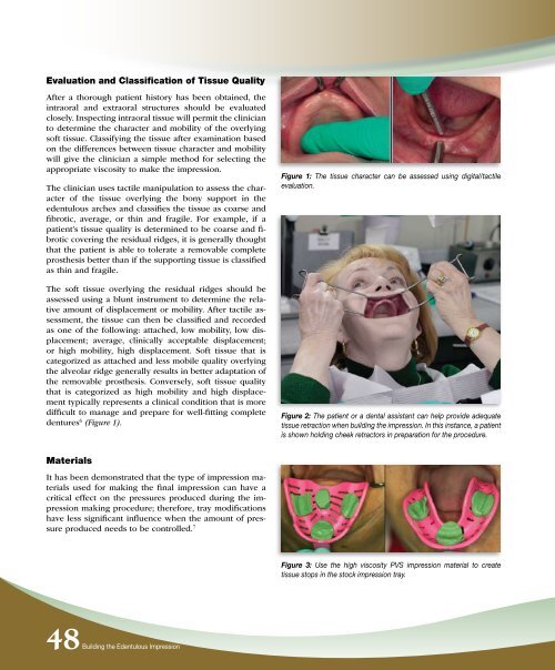
Figure 1: The tissue character can be assessed using digital/tactile<br />
evaluation.<br />
Figure 2: The patient or a dental assistant can help provide adequate<br />
tissue retraction when building the impression. In this instance, a patient<br />
is shown holding cheek retractors in preparation for the procedure.<br />
Figure 3: Use the high viscosity PVS impression material to create<br />
tissue stops in the stock impression tray.


