SCIENTIFIC REPORT 2010 - 2011 - IOV
SCIENTIFIC REPORT 2010 - 2011 - IOV
SCIENTIFIC REPORT 2010 - 2011 - IOV

You also want an ePaper? Increase the reach of your titles
YUMPU automatically turns print PDFs into web optimized ePapers that Google loves.
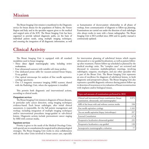
Mission<br />
The Breast Imaging Unit mission is manifested in the Diagnostic<br />
service for breast disease for the population of Padova, the Veneto<br />
Region and Italy, and in the specialist support given to the medical<br />
and surgical units of the <strong>IOV</strong>. The Breast Imaging Unit has been<br />
organized to provide tailored diagnostic paths, on the basis of<br />
individual patient needs, using multiple imaging techniques,<br />
and ensuring the integration of all diagnostic information, as well<br />
Clinical Activity<br />
The Breast Imaging Unit is equipped with all standard<br />
modalities used in breast imaging:<br />
Three direct digital mammography units, including review<br />
workstations;<br />
Four ultrasound scanners with suitable soft tissue probes;<br />
One dedicated prone table for vacuum-assisted breast biopsy,<br />
X-ray guided;<br />
One optical microscope for analysis of fine needle aspiration<br />
cytology specimens;<br />
One 1.5 magnetic resonance imaging (MRI) scanner, shared<br />
with the Radiology Unit, where the equipment is installed.<br />
This permits both diagnostic and interventional actions,<br />
according to clinical needs.<br />
Outpatient services<br />
The Breast Imaging Unit mission is diagnosis of breast diseases,<br />
in particular early cancer detection, using imaging techniques<br />
evidence-based. Each breast radiologist, after initial clinical<br />
assessment, is responsible, for the full patient management and<br />
workup, including integration of multiple imaging techniques<br />
and/or interventional procedures image-guided in sub-clinical<br />
lesions. Diagnostic actions include pretreatment cancer staging<br />
by MRI with contrast media.<br />
Inpatient services<br />
Priority is given to the needs of the Medical Oncology Units,<br />
with special care to consultations for surgical and/or pharmacological<br />
strategies. The Breast Imaging Unit works in close collaboration<br />
with all the other Units involved in breast cancer care, especially<br />
as humanization of doctor-patient relationship in all phases of<br />
workup, from communication of diagnosis to follow-up planning.<br />
Examinations are carried out under the direction of each radiologist<br />
who always works in team with a breast radiographer. The Breast<br />
Imaging Unit is ISO-certified since 2004 and its quality manual is<br />
continuously updated.<br />
for intervention planning of subclinical lesions which require<br />
ultrasound-or x-ray-guided localizations, as well as patient followup<br />
after treatment. Patient follow-up scheduled as planned by the<br />
medical oncology units. The “complex cases” are reviewed and<br />
discussed in consensus multidisciplinary meetings, involving<br />
radiologists, surgeons, oncologists, and any other specialist who<br />
is part of the Breast Unit. The Breast Imaging Unit represents<br />
an area of excellence for diagnosis of subclinical lesions, in both<br />
diagnostic and preoperative phases. The Breast Imaging Unit also<br />
represents a possible diagnostic reference during patient follow-up<br />
in detection of recurrences or in evaluation of breast reconstruction<br />
with implants and/or biological tissues.<br />
Types and amounts of examinations performed in <strong>2010</strong><br />
Clinical and instrumental breast examinations (breast<br />
examination, ultrasounds, and mammography)<br />
THE DEPARTMENTS - DEPARTMENT OF IMAGING, RADIOLOGY AND PATHOLOGY<br />
98<br />
9.975<br />
MRI of the breast with and without contrast media 550<br />
Fine needle aspiration cytology 1.165<br />
Tru-cut needle aspiration biopsy (microbiopsy) 297<br />
External Consultation 474<br />
Preoperative localization ultrasound-guided 235<br />
Preoperative localization x-ray-guided 120<br />
Intraoperative mammographic evaluation 77