Graphite pseudomorphs after diamond? A carbon isotope and ...
Graphite pseudomorphs after diamond? A carbon isotope and ...
Graphite pseudomorphs after diamond? A carbon isotope and ...

Create successful ePaper yourself
Turn your PDF publications into a flip-book with our unique Google optimized e-Paper software.
Pergamon<br />
PII S0016-7037(98)00142-2<br />
<strong>Graphite</strong> <strong>pseudomorphs</strong> <strong>after</strong> <strong>diamond</strong>? A <strong>carbon</strong> <strong>isotope</strong> <strong>and</strong> spectroscopic study of<br />
graphite cuboids from the Maksyutov Complex, south Ural Mountains, Russia<br />
MARY L. LEECH <strong>and</strong> W. G. ERNST<br />
Department of Geological <strong>and</strong> Environmental Sciences, Stanford University,<br />
Stanford, California 94305-2115, USA<br />
(Received November 12, 1997; accepted in revised form March 26, 1998)<br />
ABSTRACT—Unusual cuboid graphite aggregates (up to 13 mm edge length) from the eclogitic gneiss unit<br />
of the Maksyutov Complex deflect a foliation defined by groundmass graphite <strong>and</strong> phengite, <strong>and</strong> pressure<br />
shadows have developed around these blocky aggregates. Carbon <strong>isotope</strong> ratios, 13 C/ 12 C, for the cuboid<br />
graphite range from about 24 to 42‰, demonstrating that these rocks have retained an original biogenic<br />
<strong>carbon</strong> signature. X-ray diffraction, laser Raman spectroscopy, infrared spectroscopy, <strong>and</strong> transmission<br />
electron microscopy indicate that graphite is well-crystallized with minor defects; no relict organic compounds<br />
were detected. Comparisons of these cuboid aggregates with thin sections <strong>and</strong> scanning electron microscope<br />
images of proven graphitized <strong>diamond</strong>s from the Beni Bousera peridotite massif show that Maksyutov<br />
graphite is similar. Laboratory experiments by other workers on graphite demonstrate that this intriguing<br />
morphology could not be the result of deformation, because graphite returns to its original shape <strong>and</strong> size on<br />
stress release. Existing experiments on <strong>diamond</strong> graphitization do not adequately replicate the conditions of<br />
natural rocks being exhumed from subduction zones characterized by ultrahigh pressures, <strong>and</strong> thus cannot be<br />
applied with confidence to the Maksyutov Complex. Our spectroscopic <strong>and</strong> microscopic studies suggest that<br />
these cuboid aggregates probably are <strong>diamond</strong> <strong>pseudomorphs</strong>. Copyright © 1998 Elsevier Science Ltd<br />
1. INTRODUCTION<br />
<strong>Graphite</strong> <strong>pseudomorphs</strong> <strong>after</strong> <strong>diamond</strong> have been recognized in<br />
the Beni Bousera peridotite massif in northern Morocco <strong>and</strong> the<br />
Ronda peridotite massif in southern Spain (e.g., Pearson et al.,<br />
1989; Davies et al., 1993). Scanning electron microscope<br />
(SEM) imagery of graphite from these massifs revealed octahedral<br />
<strong>and</strong> cubic faces with corresponding depressions within<br />
graphite <strong>and</strong> showed that thin coatings of differently-oriented<br />
graphite surround the <strong>pseudomorphs</strong>.<br />
Thus far, only a few crustal terranes subjected to ultrahigh<br />
pressure (UHP) have been recognized worldwide: the<br />
Kokchetav Massif in northern Kazakhstan, the Sulu-Dabie belt<br />
in east-central China, the Dora Maira Massif in the Italian Alps,<br />
<strong>and</strong> the Western Gneiss Region in coastal Norway are examples<br />
of well-studied UHP continental terranes with confirmed<br />
coesite, coesite <strong>pseudomorphs</strong>, <strong>and</strong>, in the case of the<br />
Kokchetav occurrence, <strong>diamond</strong>. Rare occurrences of micro<strong>diamond</strong><br />
inclusions have also been described from the Dabie<br />
Shan (Xu et al., 1992; Okay, 1993) <strong>and</strong> from the Western<br />
Gneiss region (Dobrzhinetskaya et al., 1995). Reports of coesite<br />
<strong>pseudomorphs</strong> in the eclogitic unit of the Maksyutov Complex<br />
(Chesnokov <strong>and</strong> Popov, 1965; Dobretsov <strong>and</strong> Dobretsova,<br />
1988) suggest that these rocks, too, may have been metamorphosed<br />
at ultrahigh pressures. Although thermobarometric calculations<br />
have demonstrated minimum conditions of about<br />
600°C, 1.5 GPa for the Maksyutov Complex (Beane et al.,<br />
1995; Lennykh et al., 1995; Dobretsov et al., 1996; Beane,<br />
1997), cuboid graphite aggregates from host mica schist support<br />
the earlier suggestion of UHP metamorphism proposed by<br />
Chesnokov <strong>and</strong> Popov (1965) <strong>and</strong> Dobretsov <strong>and</strong> Dobretsova<br />
(1988).<br />
Reports of coesite have not been independently confirmed,<br />
but if the cuboid graphite is pseudomorphic <strong>after</strong> <strong>diamond</strong>, it<br />
2143<br />
Geochimica et Cosmochimica Acta, Vol. 62, No. 12, pp. 2143–2154, 1998<br />
Copyright © 1998 Elsevier Science Ltd<br />
Printed in the USA. All rights reserved<br />
0016-7037/98 $19.00 .00<br />
would indicate even higher pressures than previously thought.<br />
<strong>Graphite</strong> aggregates pseudomorphic <strong>after</strong> <strong>diamond</strong> may occur<br />
unrecognized elsewhere in continental collision zones; if our<br />
speculations about the Maksyutov paragenesis are correct, this<br />
example could encourage further study of <strong>carbon</strong>aceous matter.<br />
2. GEOLOGIC SETTING<br />
The Maksyutov Complex trends north-south in the south<br />
Ural Mountains of central Russia (Fig. 1). The complex consists<br />
of two main units tectonically juxtaposed in this continental<br />
collision suture zone (Zonenshain et al., 1990): an eclogitebearing<br />
gneiss, called Unit #1; <strong>and</strong> a meta-ophiolite, termed<br />
Unit #2. Unit #1 contains boudins of eclogite, layers of<br />
eclogitic gneiss, <strong>and</strong> rare ultramafic bodies within host metasedimentary<br />
mica schist <strong>and</strong> quartzite; Unit #2 consists of<br />
lenses of serpentinite melange <strong>and</strong> blocks of metasomatic rock<br />
(rodingite), metabasalt, <strong>and</strong> marble within mica schist <strong>and</strong><br />
graphite quartzite host rock (Fig. 2). Unit #1 protoliths are<br />
Middle to Late Proterozoic; Unit #2 is Late Proterozoic with<br />
blocks of Ordovician to Silurian marble (Dobretsov et al.,<br />
1996). Unit #2, considered to tectonically overlie Unit #2<br />
(Lennykh et al., 1995), was probably thrust over Unit #1 <strong>after</strong><br />
the Middle Devonian HP-UHP metamorphic event that affected<br />
Unit #1 <strong>and</strong> the Early Carboniferous retrograde blueschist- to<br />
high-pressure greenschist-facies metamorphism that affected<br />
Unit #2 (Matte et al., 1993; Beane, 1997; Beane et al., 1995).<br />
Both units were overprinted by a late, low-pressure, greenschist-facies<br />
metamorphism <strong>and</strong> subsequently folded together<br />
about NE-SW trending axes.<br />
Fe-Mg exchange geothermometry calculated for garnet <strong>and</strong><br />
clinopyroxene (<strong>after</strong> Powell, 1985) yields an equilibrium temperature<br />
ranging from 594 to 637°C. Minimum pressure estimates<br />
using the jadeite component of clinopyroxene (<strong>after</strong>
2144 M. L. Leech <strong>and</strong> W. G. Ernst<br />
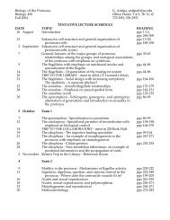
Fig. 1. Tectonic map of the south Ural Mountains, Russia, the collisional zone between the East European platform <strong>and</strong><br />
the Siberian craton. The inset map shows the location of the figure. After Beane et al. (1995).<br />
Holl<strong>and</strong>, 1980) range from 1.5 to 1.7 GPa (Beane et al., 1995;<br />
Lennykh et al., 1995; this study), but may be as high as 2.7 GPa<br />
if coesite <strong>pseudomorphs</strong> described in eclogite (Chesnokov <strong>and</strong><br />
Popov, 1965) <strong>and</strong> jadeite quartzite (Dobretsov <strong>and</strong> Dobretsova,<br />
1988) are present (Bohlen <strong>and</strong> Boettcher, 1982). Thermobarometric<br />
estimates may represent annealing during exhumation in<br />
which minerals reequilibrated at lower P-T conditions, leaving<br />
little evidence of UHP metamorphism (Fig. 3).<br />
3. CUBOID GRAPHITE, PSEUDOMORPHS<br />
AFTER DIAMOND?<br />
3.1. Petrography<br />
<strong>Graphite</strong>-phengite schist (sample M-16-94) from near the<br />
former village of Karayanova contains 40% phengite, 38%<br />
graphite, 19% quartz, 1% rutile, 1% zircon, <strong>and</strong> 1% iron<br />
oxide minerals. A single cleavage is defined by oriented phengite<br />
<strong>and</strong> graphite flakes. The flakes wrap around large, subangular,<br />
blocky graphite aggregates; pressure shadows containing<br />
quartz <strong>and</strong> coarse-grained phengite have developed around the<br />
aggregates, suggesting that during deformation, the aggregates<br />
behaved as coherent, rigid blocks within a more ductile matrix<br />
(Fig. 4a). Aligned inclusions of phengite <strong>and</strong> rutile in the<br />
graphite aggregates are subparallel to the foliation. <strong>Graphite</strong><br />
aggregates have an angular to subrounded cross-sectional morphology<br />
(Fig. 4b); this is significant because these aggregates<br />
may be <strong>pseudomorphs</strong> <strong>after</strong> <strong>diamond</strong> indicating ultrahigh-pressure<br />
metamorphism at a minimum pressure of about 3.2 GPa<br />
(Kennedy <strong>and</strong> Kennedy, 1976).<br />
<strong>Graphite</strong> is abundant throughout the Maksyutov Complex,<br />
with volumetric modes in the mica schists locally ranging up to
38% graphite. Disseminated tabular graphite occurs parallel to<br />
the dominant foliation in quartzites of both Units #1 <strong>and</strong> #2 <strong>and</strong><br />
in metasomatic rocks of Unit #2. Cuboid graphite aggregates<br />
(most 5 mm long, but up to 13 10 mm), made up of flakes<br />
ranging in size from about 4 to 100 m, are found in mica<br />
schist near Karayanova, <strong>and</strong> occur with graphite flakes aligned<br />
in the foliation. Scanning electron microscope images show<br />
cubic forms of the graphite aggregates (Fig. 5). <strong>Graphite</strong> is also<br />
found as inclusions in garnet in eclogitic gneisses near Antingan<br />
village.<br />
3.2. <strong>Graphite</strong> Deformation<br />
Are these cuboid graphite aggregates a result of a specific<br />
deformation mechanism acting on graphite? Indeed, how does<br />
graphite deform under the P-T conditions <strong>and</strong> differential stress<br />
these rocks experienced? Edmond <strong>and</strong> Paterson (1971) described<br />
stress-strain experiments for graphite samples (10 20<br />
mm) under confining pressures up to 0.8 or 1.0 GPa <strong>and</strong> room<br />
temperature. Under these conditions, they found that graphite<br />
specimens returned almost to their original dimensions when<br />
both the differential stress <strong>and</strong> confining pressure were released,<br />
even <strong>after</strong> 20% shortening at 0.4 GPa; other samples<br />
<strong>Graphite</strong> <strong>pseudomorphs</strong> <strong>after</strong> <strong>diamond</strong><br />
Fig. 2. Geologic map of the Maksyutov Complex showing exposure of Units #1 <strong>and</strong> #2 along the Sakmara River near<br />
Karayanova (see Fig. 1). Sample M-16-94 indicates where cuboid aggregates occur. After Lennykh et al. (1995).<br />
were shortened 20% under confining pressures of 0.2–0.8 GPa<br />
<strong>and</strong> gave similar recoveries. Later experiments by Edmond <strong>and</strong><br />
Paterson (1972) showed that, in addition to an almost complete<br />
recovery of volume, the initial shape of the graphite was<br />
retained as well, with most of the recovery occurring below<br />
0.05 GPa; graphite specimens appeared to be undeformed.<br />
Kretz (1996) described graphite in high-grade marble (T <br />
650–700°C; P 0.65–0.70 GPa) occurring as both undeformed<br />
<strong>and</strong> deformed tabular grains <strong>and</strong> suggested that the<br />
deformational behavior of graphite is similar to that of biotite.<br />
<strong>Graphite</strong> deforms by cleavage separation, kink-b<strong>and</strong> formation,<br />
<strong>and</strong> folding; breaking across the (0001) basal plane of strong<br />
<strong>carbon</strong> bonds occurred only in mylonitic marble where strain<br />
rates were very high.<br />
3.3. Graphitization of Organic Matter<br />
2145<br />
Is this poorly crystallized graphite? Graphitization of organic<br />
matter is primarily dependent on metamorphic temperature, but<br />
the process is facilitated at lower temperatures by shear strain<br />
in combination with elevated pressure (L<strong>and</strong>is, 1971; Teichmuller,<br />
1987; Ross <strong>and</strong> Bustin, 1990; Ross et al., 1991). Temperatures<br />
<strong>and</strong> pressures suggested for the formation of true,
2146 M. L. Leech <strong>and</strong> W. G. Ernst<br />
Fig. 3. Possible retrograde P-T paths for the Maksyutov Complex: The solid curve is based on Russian reports of coesite<br />
(Chesnokov <strong>and</strong> Popov, 1965; Dobretsov <strong>and</strong> Dobretsova, 1988) <strong>and</strong> possible graphite <strong>pseudomorphs</strong> <strong>after</strong> <strong>diamond</strong> (this<br />
study); a second possible P-T path is shown as a dashed curve in accordance with the thermobarometric calculations <strong>and</strong><br />
petrographic studies conducted on eclogites <strong>and</strong> related rocks from Unit #1 (Beane et al., 1995). The dotted line shows the<br />
probable path for Unit #2 rocks. After Lennykh et al. (1995).<br />
well-ordered graphite in nature range from about 350° to 700°C<br />
<strong>and</strong> from 0.2 to 0.6 GPa (L<strong>and</strong>is, 1971; Grew, 1974; Diessel et<br />
al., 1978; Tagiri <strong>and</strong> Oba, 1986).<br />
Well-ordered graphite develops over a wide range of metamorphic<br />
conditions. Moderately high temperatures (sillimanite<br />
zone or amphibolite-facies) are required for near complete<br />
graphitization (Grew, 1974; Armstrong, 1982), although<br />
Buseck <strong>and</strong> Huang (1985) described well-ordered graphite in<br />
the chlorite zone (greenschist-facies). It is clear that increasing<br />
crystallinity is roughly proportional to metamorphic grade<br />
within a specific terrane, but not necessarily between terranes.<br />
However, higher pressures such as those calculated for the<br />
Maksyutov Complex may retard the graphitization process<br />
(Dalla Torre et al., 1996, 1997) even if metamorphic temperatures<br />
are above those required for complete graphitization at<br />
low pressures. With continued progressive metamorphism, the<br />
long-range order increases, the <strong>carbon</strong> layers lengthen <strong>and</strong><br />
become more planar, interlayer spacing decreases, <strong>and</strong> the<br />
number of defects decreases (Grew, 1974; Buseck <strong>and</strong> Huang,<br />
1985; Teichmuller, 1987).<br />
3.4. Diamond Pseudomorphs from the Beni Bousera <strong>and</strong><br />
Ronda Peridotite Massifs<br />
An octahedral or cubic morphology of graphite is the most<br />
convincing evidence for graphitized <strong>diamond</strong>. <strong>Graphite</strong><br />
<strong>pseudomorphs</strong> <strong>after</strong> <strong>diamond</strong> have been recognized in the Beni<br />
Bousera peridotite massif in northern Morocco <strong>and</strong> the Ronda<br />
peridotite massif in southern Spain (e.g., Pearson et al., 1989;<br />
Davies et al., 1993). Most graphite from both locations has
<strong>Graphite</strong> <strong>pseudomorphs</strong> <strong>after</strong> <strong>diamond</strong><br />
Fig. 4. Thin sections (in unpolarized light) of graphite schist from Unit #1 cut perpendicular to the foliation: (a)<br />
subangular graphite aggregate (13 10 mm) with phengite <strong>and</strong> rutile inclusions. The foliation is defined by graphite <strong>and</strong><br />
phengite flakes that wrap around the graphite aggregate. Note the pressure shadows of quartz <strong>and</strong> coarse-grained phengite<br />
that have formed around the aggregate. Vertical black lines <strong>and</strong> rings of dots in lower right-h<strong>and</strong> <strong>and</strong> upper left-h<strong>and</strong> corners<br />
are from marking thin section for electron microprobe analysis; (b) cuboid graphite aggregate (about 6 mm long) typical<br />
of those in graphite schist in Unit #1.<br />
2147
2148 M. L. Leech <strong>and</strong> W. G. Ernst<br />
Fig. 5. SEM images showing the external morphology of graphite aggregates that display rough or perhaps slightly<br />
deformed cubic forms; dashed white lines trace the edges of cubes on the right side of the figure. These images are similar<br />
to those described by Pearson <strong>and</strong> Nixon (1996) for aggregates from the Beni Bousera massif. Aggregates have about a 4<br />
mm edge length.<br />
cubic symmetry, but more than 30% of the graphite occurs as<br />
aggregates with no obvious external morphology. <strong>Graphite</strong><br />
cubes <strong>and</strong> octahedra are commonly 2–8 mm in diameter, but<br />
can be up to 12–20 mm (Pearson et al., 1989; Davies et al.,<br />
1991, 1993; Pearson <strong>and</strong> Nixon, 1996). Dissolution of silicates<br />
surrounding the graphite with hydrofluoric acid reveals graphite<br />
cubes <strong>and</strong> octahedra, but more commonly, graphite with an<br />
ovoid form which is due to a 0.01–3 mm thick fibrous graphite<br />
shell that coats most of the octahedra; crushing samples separates<br />
some of the graphite cubes <strong>and</strong> octahedra from the shell<br />
graphite (Pearson et al., 1989). Pearson <strong>and</strong> Nixon (1996)<br />
described graphite aggregates occurring as octahedra <strong>and</strong> other<br />
forms of cubic symmetry in the Beni Bousera massif. The<br />
rounded, coated graphite aggregates shown in thin section in<br />
their Fig.10a,b <strong>and</strong> in an SEM image in their Fig. 11a are<br />
strikingly similar to graphite aggregates from the Maksyutov<br />
Complex.<br />
Diamond crystal morphology is largely controlled by the<br />
temperature <strong>and</strong> pressure conditions <strong>and</strong>/or oxygen fugacity at<br />
the time of crystallization. Octahedral <strong>diamond</strong>s result from<br />
crystallization at either lower relative temperatures <strong>and</strong>/or<br />
higher pressures, or an oxygen fugacity near magnetite-wustite<br />
(Robinson et al., 1978; Taylor, 1985; Sobolev <strong>and</strong> Shatsky,<br />
1990; Deines et al., 1993). The morphology of the resulting<br />
graphite aggregates is most likely dictated by the original<br />
crystal form of the <strong>diamond</strong> (Pearson <strong>and</strong> Nixon, 1996).<br />
Octahedral forms of <strong>diamond</strong> predominate in most localities<br />
worldwide (Taylor, 1985), but the entire range of morphologies<br />
from cubic to octahedral is represented in peridotite massifs<br />
(e.g., Beni Bousera), kimberlites <strong>and</strong> associated mantle xenoliths<br />
(e.g., Orapa, Botswana; Roberts Victor, South Africa; <strong>and</strong><br />
Yakutia, Siberia), <strong>and</strong> ultrahigh-pressure metamorphic terranes<br />
(e.g., the Kokchetav Massif, Kazakhstan; the Western Gneiss<br />
region, Norway; <strong>and</strong> Dabie Shan, China) (Sobolev <strong>and</strong> Shatsky,<br />
1990; Shatsky et al., 1991; Viljoen et al., 1991; Xu et al., 1992;<br />
Deines et al., 1993; Jerde et al., 1993; Okay, 1993; Dobrzhinetskaya<br />
et al., 1994, 1995). Most <strong>diamond</strong>s with a cubic<br />
morphology are found in rocks with an eclogitic affinity (Spetius,<br />
1995), as is the case for the Maksyutov Complex.<br />
4. SAMPLE SEPARATION<br />
Three techniques were used to separate graphite from<br />
Maksyutov rock samples for both isotopic analyses <strong>and</strong> spectroscopic<br />
studies: flotation of graphite from crushed rock samples,<br />
acid dissolution of silicates surrounding graphite, <strong>and</strong><br />
h<strong>and</strong>-picking graphite directly from the samples. For finely<br />
disseminated graphite, manual separation from dry, crushed
ock was tedious, so graphite was floated in water; because of<br />
the hydrophobic character of graphite, other phases settled out<br />
<strong>and</strong> left a film of graphite on the surface of the water to be<br />
skimmed off.<br />
Several samples, including cuboid aggregates, were dissolved<br />
in a solution of hydrofluoric <strong>and</strong> hydrochloric acid to<br />
separate graphite from the silicates, in an attempt to preserve<br />
any relict cubes or octahedra (shown in Fig. 5). A 1:1 solution<br />
of 48.8–49.2% HF <strong>and</strong> 36.5–38.0% HCl was used to dissolve<br />
the silicates surrounding the graphite aggregates in cm-sized<br />
samples. About half of the mixture was decanted every 48 h<br />
<strong>and</strong> refreshed with another 1:1 solution; this process was repeated<br />
at least five times. The procedure left some insoluble<br />
fluorides, but did not interfere with the final graphite separation<br />
by h<strong>and</strong>.<br />
For <strong>carbon</strong> <strong>isotope</strong> analyses, graphite was h<strong>and</strong>-picked from<br />
several samples of the graphite schist. Aggregate graphite was<br />
readily separated by h<strong>and</strong> from the graphite occurring in the<br />
foliation to allow analyses of both forms.<br />
5. CARBON ISOTOPE GEOCHEMISTRY<br />
Carbon <strong>isotope</strong> measurements were performed at the University<br />
of California, Davis <strong>and</strong> the Geophysical Laboratory,<br />
Carnegie Institution in Washington, D. C. to establish 13 C vs.<br />
PDB for graphite from the Maksyutov Complex. An elemental<br />
analyzer was used at UC Davis <strong>and</strong> samples were compared to<br />
<strong>Graphite</strong> <strong>pseudomorphs</strong> <strong>after</strong> <strong>diamond</strong><br />
Fig. 6. Carbon <strong>isotope</strong> composition of graphite-bearing rocks <strong>and</strong> marble from the Maksyutov Complex (Table 1 contains<br />
more detailed information). Ranges of typical <strong>carbon</strong> <strong>isotope</strong> values for marine <strong>carbon</strong>ates, mantle <strong>carbon</strong>, <strong>and</strong> biogenic<br />
<strong>carbon</strong> from Javoy et al. (1986).<br />
2149<br />
the USGS graphite st<strong>and</strong>ard #24 (16.0‰ vs. PDB), while at<br />
the Geophysical Laboratory, samples were analyzed on a mass<br />
spectrometer <strong>and</strong> compared to NBS-21 st<strong>and</strong>ard (28.1‰ vs.<br />
PDB); several samples were analyzed at both facilities to<br />
provide an interlaboratory calibration. The isotopic composition<br />
of <strong>carbon</strong> is expressed in terms of delta notation (Hoefs,<br />
1987):<br />
13 C(‰) [( 13 C/ 12 C) spl ( 13 C/ 12 C) std]/( 13 C/ 12 C) std 10 3 .<br />
The reference st<strong>and</strong>ard is CO 2 gas released from belemnites of<br />
the Peedee Formation (PDB).<br />
Comparison of these values with <strong>carbon</strong> <strong>isotope</strong> compositions<br />
for <strong>diamond</strong>s in other continent-continent collision zones,<br />
kimberlites, <strong>and</strong> peridotites with graphite <strong>pseudomorphs</strong> <strong>after</strong><br />
<strong>diamond</strong> establishes the source of <strong>carbon</strong> in these high-grade<br />
rocks <strong>and</strong> perhaps will lead to a fuller underst<strong>and</strong>ing regarding<br />
<strong>carbon</strong> cycling <strong>and</strong> the geochemistry of the mantle.<br />
Carbon <strong>isotope</strong> ratios for Unit #1 mica schist range from<br />
about 21 to 42‰, with a pronounced frequency peak<br />
around 28‰ (Fig. 6). The range of <strong>carbon</strong> <strong>isotope</strong> values for<br />
the Maksyutov Complex is comparable to that in micro<strong>diamond</strong>s<br />
from the UHP Kokchetav Massif, Kazakhstan (Sobolev<br />
et al., 1979; Sobolev <strong>and</strong> Shatsky, 1990), <strong>diamond</strong>s with<br />
eclogitic inclusions in kimberlitic rocks <strong>and</strong> associated mantle<br />
xenoliths (e.g., Milledge et al., 1983; Galimov, 1988) <strong>and</strong>
2150 M. L. Leech <strong>and</strong> W. G. Ernst<br />
graphite <strong>pseudomorphs</strong> <strong>after</strong> <strong>diamond</strong> in the Beni Bousera<br />
peridotite massif, Morocco (Slodkevich, 1983; Pearson et al.,<br />
1989, 1995). However, kimberlitic <strong>and</strong> peridotitic <strong>diamond</strong>s<br />
have a wider range of <strong>carbon</strong> <strong>isotope</strong> values, from about 2 to<br />
34‰, <strong>and</strong> a large frequency peak at 5 to6‰ (Milledge,<br />
1983; Kirkley <strong>and</strong> Gurney, 1991; Kirkley et al., 1991) indicating<br />
a mantle source for the <strong>carbon</strong> (see Mattey, 1987; Galimov,<br />
1988).<br />
Very negative <strong>carbon</strong> <strong>isotope</strong> values might represent <strong>carbon</strong><br />
from a deep mantle source, perhaps a primitive mantle <strong>carbon</strong><br />
reservoir (Javoy et al., 1986; Deines et al., 1987, 1993; Galimov,<br />
1988), but more likely these rocks have retained their<br />
original biogenic <strong>carbon</strong> signature (Milledge et al., 1983; Kirkley<br />
et al., 1991). Javoy et al. (1986) <strong>and</strong> Mattey (1987) give<br />
ranges for various <strong>carbon</strong> sources: Organic <strong>carbon</strong> has a 13 C<br />
signature ranging from about 15 to 30‰ (mean 13 C <br />
25‰), whereas marine <strong>carbon</strong>ates have a mean value around<br />
0‰, <strong>and</strong> mantle <strong>carbon</strong> ranges from 5 to8‰. Intermediate<br />
values in kimberlites <strong>and</strong> associated rocks probably result from<br />
the mixing of subducted biogenic <strong>carbon</strong> <strong>and</strong> mantle <strong>carbon</strong> in<br />
the uppermost mantle (Javoy et al., 1986).<br />
In the Maksyutov Complex, there are no apparent differences<br />
in isotopic signature according to the type of graphite sampled;<br />
blocky aggregates, graphite aligned in the foliation, <strong>and</strong> inclusions<br />
in other minerals all occupy the same general isotopic<br />
Fig. 7. TEM images of a graphite aggregate. (a) shows fairly straight<br />
lattice fringes; graphite contains many lattice fringe terminations. The<br />
inset box is a selected area diffraction pattern for the graphite. (b)<br />
shows the anastamosing character of some crystallites.<br />
range for both tectonic units. Marble is found only rarely as<br />
small blocks in Unit #2 <strong>and</strong> has a mean <strong>carbon</strong> <strong>isotope</strong> value of<br />
about 1‰ vs. PDB, which corresponds to typical marine<br />
<strong>carbon</strong>ates (Table 1). Nitrogen analyses for graphite yield values<br />
which indicate atmospheric contamination.<br />
6. SPECTROSCOPIC AND MICROSCOPIC STUDIES<br />
Transmission electron microscopy (TEM), x-ray diffraction<br />
(XRD), <strong>and</strong> both infrared <strong>and</strong> laser Raman spectroscopic techniques<br />
were used to investigate the structure of the graphite<br />
aggregates in order to gain insight into their origin <strong>and</strong> subsequent<br />
history. Infrared spectroscopy shows whether or not relict<br />
organic compounds remain within the cuboid aggregates. X-ray<br />
diffraction characterizes a bulk sample (up to a cm-size area)<br />
which is thought to be useful in the determination of graphite<br />
crystallization state, although heterogeneity within the sample<br />
may be missed because of the scale <strong>and</strong> nature of the analysis.<br />
Sample preparation for XRD is destructive <strong>and</strong> may change the<br />
character of the graphite. Transmission electron microscope<br />
analyses are on the scale of a few Å <strong>and</strong> serve as an excellent<br />
tool for investigating the structure of the crystallites, but do not<br />
easily allow characterization of a sample as a whole. Laser
Raman microspectroscopy allows an intermediate-scale analysis<br />
of graphite crystallization in the cuboid aggregates, with a<br />
beam size on the order of about 100 m; it is possible to run a<br />
profile across individual graphite aggregates to check for<br />
localized differences in crystallization state within a single<br />
sample.<br />
6.1. Transmission Electron Microscopy<br />
Transmission electron microscopy samples were prepared by<br />
epoxying a copper sample holder ring directly onto graphite<br />
aggregates, removing <strong>and</strong> mounting the aggregate into a TEM<br />
sample holder, then ion milling through the center of the<br />
aggregate. Imagery of the graphite aggregates from Maksyutov<br />
show that the graphite contains minor defects such as dislocations,<br />
causing lattice-fringe terminations (Fig. 7). Study of one<br />
graphite aggregate shows fairly straight, uniformly spaced lattice<br />
fringes containing defects such as lattice-fringe terminations<br />
<strong>and</strong> crystallites that have an anastamosing character,<br />
showing a lower degree of structural order.<br />
Selected-area diffraction (SAD) patterns are diffuse <strong>and</strong> lack<br />
rings, which probably result from the defects seen in TEM<br />
imaging (Buseck <strong>and</strong> Huang, 1985). Low-magnification TEM<br />
images show that graphite crystals have a very roughly aligned<br />
orientation; graphite grains in reflected light appear to be<br />
aligned in two preferred directions subparallel to the external<br />
morphology of the aggregates. Induced effects from the ion<br />
milling process may explain some of the apparent defects, but<br />
are probably not responsible for all those seen in TEM images<br />
because the alleged milling effects continue into the thicker<br />
portions of the milled thin section.<br />
6.3. X-Ray Diffraction<br />
Samples were run employing CuK a radiation <strong>and</strong> a Philips<br />
x-ray diffractometer. X-ray diffraction spectra were used to<br />
determine the degree of crystallinity in powdered graphite<br />
aggregates <strong>after</strong> obtaining the results of the TEM. <strong>Graphite</strong><br />
samples were prepared for XRD analysis by mixing powder in<br />
distilled water, followed by drying, in order to orient graphite<br />
flakes with the (002) planes coincident with the specimen<br />
holder; other analyses were performed on dry, powdered graphite.<br />
The samples were scanned from 5 to 45° 2, scanning 0.05°<br />
2 every second. Sharp peaks on x-ray diffraction spectra from<br />
powdered, <strong>and</strong> oriented samples for the aggregates indicate that<br />
the graphite is well crystallized; no broad peaks were obtained,<br />
<strong>and</strong> all 2 angles correspond with graphite d-spacings. Interplanar<br />
d 002 spacing for the aggregates is 3.36 Å, which is the<br />
d-spacing for fully-ordered graphite according to Warneke <strong>and</strong><br />
Ernst (1984) <strong>and</strong> Tagiri <strong>and</strong> Oba (1986); peak width at halfheight<br />
(in °2) is about 0.324.<br />
6.4. Laser Raman Microspectroscopy<br />
First-order spectra were analyzed from 1200 to 1700 cm 1<br />
<strong>and</strong> second-order spectra from 2350 to 3350 cm 1 on a Lab-<br />
Ram spectrometer; these scans analyzed for the first-order<br />
single b<strong>and</strong> at about 1582 cm 1 that is characteristic of wellcrystallized<br />
graphite as well as two overlapping b<strong>and</strong>s near<br />
2700 cm 1 in second-order wave numbers (Pasteris <strong>and</strong><br />
Wopenka, 1991). Disorder in graphite appears as a broadening<br />
<strong>Graphite</strong> <strong>pseudomorphs</strong> <strong>after</strong> <strong>diamond</strong><br />
Fig. 8. Laser Raman spectra of graphite aggregates showing two<br />
large peaks, with the first-order peak at about 1582 cm 1 (O-peak), a<br />
peak at 1360 cm 1 showing disorder (D-peak), <strong>and</strong> a smaller, broad<br />
second-order peak at about 2700 cm 1 (S-peak). Note the high wavenumber<br />
shoulder on the O-peak <strong>and</strong> a suppressed peak at about 2450<br />
cm 1 .<br />
of the 1582 cm 1 b<strong>and</strong> <strong>and</strong> its shift toward higher wavenumbers<br />
as a result of the development of an additional b<strong>and</strong> near<br />
1360 cm 1 ; second-order spectra exhibit a broadening of the<br />
b<strong>and</strong> at 2700 cm 1 , the loss of resolution of the two overlapping<br />
b<strong>and</strong>s that create that peak, <strong>and</strong> the suppression of a peak<br />
at about 2450 cm 1 (Pasteris <strong>and</strong> Wopenka, 1991).<br />
Most analyses of Maksyutov graphite aggregates show two<br />
large peaks, at about 1582 cm 1 <strong>and</strong> 1360 cm 1 , <strong>and</strong> a smaller,<br />
broad peak at about 2700 cm 1 . Figure 8 shows a peak at about<br />
1360 cm 1 , a shoulder developed on the high wavenumber side<br />
of the 1582 cm 1 peak, <strong>and</strong> a suppressed peak at about 2450<br />
cm 1 which indicate minor disorder in the graphite probably<br />
resulting from the dislocations found in TEM imaging. The<br />
Raman spectra for these aggregates are similar to those shown<br />
for graphite in the chlorite to biotite zone of Barrovian metamorphism<br />
for metapelites, according to Wopenka <strong>and</strong> Pasteris<br />
(1993).<br />
6.5. Infrared Spectroscopy<br />
2151<br />
If organic C-H or C-O bonds exist, it would follow that these<br />
graphite aggregates could never have been <strong>diamond</strong>, for organic<br />
bonds would certainly have been broken during transformation<br />
to <strong>diamond</strong>. <strong>Graphite</strong> aggregates separated by acid<br />
dissolution were analyzed with an infrared spectrometer to<br />
search for organic compounds. <strong>Graphite</strong> was powdered <strong>and</strong><br />
mixed with KBr in the ratios 1:100 <strong>and</strong> 1:20. Each sample was<br />
scanned 500 times in addition to the st<strong>and</strong>ard KBr for background<br />
analyses; background KBr scans were subtracted from<br />
the graphite scans to isolate graphite peaks. The spectra show<br />
a broad hump between 2900 <strong>and</strong> 3700 cm 1 (Fig. 9); this<br />
adsorptance represents O-H bonds probably from water adsorbed<br />
by the sample. Two small peaks between 2300 <strong>and</strong> 2400<br />
cm 1 result from atmospheric CO 2. The large, broad, composite<br />
peak between 400 <strong>and</strong> 800 cm 1 is from silicates that were<br />
not dissolved during the acid dissolution process <strong>and</strong>/or from
2152 M. L. Leech <strong>and</strong> W. G. Ernst<br />
inclusions in the graphite. Two low-intensity absorption b<strong>and</strong>s<br />
were isolated in the graphite, one at about 1425 cm 1 <strong>and</strong><br />
another at about 1633 cm 1 .<br />
The wavenumbers associated with the two peaks described<br />
above might represent C-C bonds or C-O or C-H bonds (Robin<br />
<strong>and</strong> Rouxhet, 1978; Tissot <strong>and</strong> Welte, 1978). A USGS graphite<br />
st<strong>and</strong>ard (NBS-21) was run subsequent to the aggregate sample;<br />
two noisy peaks representing C-C bonds are located at<br />
approximately the same wavenumber positions as the aggregate.<br />
It is more probable that the two similar peaks are C-C<br />
bonds <strong>and</strong> not organic-group bonds.<br />
6.6. Rate of Graphitization of Diamond<br />
Experiments on rates of transformation from C Diamond 3<br />
C <strong>Graphite</strong> show that graphitization per unit time can be estimated<br />
by:<br />
(E PV)/RT,<br />
dx/dt Cexp where C is a constant proportional to graphitization rate, E is<br />
the activation energy, <strong>and</strong> V is the activation volume (Davies<br />
<strong>and</strong> Evans, 1972). The low activation volume for graphitization<br />
(10cm 3<br />
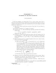
Fig. 9. Infrared spectra of acid-separated graphite aggregates. Peaks are at about 1425 cm 1 <strong>and</strong> about 1633 cm 1 , both<br />
probably representing C-C bonds.<br />
● mol 1 ) indicates that the influence of pressure is<br />
small; thus the rate of graphitization is largely dependent on<br />
temperature (Pearson et al., 1995). Experimentally determined<br />
activation energies for graphitization of <strong>diamond</strong> are lowest for<br />
{110} faces of octahedra (760 kJmol 1 ) <strong>and</strong> highest for {111}<br />
faces (1060 kJmol 1 ) under anhydrous conditions (Pearson <strong>and</strong><br />
Nixon, 1996). Using the activation energy for the graphitization<br />
of {110} faces, Pearson et al. (1995) calculated that the complete<br />
conversion of a <strong>diamond</strong> octahedron (10 mm edge<br />
length) to graphite would require about 1 m.y. at 1200°C or<br />
1 b.y. at 1000°C.<br />
Rate studies for <strong>diamond</strong> graphitization require a much<br />
slower exhumation rate <strong>and</strong> higher temperature than is thought<br />
to have occurred in the evolution of the Maksyutov Complex.<br />
However, these dry graphitization experiments do not take into<br />
account the presence of fluids <strong>and</strong> other rate-enhancing constituents<br />
(Tagiri <strong>and</strong> Oba, 1986) that increase the kinetics of<br />
graphitization during return to the Earth’s surface. Reequilibration<br />
that occurred during the exhumation history of the<br />
Maksyutov Complex evidently was sufficient for complete<br />
graphitization of any <strong>diamond</strong> that had been present. Diamond<br />
is preserved in the Kokchetav Massif under only very special<br />
conditions, chiefly as armored micro-inclusions in garnet <strong>and</strong><br />
zircon, which acted as pressure vessels (Sobolev <strong>and</strong> Shatsky,<br />
1990; Sobolev et al., 1994).<br />
The rate of transformation for silicates is faster than for<br />
<strong>diamond</strong> 3 graphite. According to Poirier (1981), the activation<br />
energy for the olivine 3 spinel transition is 259 kJmol 1 ,<br />
which is similar to the coesite 3 quartz reaction (Mosenfelder<br />
<strong>and</strong> Bohlen, 1997). The correspondence of these activation<br />
energies probably reflects the reactions being controlled by<br />
diffusion of Si across an interface, which is likely to be analogous<br />
in many silicate structures (J. L. Mosenfelder, pers.<br />
commun., 1997). Therefore, if <strong>diamond</strong> has been completely<br />
back-reacted to graphite, the possibility that coesite has survived<br />
becomes very unlikely.<br />
7. DISCUSSION<br />
According to the results of graphite deformation experiments,<br />
deformation would be incapable of producing the<br />
cuboid morphologies seen in Maksyutov graphite aggregates.<br />
Cross-sections of graphite aggregates from Maksyutov in thin<br />
sections taken perpendicular to the foliation show a subangular,<br />
cubic morphology (Fig. 4), <strong>and</strong> display prominent pressure<br />
shadows, indicative of significant strength during deformation<br />
<strong>and</strong> recrystallization. Although SEM imagery was limited by<br />
the large size of the aggregates, rounded, or perhaps slightly
deformed cubic forms seem probable (Fig. 5). These images are<br />
very similar to thin sections <strong>and</strong> SEM images described by<br />
Pearson <strong>and</strong> Nixon (1996) of graphite <strong>pseudomorphs</strong> from the<br />
Beni Bousera massif. However, Maksyutov graphite has undergone<br />
a far more complex history of metamorphism <strong>and</strong><br />
deformation than Beni Bousera, which probably affected the<br />
cuboid graphite morphology: Maksyutov graphite therefore<br />
only preserves cubic to rounded morphologies with no apparent<br />
difference between the form of the core <strong>and</strong> coating graphite.<br />
Graphitization of the inferred <strong>diamond</strong> precursor occurred synor<br />
post-deformation because of the pressure shadows that<br />
formed around some aggregates (Fig. 4a). In fact, the rounded<br />
graphite shape probably results more from the original <strong>diamond</strong><br />
form than the deformational history that these rocks underwent.<br />
Grew (1974) noted that XRD analysis provides no evidence<br />
for the presence of more than one type of <strong>carbon</strong>aceous material<br />
in a sample; TEM images commonly show considerable<br />
structural variability within a single grain <strong>and</strong> between different<br />
graphite grains in a single sample (Buseck <strong>and</strong> Huang, 1985).<br />
X-ray diffraction, laser Raman microspectroscopy, <strong>and</strong> TEM<br />
should all be in fairly good agreement in determining the<br />
structural order of graphite; apparent discrepancies between<br />
different techniques are simply a result of the nature of the<br />
analyses. With respect to all three techniques, graphite aggregates<br />
from the Maksyutov Complex are composed of wellcrystallized<br />
graphite with minor disorder probably brought on<br />
by crystallographic defects such as dislocations.<br />
There is no evidence for the survival of relict <strong>diamond</strong> at<br />
Maksyutov using any of these techniques. <strong>Graphite</strong> that is not<br />
pseudomorphic <strong>after</strong> <strong>diamond</strong> occurs with <strong>diamond</strong> in kimberlites<br />
<strong>and</strong> associated eclogite xenoliths, <strong>and</strong> in several other<br />
UHP terranes. If metamorphism took place near the equilibrium<br />
P-T field boundary between graphite <strong>and</strong> <strong>diamond</strong>, not all<br />
graphite may have initially transformed to <strong>diamond</strong>.<br />
Existing experimental data on graphitization of <strong>diamond</strong> do<br />
not adequately replicate the conditions of natural rocks being<br />
exhumed from great depth <strong>and</strong> thus cannot be applied realistically<br />
to the Maksyutov Complex. Experiments using <strong>diamond</strong><br />
in conditions duplicating those of the natural rock <strong>and</strong> under<br />
appropriate P-T <strong>and</strong> fluid-present conditions are certainly necessary<br />
to address the rate problem.<br />
8. CONCLUSIONS<br />
Carbon <strong>isotope</strong> ratios for graphite in Units #1 <strong>and</strong> #2 of the<br />
Maksyutov Complex indicate that it is biogenic <strong>carbon</strong>, except<br />
for graphite in marble which retains a marine <strong>carbon</strong>ate signature.<br />
The morphology of the cuboid graphite aggregates is<br />
probably an original growth feature; it is not a result of deformation,<br />
as indicated by the results of graphite deformation<br />
experiments of other workers. Spectroscopic studies including<br />
TEM imaging <strong>and</strong> XRD <strong>and</strong> laser Raman microspectroscopy<br />
demonstrate that the graphite is well-crystallized with only<br />
minor dislocation defects; infrared spectroscopy shows an absence<br />
of relict organic compounds in the aggregates. Comparison<br />
of thin sections through cuboid graphite aggregates <strong>and</strong><br />
SEM imagery of Maksyutov graphite aggregates with <strong>diamond</strong><br />
<strong>pseudomorphs</strong> from the Beni Bousera peridotite massif shows<br />
many similarities. Maksyutov rocks have undergone a far more<br />
complicated metamorphic <strong>and</strong> deformational history <strong>and</strong>, there-<br />
<strong>Graphite</strong> <strong>pseudomorphs</strong> <strong>after</strong> <strong>diamond</strong><br />
2153<br />
fore, preserve only deformed cubic to rounded forms <strong>and</strong> not<br />
the core/coating graphite relationship found in the unambiguous<br />
<strong>diamond</strong> <strong>pseudomorphs</strong> from Beni Bousera. Experimental<br />
data on <strong>diamond</strong> graphitization rates do not bear on the possibility<br />
that cuboid graphite aggregates represent <strong>pseudomorphs</strong><br />
<strong>after</strong> <strong>diamond</strong>.<br />
The problem of identifying the origin of these oddly shaped<br />
graphite aggregates may be unsolvable. We have tried several<br />
analytical techniques to explain the unusual character of graphite<br />
in the Maksyutov rocks, but nothing can be proven conclusively.<br />
A more thorough search of thick sections of Maksyutov<br />
eclogite <strong>and</strong> host rocks that may reveal coesite, coesite <strong>pseudomorphs</strong>,<br />
or <strong>diamond</strong> relicts is underway, but so far none has<br />
been found to substantiate earlier claims by Russian workers.<br />
This study of cuboid graphite aggregates from the Maksyutov<br />
Complex has yielded suggestive though nondefinitive results,<br />
but we believe that the preponderance of evidence supports the<br />
possibility that they are <strong>diamond</strong> <strong>pseudomorphs</strong>.<br />
Acknowledgments—We thank Howie Spero <strong>and</strong> Douglas Rumble for<br />
help analyzing the <strong>carbon</strong> <strong>isotope</strong>s presented here, Michael Dalla Torre<br />
for aid in interpreting TEM images <strong>and</strong> XRD <strong>and</strong> infrared spectra, <strong>and</strong><br />
Robert Jones for help operating the XRD. Special thanks are also due<br />
to the late Tracy Tingle for encouragement <strong>and</strong> support of this research.<br />
Partial funding for research in the Maksyutov Complex was derived<br />
from an NSF grant to R. G. Coleman (EAR 93-04480), a GSA Research<br />
grant to M. L. Leech (#6075-97), <strong>and</strong> McGee <strong>and</strong> Shell Fund<br />
grants from Stanford University (Mary L. Leech). This report was<br />
materially improved from very helpful <strong>and</strong> thorough reviews by Peter<br />
Buseck <strong>and</strong> Edward Grew.<br />
REFERENCES<br />
Armstrong L. F. (1982) Metamorphic mineral parageneses in Mesozoic<br />
<strong>and</strong> Paleogene rocks, southern east-west Cross-Isl<strong>and</strong> Highway, Taiwan.<br />
Master’s thesis, Univ. California at Los Angeles.<br />
Beane R. J. (1997) Petrologic evolution <strong>and</strong> geochronologic constraints<br />
for high-pressure metamorphism in the Maksyutov Complex, south<br />
Ural Mountains. Ph.D. dissertation, Stanford Univ.<br />
Beane R. J., Liou J. G., Coleman R. G., <strong>and</strong> Leech M. L. (1995)<br />
Mineral assemblages <strong>and</strong> retrograde P-T path for high- to ultrahighpressure<br />
metamorphism in the lower unit of the Maksyutov Complex,<br />
Southern Ural Mountains, Russia. Isl<strong>and</strong> Arc 4, 254–266.<br />
Bohlen S. R. <strong>and</strong> Boettcher A. L. (1982) The quartz 7 coesite transformation;<br />
a precise determination <strong>and</strong> the effects of other components.<br />
J. Geophys. Res. 87, 7073–7078.<br />
Buseck P. R. <strong>and</strong> Huang B.-J. (1985) Conversion of <strong>carbon</strong>aceous<br />
material to graphite during metamorphism. Geochim. Cosmochim.<br />
Acta 49, 2003–2016.<br />
Chesnokov B. V. <strong>and</strong> Popov V. A. (1965) Increasing volume of quartz<br />
grains in eclogites of the South Urals. Dokl. Akad. Nauk SSSR 162,<br />
176–178.<br />
Dalla Torre M. et al. (1996) Very low-temperature metamorphism of<br />
shales from the Diablo Range, Franciscan Complex, California: New<br />
constraints on the exhumation path. Geol. Soc. Amer. Bull. 108,<br />
578–601.<br />
Dalla Torre M., Ferreiro-Mahlmann R., <strong>and</strong> Ernst W. G. (1997) Experimental<br />
study on the pressure dependence of vitrinite maturation.<br />
Geochim. Cosmochim. Acta 61, 2921–2928.<br />
Davies G. <strong>and</strong> Evans T. (1972) Graphitization of <strong>diamond</strong> at zero<br />
pressure <strong>and</strong> at a high pressure. Proc. Roy. Soc. London 328, 413–427.<br />
Davies G. R., Nixon P. H., Pearson D. G., <strong>and</strong> Obata M. (1991)<br />
Graphitised <strong>diamond</strong>s from the Ronda peridotite massif, S. Spain.<br />
Proc. 5th Intl. Kimberlite Conf., 318–326.<br />
Davies G. R., Nixon P. H., Pearson D. G., <strong>and</strong> Obata M. (1993)<br />
Tectonic implications of graphitized <strong>diamond</strong>s from the Ronda peridotite<br />
massif, southern Spain. Geology 21, 471–474.<br />
Deines P., Harris J. W., <strong>and</strong> Gurney J. J. (1987) Carbon isotopic<br />
composition, nitrogen content <strong>and</strong> inclusion composition of dia-
2154 M. L. Leech <strong>and</strong> W. G. Ernst<br />
monds from the Roberts Victor kimberlite, South Africa: Evidence<br />
for 13 C depletion in the mantle. Geochim. Cosmochim. Acta 51,<br />
1227–1243.<br />
Deines P., Harris J. W., <strong>and</strong> Gurney J. J. (1993) Depth-related <strong>carbon</strong><br />
<strong>isotope</strong> <strong>and</strong> nitrogen concentration variability in the mantle below<br />
the Orapa kimberlite, Botswana, Africa. Geochim. Cosmochim. Acta<br />
57, 2781–2796.<br />
Diessel C. F. K., Brothers R. N., <strong>and</strong> Black P. M. (1978) Coalification<br />
<strong>and</strong> graphitization in high-pressure schists in New Caledonia. Contrib.<br />
Mineral. Petrol. 68, 63–78.<br />
Dobretsov N. L. <strong>and</strong> Dobretsova L. V. (1988) New mineralogic data on<br />
the Maksyutovo eclogite-glaucophane schist complex, southern<br />
Urals. Dokl. Akad. Nauk SSSR 300, 111–116.<br />
Dobretsov N. L et al. (1996) Tectonic setting <strong>and</strong> petrology of ultrahigh-pressure<br />
metamorphic rocks in the Maksyutov Complex, Ural<br />
Mountains, Russia. Intl. Geol. Rev. 38, 136–160.<br />
Dobrzhinetskaya L. F., Braun T. V., Sheshkel G. G., <strong>and</strong> Podkuiko<br />
Y. A. (1994) Geology <strong>and</strong> structure of <strong>diamond</strong>-bearing rocks of the<br />
Kokchetav massif (Kazakhstan). Tectonophysics 233, 293–313.<br />
Dobrzhinetskaya L. F. et al. (1995) Micro<strong>diamond</strong> in high-grade metamorphic<br />
rocks of the Western Gneiss region, Norway. Geology 23,<br />
597–600.<br />
Edmond J. M. <strong>and</strong> Paterson M. S. (1971) Strength of solid pressure<br />
media <strong>and</strong> implications for high pressure apparatus. Contrib. Mineral.<br />
Petrol. 30, 141–160.<br />
Edmond J. M. <strong>and</strong> Paterson M. S. (1972) Volume changes during the<br />
deformation of rocks at high pressures. Intl. J. Rock Mech. Mineral<br />
Sci. 9, 161–182.<br />
Galimov E. M. (1988) Carbon geochemistry. Geochem. Intl. 25, 94–110.<br />
Grew E. S. (1974) Carbonaceous material in some metamorphic rocks<br />
of New Engl<strong>and</strong> <strong>and</strong> other areas. J. Geology 82, 50–73.<br />
Hoefs J. (1987) Stable Isotope Geochemistry. Springer-Verlag.<br />
Holl<strong>and</strong> T. J. B. (1980) The reaction albite jadeite quartz determined<br />
experimentally in the range 600–1200 degrees C. Amer.<br />
Mineral. 65, 129–134.<br />
Javoy M., Pineau F., <strong>and</strong> Delorme H. (1986) Carbon <strong>and</strong> nitrogen<br />
<strong>isotope</strong>s in the mantle. Chem. Geol. 57, 41–62.<br />
Jerde E. A., Taylor L. A., Crozaz G., Sobolev N. V., <strong>and</strong> Sobolev V. N.<br />
(1993) Diamondiferous eclogites from Yakutia, Siberia: Evidence<br />
for a diversity of protoliths. Contrib. Mineral. Petrol. 114, 189–202.<br />
Kennedy C. S. <strong>and</strong> Kennedy G. C. (1976) The equilibrium boundary<br />
between graphite <strong>and</strong> <strong>diamond</strong>. J. Geophys. Res. 81, 2467–2470.<br />
Kirkley M. B. <strong>and</strong> Gurney J. J. (1991) Carbon <strong>isotope</strong> modelling of<br />
biogenic origins for <strong>carbon</strong> in eclogitic <strong>diamond</strong>s (abstr.). Extended<br />
Abstr. 5th Intl. Kimberlite Conf., 40–43.<br />
Kirkley M. B., Gurney J. J., Otter M. L., Hill S. J., <strong>and</strong> Daniels L. R. (1991)<br />
The application of C <strong>isotope</strong> measurements to the identification of the<br />
sources of <strong>carbon</strong> in <strong>diamond</strong>s: A review. Appl. Geochem. 6, 477–494.<br />
Kretz R. (1996) <strong>Graphite</strong> deformation in marble <strong>and</strong> mylonitic marble,<br />
Grenville Province, Canadian Shield. J. Meta. Geol. 14, 399–412.<br />
L<strong>and</strong>is C. A. (1971) Graphitization of dispersed <strong>carbon</strong>aceous material<br />
in metamorphic rocks. Contrib. Mineral. Petrol. 30, 34–45.<br />
Lennykh V. I., Valizer P. M., Beane R. J., Leech M. L., <strong>and</strong> Ernst<br />
W. G. (1995) Petrotectonic evolution of the Maksyutov Complex,<br />
south Urals, Russia: Implications for ultrahigh-pressure metamorphism.<br />
Intl. Geol. Rev. 37, 584–600.<br />
Matte P., Maluski H., Caby R., Nicholas A., Kepezhinkskas P., <strong>and</strong><br />
Sobolev S. (1993) Geodynamic model <strong>and</strong> 39 Ar/ 40 Ar dating for the<br />
generation <strong>and</strong> emplacement of the high pressure metamorphic rocks<br />
in SW Urals. C. R. Acad. Sci. Paris 317, 1667–1674.<br />
Mattey D. P. (1987) Carbon <strong>isotope</strong>s in the mantle. Terra Cognita 7,<br />
31–37.<br />
Milledge H. J., Mendelssohn M. J., Seal M., Rouse J. E., Swart P. K.,<br />
<strong>and</strong> Pillinger P. T. (1983) Carbon <strong>isotope</strong> variation in spectral type<br />
II <strong>diamond</strong>s. Nature 303, 791–792.<br />
Mosenfelder J. L. <strong>and</strong> Bohlen S. R. (1997) Kinetics of the coesite to<br />
quartz transformation. Earth Planet. Sci. Lett. 153, 133–147.<br />
Okay A. I. (1993) Petrology of a <strong>diamond</strong> <strong>and</strong> coesite-bearing metamorphic<br />
terrain: Dabie Shan, China Eur. J. Mineral. 5, 659–675.<br />
Pasteris J. D. <strong>and</strong> Wopenka B. (1991) Raman spectra of graphite as<br />
indicators of degree of metamorphism. Canadian Mineral. 29, 1–9.<br />
Pearson D. G. <strong>and</strong> Nixon P. H. (1996) Diamonds in young orogenic<br />
belts: graphitised <strong>diamond</strong> from Beni Bousera, N. Morocco, a com-<br />
parison with kimberlite-derived <strong>diamond</strong> occurrences <strong>and</strong> implications<br />
for <strong>diamond</strong> genesis <strong>and</strong> exploration. Africa Geosci. Rev. 3,<br />
295–316.<br />
Pearson D. G., Davies G. R., <strong>and</strong> Nixon P. H. (1989) Graphitized<br />
<strong>diamond</strong>s from a peridotite massif in Morocco <strong>and</strong> implications for<br />
anomalous <strong>diamond</strong> occurrences. Nature 338, 60–62.<br />
Pearson D. G., Davies G. R., <strong>and</strong> Nixon P. H. (1995) Orogenic<br />
ultramafic rocks of UHP (<strong>diamond</strong> facies) origin. In Ultrahigh Pressure<br />
Metamorphism (ed. R. G. Coleman <strong>and</strong> X. Wang), pp. 456–<br />
510. Cambridge Univ. Press.<br />
Poirier J. P. (1981) On the kinetics of olivine-spinel transition. Phys.<br />
Earth Planet. Int. 26, 179–187.<br />
Powell R. (1985) Regression diagnostics <strong>and</strong> robust regression in<br />
geothermometer/geobarometer calibration: the garnet-clinopyroxene<br />
geothermometer revisited. J. Metam. Geol. 3, 231–243.<br />
Robin P. L. <strong>and</strong> Rouxhet P. G. (1978) Characterization of kerogens <strong>and</strong><br />
study of their evolution by infrared spectroscopy: Carbonyl <strong>and</strong><br />
carboxyl groups. Geochim. Cosmochim. Acta 42, 1341–1349.<br />
Robinson D. N., Gurney J. J., <strong>and</strong> Shee S. R. (1978) Diamond eclogite<br />
<strong>and</strong> graphite eclogite xenoliths from Orapa, Botswana In Kimberlites.<br />
II. The Mantle <strong>and</strong> Crust-Mantle Relationships (ed. J. Kornprobst),<br />
pp. 11–24. Elsevier Sci. Publ.<br />
Ross J. V. <strong>and</strong> Bustin R. M. (1990) The role of strain energy in creep<br />
graphitization of anthracite. Nature 343, 58–60.<br />
Ross J. V., Bustin R. M., <strong>and</strong> Rouzaud J. N. (1991) Graphitization of<br />
high rank coals the role of shear stain: experimental considerations.<br />
Organic Geochemistry 17, 585–596.<br />
Shatsky V. S., Sobolev N. V., <strong>and</strong> Yefimova E. S. (1991) Morphological<br />
features of accessory micro<strong>diamond</strong>s from metamorphic rocks of the<br />
Earth’s crust. Ext. Abstr. 5th Intl. Kimberlite Conf., 94–94. (abstr.).<br />
Slodkevich V. V. (1983) <strong>Graphite</strong> <strong>pseudomorphs</strong> <strong>after</strong> <strong>diamond</strong>. Intl.<br />
Geol. Rev. 25, 497–514.<br />
Sobolev N. V. <strong>and</strong> Shatsky V. S. (1990) Diamond inclusions in garnets<br />
from metamorphic rocks. Nature 343, 742–746.<br />
Sobolev N. V., Galimov E. M., Ivanovskaya I. N., <strong>and</strong> Efimova E. S.<br />
(1979) Isotopic composition of <strong>carbon</strong> from <strong>diamond</strong>s containing crystalline<br />
inclusions. Dokl. Akad. Nauk SSSR 249, 1217–1220 (in Russian).<br />
Sobolev N. V., Shatsky V. S., Valivov M. A., <strong>and</strong> Goryainov S. V.<br />
(1994) Zircon from ultra high pressure metamorphic rocks of folded<br />
regions as an unique container of inclusions of <strong>diamond</strong>, coesite, <strong>and</strong><br />
coexisting minerals. Dokl. Akad. Nauk 334, 488–492 (in Russian).<br />
Sobolev V. S. <strong>and</strong> Sobolev N. V. (1980) New proof on very deep<br />
subsidence of eclogitized crustal rocks. Dokl. Akad. Nauk SSSR 250,<br />
88–90.<br />
Spetius Z. V. (1995) Occurrence of <strong>diamond</strong> in the mantle: A case<br />
study from the Siberian Platform. J. Geochem. Explor. 53, 25–39.<br />
Tagiri M. <strong>and</strong> Oba T. (1986) Hydrothermal syntheses of graphite from<br />
bituminous coal at 0.5–5 kbar water vapor pressure <strong>and</strong> 300–600°C.<br />
J. Japan. Assoc. Mineral. Petrol. Econ. Geol. 81, 260–271.<br />
Taylor W. R. (1985) A reappraisal of the nature of fluids included by<br />
<strong>diamond</strong>—a window to deep-seated mantle fluids <strong>and</strong> redox conditions.<br />
In Stable Isotopes <strong>and</strong> Fluid Processes in Mineralization (ed.<br />
H. K. Herbert <strong>and</strong> S. E. Ho), pp. 333–349. Univ. Western Australia<br />
Pub 23.<br />
Teichmuller M. (1987) Organic material <strong>and</strong> very low-grade metamorphism.<br />
In Low Temperature Metamorphism (ed. M. Frey), pp. 114–<br />
161. Blackie.<br />
Tissot B. P. <strong>and</strong> Welte D. H., ed. (1978) Petroleum Formation <strong>and</strong><br />
Occurrence: A New Approach to Oil <strong>and</strong> Gas Exploration. Springer-<br />
Verlag.<br />
Viljoen K. S. et al. (1991) Diamond- <strong>and</strong> graphite-bearing peridotite<br />
xenoliths from the Roberts Victor kimberlite, South Africa. Proc. 5 th<br />
Intl. Kimberlite Conf., 318–326.<br />
Warneke L. A. <strong>and</strong> Ernst W. G. (1984) Progressive Cenozoic metamorphism<br />
of rocks cropping out along the southern east-west Cross-<br />
Isl<strong>and</strong> Highway, Taiwan. Mem. Geol. Soc. Amer. 6, 105–132.<br />
Wopenka B. <strong>and</strong> Pasteris J. D. (1993) Structural characterization of<br />
kerogens to granulite-facies graphite: Applicability of Raman microprobe<br />
spectroscopy. Amer. Mineral. 78, 533–557.<br />
Xu S. et al. (1992) Diamond from the Dabie Shan metamorphic rocks<br />
<strong>and</strong> its implication for tectonic setting. Science 256, 80–82.<br />
Zonenshain L. P., Kuzmin M. I., <strong>and</strong> Natavov L. M. (1990) Geology of<br />
the USSR: A Plate Tectonic Synthesis. AGU Geodyn. Ser. 21.