The exceptional brain of Albert Einstein
The exceptional brain of Albert Einstein
The exceptional brain of Albert Einstein

Create successful ePaper yourself
Turn your PDF publications into a flip-book with our unique Google optimized e-Paper software.
DEPARTMENT OF MEDICAL HISTORY<br />
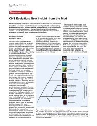
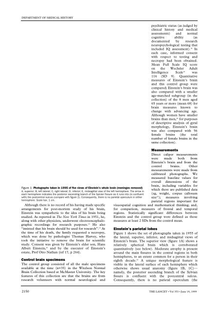
Figure 1: Photographs taken in 1995 <strong>of</strong> five views <strong>of</strong> <strong>Einstein</strong>’s whole <strong>brain</strong> (meninges removed)<br />
A, superior; B, left lateral; C, right lateral; D, inferior; E, midsagittal view <strong>of</strong> the left hemisphere. <strong>The</strong> arrow in<br />
each hemisphere indicates the posterior ascending branch <strong>of</strong> the Sylvian fissure as it runs into (is confluent<br />
with) the postcentral sulcus (compare with figure 2). Consequently, there is no parietal operculum in either<br />
hemisphere. Scale bar, 1 cm.<br />
Although there is no record <strong>of</strong> his having made specific<br />
arrangements for post-mortem study <strong>of</strong> his <strong>brain</strong>,<br />
<strong>Einstein</strong> was sympathetic to the idea <strong>of</strong> his <strong>brain</strong> being<br />
studied. As reported in <strong>The</strong> New York Times in 1951, he,<br />
along with other physicists, underwent electroencephalographic<br />
recordings for research purposes. 1 4 He also<br />
“insisted that his <strong>brain</strong> should be used for research”. 1 5 A t<br />
the time <strong>of</strong> his death, the family requested a necropsy,<br />
which was done by pathologist Thomas Harvey, who<br />
took the initiative to remove the <strong>brain</strong> for scientific<br />
study. Consent was given by <strong>Einstein</strong>’s elder son, Hans<br />
<strong>Albert</strong> <strong>Einstein</strong>, 1 6 and by the executor <strong>of</strong> <strong>Einstein</strong>’s<br />
estate, Pr<strong>of</strong> Otto Nathan (ref 17, p 264).<br />
Control <strong>brain</strong> specimens<br />
<strong>The</strong> control group consisted <strong>of</strong> all the male specimens<br />
available at the time (n=35) in the Witelson Normal<br />
Brain Collection based at McMaster University. <strong>The</strong> key<br />
features <strong>of</strong> this collection are that the <strong>brain</strong>s are from<br />
research volunteers with normal neurological and<br />
psychiatric status (as judged by<br />
clinical history and medical<br />
assessments) and normal<br />
cognitive ability (as<br />
documented by research<br />
neuropsychological testing that<br />
included IQ assessment). 1 8 I n<br />
each case, informed consent<br />
with respect to testing and<br />
necropsy had been obtained.<br />
Mean Full Scale IQ score<br />
o n the Wechsler Adult<br />
Intelligence Scale 1 9 w a s<br />
1 1 6 (SD 9). Quantitative<br />
measures <strong>of</strong> <strong>Einstein</strong>’s <strong>brain</strong><br />
and this control group were<br />
compared; <strong>Einstein</strong>’s <strong>brain</strong> was<br />
also compared with a smaller<br />
age-matched subgroup (in the<br />
collection) <strong>of</strong> the 8 men aged<br />
65 years or more (mean 68) for<br />
<strong>brain</strong> measures known to<br />
change with advancing age.<br />
Although women have smaller<br />
<strong>brain</strong>s than men, 2 0 for purposes<br />
<strong>of</strong> descriptive analysis <strong>of</strong> gyral<br />
morphology, <strong>Einstein</strong>’s <strong>brain</strong><br />
was also compared with 56<br />
female <strong>brain</strong>s (the total<br />
number <strong>of</strong> female <strong>brain</strong>s in the<br />
same collection).<br />
M e a s u r e m e n t s<br />
Direct caliper measurements<br />
were made both from<br />
<strong>Einstein</strong>’s <strong>brain</strong> and from the<br />
control <strong>brain</strong>s. Other<br />
measurements were made from<br />
calibrated photographs. We<br />
measured baseline values for<br />
overall dimensions <strong>of</strong> the<br />
<strong>brain</strong>, including variables for<br />
which there are published data<br />
(eg, weight, corpus callosum<br />
s i z e 2 1 ); measures involving<br />
parietal regions important for<br />
visuospatial cognition and mathematical thinking; and,<br />
for comparison, measures <strong>of</strong> frontal and temporal<br />
regions. Statistically significant differences between<br />
<strong>Einstein</strong> and the control group were defined as those<br />
measures at least 2 SDs from the control mean.<br />
<strong>Einstein</strong>’s parietal lobes<br />
Figure 1 shows the set <strong>of</strong> photographs taken in 1955 <strong>of</strong><br />
the lateral, superior, inferior, and midsagittal views <strong>of</strong><br />
<strong>Einstein</strong>’s <strong>brain</strong>. <strong>The</strong> superior view (figure 1A) shows a<br />
relatively spherical <strong>brain</strong> which is corroborated<br />
quantitatively (see below). Moderate atrophy is present<br />
around the main fissures in the central regions in both<br />
hemispheres, to an extent common for a person in their<br />
eighth decade. 2 2 A unique morphological feature is<br />
visible in the lateral surface <strong>of</strong> each hemisphere which<br />
otherwise shows usual anatomy (figure 1B, 1C)—<br />
namely, the posterior ascending branch <strong>of</strong> the Sylvian<br />
fissure is confluent with the postcentral sulcus.<br />
Consequently, there is no parietal operculum (the<br />
2150 THE LANCET • Vol 353 • June 19, 1999