U. Bellugi et al. (1999) - Duke-UNC Brain Imaging and Analysis Center
U. Bellugi et al. (1999) - Duke-UNC Brain Imaging and Analysis Center
U. Bellugi et al. (1999) - Duke-UNC Brain Imaging and Analysis Center
- No tags were found...

You also want an ePaper? Increase the reach of your titles
YUMPU automatically turns print PDFs into web optimized ePapers that Google loves.
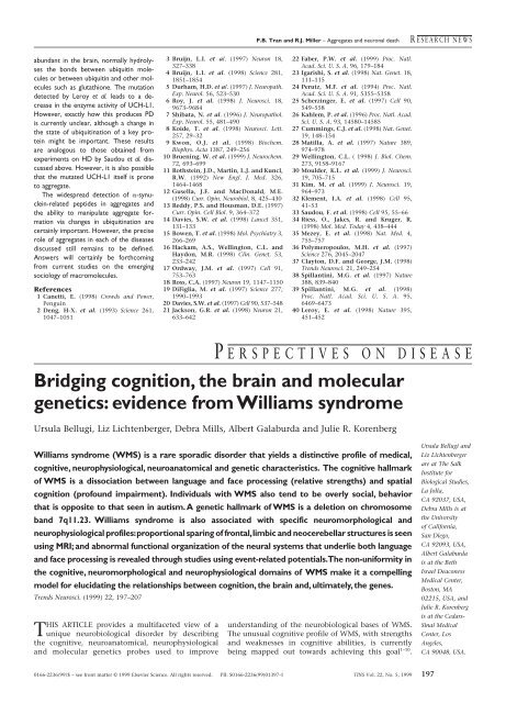
P.B. Tran <strong>and</strong> R.J. Miller – Aggregates <strong>and</strong> neuron<strong>al</strong> deathR ESEARCH NEWSabundant in the brain, norm<strong>al</strong>ly hydrolysesthe bonds b<strong>et</strong>ween ubiquitin moleculesor b<strong>et</strong>ween ubiquitin <strong>and</strong> other moleculessuch as glutathione. The mutationd<strong>et</strong>ected by Leroy <strong>et</strong> <strong>al</strong>. leads to a decreasein the enzyme activity of UCH-L1.However, exactly how this produces PDis currently unclear, <strong>al</strong>though a change inthe state of ubiquitination of a key proteinmight be important. These resultsare an<strong>al</strong>ogous to those obtained fromexperiments on HD by Saudou <strong>et</strong> <strong>al</strong>. discussedabove. However, it is <strong>al</strong>so possibl<strong>et</strong>hat the mutated UCH-L1 itself is pron<strong>et</strong>o aggregate.The widespread d<strong>et</strong>ection of -synuclein-relatedpeptides in aggregates <strong>and</strong>the ability to manipulate aggregate formationvia changes in ubiquitination arecertainly important. However, the preciserole of aggregates in each of the diseasesdiscussed still remains to be defined.Answers will certainly be forthcomingfrom current studies on the emergingsociology of macromolecules.References1 Can<strong>et</strong>ti, E. (1998) Crowds <strong>and</strong> Power,Penguin2 Deng, H-X. <strong>et</strong> <strong>al</strong>. (1993) Science 261,1047–10513 Bruijn, L.I. <strong>et</strong> <strong>al</strong>. (1997) Neuron 18,327–3384 Bruijn, L.I. <strong>et</strong> <strong>al</strong>. (1998) Science 281,1851–18545 Durham, H.D. <strong>et</strong> <strong>al</strong>. (1997) J. Neuropath.Exp. Neurol. 56, 523–5306 Roy, J. <strong>et</strong> <strong>al</strong>. (1998) J. Neurosci. 18,9673–96847 Shibata, N. <strong>et</strong> <strong>al</strong>. (1996) J. Neuropathol.Exp. Neurol. 55, 481–4908 Koide, T. <strong>et</strong> <strong>al</strong>. (1998) Neurosci. L<strong>et</strong>t.257, 29–329 Kwon, O.J. <strong>et</strong> <strong>al</strong>. (1998) Biochem.Biophys. Acta 1387, 249–25610 Bruening, W. <strong>et</strong> <strong>al</strong>. (<strong>1999</strong>) J. Neurochem.72, 693–69911 Rothstein, J.D., Martin, L.J. <strong>and</strong> Kuncl,R.W. (1992) New Engl. J. Med. 326,1464–146812 Gusella, J.F. <strong>and</strong> MacDon<strong>al</strong>d, M.E.(1998) Curr. Opin. Neurobiol. 8, 425–43013 Reddy, P.S. <strong>and</strong> Housman, D.E. (1997)Curr. Opin. Cell Biol. 9, 364–37214 Davies, S.W. <strong>et</strong> <strong>al</strong>. (1998) Lanc<strong>et</strong> 351,131–13315 Bowen, T. <strong>et</strong> <strong>al</strong>. (1998) Mol. Psychiatry 3,266–26916 Hackam, A.S., Wellington, C.L. <strong>and</strong>Haydon, M.R. (1998) Clin. Gen<strong>et</strong>. 53,233–24217 Ordway, J.M. <strong>et</strong> <strong>al</strong>. (1997) Cell 91,753–76318 Ross, C.A. (1997) Neuron 19, 1147–115019 DiFiglia, M. <strong>et</strong> <strong>al</strong>. (1997) Science 277,1990–199320 Davies, S.W. <strong>et</strong> <strong>al</strong>. (1997) Cell 90, 537–54821 Jackson, G.R. <strong>et</strong> <strong>al</strong>. (1998) Neuron 21,633–64222 Faber, P.W. <strong>et</strong> <strong>al</strong>. (<strong>1999</strong>) Proc. Natl.Acad. Sci. U. S. A. 96, 179–18423 Igarishi, S. <strong>et</strong> <strong>al</strong>. (1998) Nat. Gen<strong>et</strong>. 18,111–11524 Perutz, M.F. <strong>et</strong> <strong>al</strong>. (1994) Proc. Natl.Acad. Sci. U. S. A. 91, 5355–535825 Scherzinger, E. <strong>et</strong> <strong>al</strong>. (1997) Cell 90,549–55826 Kahlem, P. <strong>et</strong> <strong>al</strong>. (1996) Proc. Natl. Acad.Sci. U. S. A. 93, 14580–1458527 Cummings, C.J. <strong>et</strong> <strong>al</strong>. (1998) Nat. Gen<strong>et</strong>.19, 148–15428 Matilla, A. <strong>et</strong> <strong>al</strong>. (1997) Nature 389,974–97829 Wellington, C.L. ( 1998) J. Biol. Chem.273, 9158–916730 Moulder, K.L. <strong>et</strong> <strong>al</strong>. (<strong>1999</strong>) J. Neurosci.19, 705–71531 Kim, M. <strong>et</strong> <strong>al</strong>. (<strong>1999</strong>) J. Neurosci. 19,964–97332 Klement, I.A. <strong>et</strong> <strong>al</strong>. (1998) Cell 95,41–5333 Saudou, F. <strong>et</strong> <strong>al</strong>. (1998) Cell 95, 55–6634 Riess, O., Jakes, R. <strong>and</strong> Kruger, R.(1998) Mol. Med. Today 4, 438–44435 Mezey, E. <strong>et</strong> <strong>al</strong>. (1998) Nat. Med. 4,755–75736 Polymeropoulos, M.H. <strong>et</strong> <strong>al</strong>. (1997)Science 276, 2045–204737 Clayton, D.F. <strong>and</strong> George, J.M. (1998)Trends Neurosci. 21, 249–25438 Spillantini, M.G. <strong>et</strong> <strong>al</strong>. (1997) Nature388, 839–84039 Spillantini, M.G. <strong>et</strong> <strong>al</strong>. (1998)Proc. Natl. Acad. Sci. U. S. A. 95,6469–647340 Leroy, E. <strong>et</strong> <strong>al</strong>. (1998) Nature 395,451–452P ERSPECTIVES ON DISEASEBridging cognition, the brain <strong>and</strong> moleculargen<strong>et</strong>ics: evidence from Williams syndromeUrsula <strong>Bellugi</strong>, Liz Lichtenberger, Debra Mills, Albert G<strong>al</strong>aburda <strong>and</strong> Julie R. KorenbergWilliams syndrome (WMS) is a rare sporadic disorder that yields a distinctive profile of medic<strong>al</strong>,cognitive, neurophysiologic<strong>al</strong>, neuroanatomic<strong>al</strong> <strong>and</strong> gen<strong>et</strong>ic characteristics. The cognitive h<strong>al</strong>lmarkof WMS is a dissociation b<strong>et</strong>ween language <strong>and</strong> face processing (relative strengths) <strong>and</strong> spati<strong>al</strong>cognition (profound impairment). Individu<strong>al</strong>s with WMS <strong>al</strong>so tend to be overly soci<strong>al</strong>, behaviorthat is opposite to that seen in autism. A gen<strong>et</strong>ic h<strong>al</strong>lmark of WMS is a del<strong>et</strong>ion on chromosomeb<strong>and</strong> 7q11.23. Williams syndrome is <strong>al</strong>so associated with specific neuromorphologic<strong>al</strong> <strong>and</strong>neurophysiologic<strong>al</strong> profiles:proportion<strong>al</strong> sparing of front<strong>al</strong>,limbic <strong>and</strong> neocerebellar structures is seenusing MRI; <strong>and</strong> abnorm<strong>al</strong> function<strong>al</strong> organization of the neur<strong>al</strong> systems that underlie both language<strong>and</strong> face processing is reve<strong>al</strong>ed through studies using event-related potenti<strong>al</strong>s.The non-uniformity inthe cognitive, neuromorphologic<strong>al</strong> <strong>and</strong> neurophysiologic<strong>al</strong> domains of WMS make it a compellingmodel for elucidating the relationships b<strong>et</strong>ween cognition, the brain <strong>and</strong>, ultimately, the genes.Trends Neurosci. (<strong>1999</strong>) 22, 197–207THIS ARTICLE provides a multifac<strong>et</strong>ed view of aunique neurobiologic<strong>al</strong> disorder by describingthe cognitive, neuroanatomic<strong>al</strong>, neurophysiologic<strong>al</strong><strong>and</strong> molecular gen<strong>et</strong>ics probes used to improveunderst<strong>and</strong>ing of the neurobiologic<strong>al</strong> bases of WMS.The unusu<strong>al</strong> cognitive profile of WMS, with strengths<strong>and</strong> weaknesses in cognitive abilities, is currentlybeing mapped out towards achieving this go<strong>al</strong> 1–10 .Ursula <strong>Bellugi</strong> <strong>and</strong>Liz Lichtenbergerare at The S<strong>al</strong>kInstitute forBiologic<strong>al</strong> Studies,La Jolla,CA 92037, USA,Debra Mills is atthe Universityof C<strong>al</strong>ifornia,San Diego,CA 92093, USA,Albert G<strong>al</strong>aburdais at the B<strong>et</strong>hIsrael DeaconessMedic<strong>al</strong> <strong>Center</strong>,Boston, MA02215, USA, <strong>and</strong>Julie R. Korenbergis at the Cedars-Sinai Medic<strong>al</strong><strong>Center</strong>, LosAngeles,CA 90048, USA.0166-2236/99/$ – see front matter © <strong>1999</strong> Elsevier Science. All rights reserved. PII: S0166-2236(99)01397-1 TINS Vol. 22, No. 5, <strong>1999</strong> 197
P ERSPECTIVES ON DISEASEU. <strong>Bellugi</strong> <strong>et</strong> <strong>al</strong>. – Linking cognition <strong>and</strong> the brainWilliams syndrome is rare, occurring in an estimatedone in 20 000 to one in 30 000 live births. Diagnosticcharacteristics include specific faci<strong>al</strong> <strong>and</strong> physic<strong>al</strong> features:a constellation of cardiovascular difficulties,which include suprav<strong>al</strong>vular aortic stenosis (SVAS);failure to thrive in infancy; transient neonat<strong>al</strong> hyperc<strong>al</strong>cemia;delayed language <strong>and</strong> motor milestones,<strong>and</strong> abnorm<strong>al</strong> sensitivities to classes of sounds (hyperacusis).It has been recently found that in well over95% of the individu<strong>al</strong>s who have been diagnosed clinic<strong>al</strong>lywith WMS, there is a submicroscopic del<strong>et</strong>ion ofone copy of perhaps 20 contiguous genes, whichinclude the gene for elastin, among others, on chromosome7. The del<strong>et</strong>ion of one copy of elastin hasprovided a new gen<strong>et</strong>ic marker for WMS (Refs 11,12).Cognitive profile of WMSCognitive deficitsA h<strong>al</strong>lmark of WMS is the dissociation b<strong>et</strong>ween language(which is a strength in adolescents <strong>and</strong> adults)<strong>and</strong> spati<strong>al</strong> cognition (which is profoundly impaired),as shown in Fig. 1A. There are consistent cognitivedeficits in WMS: in gener<strong>al</strong>, st<strong>and</strong>ard Full Sc<strong>al</strong>e IQscores range from 40–100, with means of around 60(Refs 2,13; Fig. 1B). Many individu<strong>al</strong>s with WMS findaspects of gener<strong>al</strong> problem solving difficult; most arenot able to achieve independent living 14 <strong>and</strong> typic<strong>al</strong>lyexperience difficulty when de<strong>al</strong>ing with money or b<strong>al</strong>ancinga checkbook. However, within the context oftheir intellectu<strong>al</strong> impairments, individu<strong>al</strong>s with WMSdisplay characteristic ‘peaks’ <strong>and</strong> ‘v<strong>al</strong>leys’ in specificcognitive abilities. Complex expressive language abilitiesare relatively strong, spati<strong>al</strong> cognition is disproportionatelyimpaired (particularly at the level of glob<strong>al</strong>organization) <strong>and</strong> face-processing abilities are remarkablystrong. From studies across different populations, acharacteristic WMS cognitive profile is emerging 2,6,8,15 .Expressive language abilitiesOne striking aspect of the WMS profile is thestrength in language abilities in adolescence <strong>and</strong>adulthood, in contrast to the over<strong>al</strong>l impairment seenin cognitive abilities. Although in the earliest stages oflanguage development, children with WMS show significantdelay 16 , once language is acquired, this abilitytends to become a relative strength in their cognitiveprofile. When adolescents <strong>and</strong> adults with WMS <strong>and</strong>Down syndrome (DNS), both syndromes of ment<strong>al</strong>r<strong>et</strong>ardation that are gen<strong>et</strong>ic<strong>al</strong>ly based, are matched inage <strong>and</strong> Full Sc<strong>al</strong>e IQ, the differences in language skillsare evident at <strong>al</strong>l levels (phonologic<strong>al</strong>, lexic<strong>al</strong>, morphologic<strong>al</strong><strong>and</strong> syntactic, as well as at levels ofprosody, discourse <strong>and</strong> narrative). For example, adolescentswith WMS score significantly higher on measuresof receptive word knowledge than on measures ofgener<strong>al</strong> cognitive functioning, <strong>and</strong> perform dramatic<strong>al</strong>lyb<strong>et</strong>ter than their counterparts with DNS(Ref. 17). On a word-fluency test, which asks subjectsto name <strong>al</strong>l the anim<strong>al</strong>s they can in 60 seconds, adolescents<strong>and</strong> adults with WMS score similarly to norm<strong>al</strong>individu<strong>al</strong>s at the same ment<strong>al</strong> age. Moreover, theresponses of the WMS group might include manyatypic<strong>al</strong> examples (for example, chihuahua, ibex, condor)as well as typic<strong>al</strong> ones (Fig. 1C). As part of a basisfor their language strengths, short-term memory forspeech sounds or phonologic<strong>al</strong> working memory, aform of memory that is relevant to language learning<strong>and</strong> comprehension, is relatively preserved in theWMS population 6,18,19 . The strength in verb<strong>al</strong> memoryseen in WMS is apparent when contrasted withanother domain, that of spati<strong>al</strong> memory. Individu<strong>al</strong>swith WMS show far b<strong>et</strong>ter verb<strong>al</strong> memory than spati<strong>al</strong>memory, whereas individu<strong>al</strong>s with DNS exhibit theopposite pattern (Fig. 1D) 2,20 .In gener<strong>al</strong>, compared with age-matched <strong>and</strong> FullSc<strong>al</strong>e IQ-matched subjects with DNS, the subjects withWMS perform far b<strong>et</strong>ter on a wide vari<strong>et</strong>y of grammarprobes (reversible passives, negation, tag questions,sentence rep<strong>et</strong>ition, sentence compl<strong>et</strong>ion, sentencecorrection, condition<strong>al</strong>s, <strong>et</strong>c.; see Fig. 1E) 2,3,17 . Thus,language at <strong>al</strong>l levels in older individu<strong>al</strong>s with WMSis a remarkable strength, in light of the level of cognitivedeficits that they have gener<strong>al</strong>ly 17 . In fact, becaus<strong>et</strong>heir language abilities are often at a level that ishigher than their over<strong>al</strong>l cognitive abilities, individu<strong>al</strong>swith WMS might be perceived to be more capabl<strong>et</strong>han they re<strong>al</strong>ly are. Although the domain of languageis a strength of individu<strong>al</strong>s with WMS, there are cluesthat language might develop in somewhat differentways than in norm<strong>al</strong> children. Morphologic<strong>al</strong> errorsabate more slowly in children with WMS than in norm<strong>al</strong>children (but far more rapidly than in DNS) 21 <strong>and</strong>errors show some differences from those produced bynorm<strong>al</strong> children 6,15,22,23 . Studies are now showing thatthere are proportionately far more errors in the use ofspati<strong>al</strong> prepositions <strong>and</strong> in the use of language aboutspace by WMS individu<strong>al</strong>s than found in the norm<strong>al</strong>development (for example, when describing a pictureof an apple in a bowl during an experiment<strong>al</strong> task,WMS individu<strong>al</strong>s made errors such as ‘The apple isaround the bowl’, ‘The bowl is in the apple’ or ‘Theapple without the bowl’) 23 .There are controversi<strong>al</strong> issues raised by the researchon WMS concerning the relationship b<strong>et</strong>ween language<strong>and</strong> cognition, which are still a matter ofdebate 10,22–25 . Some researchers consider the syndrom<strong>et</strong>o be a remarkable example of the modularity of languageas a system that is separate from gener<strong>al</strong> cognitiveabilities. Others argue that as adults with WMS aresaid to function in some ways at the five- to sevenyear-oldlevel, there is a sufficient substrate of cognitiveabilities for the development of complex syntax,<strong>and</strong> that, thus, WMS does not represent a dissociationb<strong>et</strong>ween language <strong>and</strong> gener<strong>al</strong> cognitive functions.There are unresolved issues about the relationshipb<strong>et</strong>ween syntax <strong>and</strong> semantics, <strong>and</strong> about the intactnessof levels of language in WMS (Ref. 18), y<strong>et</strong>researchers agree gener<strong>al</strong>ly that language (for example,morphology <strong>and</strong> syntax) is a relative strength inWMS, which is apparently different from other syndromesthat can involve ment<strong>al</strong> r<strong>et</strong>ardation 2,6,17,18,24 .Abnorm<strong>al</strong>ly high linguistic affectA distinctive fac<strong>et</strong> of the language abilities of individu<strong>al</strong>swith WMS is their ability to use their heightened linguisticskills to engage others soci<strong>al</strong>ly. Many individu<strong>al</strong>swith WMS display a strong impulse towards soci<strong>al</strong> contact21,26 <strong>and</strong> affective expression, <strong>al</strong>though their soci<strong>al</strong>behavior is not <strong>al</strong>ways appropriate 27,28 . The intersectionof language behavior <strong>and</strong> soci<strong>al</strong> engagement in individu<strong>al</strong>swith WMS has been investigated through a series ofnarrative tasks in which subjects are asked to tell a storyfrom a wordless picture book 21,26 . Figure 1F shows examplesof the expressive language found in subjects withWMS compared to subjects with DNS (Ref. 2). The mostobvious distinction b<strong>et</strong>ween subjects with WMS, <strong>and</strong>198 TINS Vol. 22, No. 5, <strong>1999</strong>
U. <strong>Bellugi</strong> <strong>et</strong> <strong>al</strong>. – Linking cognition <strong>and</strong> the brain P ERSPECTIVES ON DISEASEAeareye headnosemouthElephantdrawingbodyElephant descriptionAnd what an elephant is it is one of theanim<strong>al</strong>s. And what the elphant does, it livesin the jungle. It can <strong>al</strong>so live in the zoo. Andwhat it has, it has long gray ears, fan ears,ears that can blow in the wind. It has a longtrunk that can pick up grass, or pick up hay...If they're in a bad mood it can be terrible... Ifthe elephant g<strong>et</strong>s mad it could stomp; itcould charge. Som<strong>et</strong>imes elephants cancharge. They have long tusks. You don'twant an elephant as a p<strong>et</strong>. You want a cat ora dog or a bird...BCount20161284020 40 60 80 100 120 140 160Wechsler Full Sc<strong>al</strong>e IQCWords produced12840WMS DNS NCUncommonwordsCommonwordsWord fluency (60 s test): 'Name <strong>al</strong>l the anim<strong>al</strong>s you can'WMS: brontosaurus, `tryr<strong>and</strong>on',dinosaurs, ibex, brontosaurusrex, elephant, dog, cat, lion,baby hippopotamus, wh<strong>al</strong>e, bull,yak, zebra, puppy, kitten, tiger,ko<strong>al</strong>a, dragonDNS: dogs, cats, fish, bird, fishDFree rec<strong>al</strong>l6420DigitspanCorsiblocksCorsi-blocksapparatusWMSDNSE% CorrectGrammar Context100806040200WMS DNS WMS DNSExperimenter question:'What if you were a bird?'DNS 1: Bird seeds.DNS 2: You'd be strong.DNS 3: I don't fly.DNS 4: I not a bird, you have wing.DNS 5: Fly in the air.WMS 1: You could fly, you could havebabies, fly north or south, east or west.WMS 2: Good question. I'd fly through theair being free.WMS 3: I would fly through the air <strong>and</strong> soarlike an airplane <strong>and</strong> dive through trees likea bird <strong>and</strong> l<strong>and</strong> like a bird.WMS 4: I would fly through the air wheremy parents would never find me. Birds wantto be independent.WMS 5: I would fly <strong>and</strong> if I liked a boy, Iwould l<strong>and</strong> on his head <strong>and</strong> start chirping.FWMS age 17, Full Sc<strong>al</strong>e IQ = 50Once upon a time when it was dark at night...theboy had a frog. The boy was looking at thefrog...sitting on the chair, on the table, <strong>and</strong> the dogwas looking through...looking up to the frog in a jar.That night he sleeped <strong>and</strong> slept for a long time, thedog did. But, the frog was not gonna go to sleep.And when the frog went out...the boy <strong>and</strong> the dogwere still sleeping. The next morning it wasbeautiful in the morning. It was bright <strong>and</strong> the sunwas nice <strong>and</strong> warm. Then suddenly when heopened his eyes...he looked at the jar <strong>and</strong> then suddenly the frog wasnot there. The jar was empty. There was no frog to found (whispered).DNS age 18, Full Sc<strong>al</strong>e IQ = 55The frog is in the jar. The jar is on the floor. The jar on the floor. That' sit. The stool is broke. The clothes is laying there.Fig. 1. Unusu<strong>al</strong> language <strong>and</strong> cognitive profiles in Williams syndrome (WMS). A characteristic of WMS is the dissociation b<strong>et</strong>ween language<strong>and</strong> space. (A) shows a drawing <strong>and</strong> description of an elephant by a teenager with WMS, Full Sc<strong>al</strong>e IQ of 49, Verb<strong>al</strong> IQ of 52 <strong>and</strong> PerformanceIQ of 54. Note the difference b<strong>et</strong>ween the impoverished drawing <strong>and</strong> grammatic<strong>al</strong>ly compl<strong>et</strong>e language. (B) Wechsler Full Sc<strong>al</strong>e IQs range from40 to 100 in WMS <strong>and</strong> are reasonably norm<strong>al</strong>ly distributed, with a mean IQ of approximately 60 (solid line; SD 11). Broken line shows populationdistribution of Full Sc<strong>al</strong>e IQs (mean 100; SD 11). (C) On a semantic fluency task, subjects with WMS give the same number of commonresponses but significantly more uncommon infrequent responses (for example, ibex, yak, dragon) than either matched subjects with Downsyndrome (DNS) or norm<strong>al</strong> subjects matched for ment<strong>al</strong> age (NC; n 10 in each group). The double dissociation b<strong>et</strong>ween verb<strong>al</strong> <strong>and</strong> spati<strong>al</strong>short-term memory is shown in (D): age-matched <strong>and</strong> Full Sc<strong>al</strong>e IQ-matched individu<strong>al</strong>s with WMS (n 10) <strong>and</strong> DNS (n 9) demonstrate adifferenti<strong>al</strong> ability to remember a list of numbers in order (Digit Span) versus remembering location <strong>and</strong> order of blocks (Corsi blocks) 2 . (E)Individu<strong>al</strong>s with WMS perform significantly b<strong>et</strong>ter than individu<strong>al</strong>s with DNS (age-matched <strong>and</strong> Full Sc<strong>al</strong>e IQ-matched) in syntactic processingtasks (for example, condition<strong>al</strong> sentences) on both grammar <strong>and</strong> content (for example, ‘Good question. I’d fly through the air being free.’). Norm<strong>al</strong>control results are shown by the broken line. (F) Individu<strong>al</strong>s with WMS use more affective prosody, more audience ‘hookers’ <strong>and</strong> more linguisticaffective devices than do norm<strong>al</strong>s or individu<strong>al</strong>s with DNS at any age. Subjects are asked to tell a story from a wordless picture book <strong>and</strong> the WMSsubjects tend to be dramatic story tellers. The figure shows samples of a story-telling task by age-matched <strong>and</strong> Full Sc<strong>al</strong>e IQ-matched individu<strong>al</strong>swith WMS <strong>and</strong> DNS. The WMS individu<strong>al</strong> shown is 17 years old, Full Sc<strong>al</strong>e IQ 50, Verb<strong>al</strong> IQ 54 <strong>and</strong> Performance IQ 55; the DNS individu<strong>al</strong>shown is 18 years old, Full Sc<strong>al</strong>e IQ 54, Verb<strong>al</strong> IQ 59 <strong>and</strong> Performance IQ 53 (Ref. 2). (D) <strong>and</strong> (F) reproduced, with permission,from Ref. 2.subjects with DNS <strong>and</strong> age-matched norm<strong>al</strong> controls, isin the use of narrative enrichment devices during thisstory-telling task by subjects with WMS. The enrichmentdevices (addition of affective qu<strong>al</strong>ities) are not found inthe pictures themselves, but are added to the narrative bythe subject as linguistic affect. Individu<strong>al</strong>s with WMSshow an abundance of affectivity in both prosody <strong>and</strong>lexic<strong>al</strong> devices <strong>and</strong> appear to be able to manipulate affectivelinguistic devices for the purposes of storytelling 21,26,29,30 . Affective prosody was measured by notinghow frequently par<strong>al</strong>inguistic affective expression wasused, including pitch change, voc<strong>al</strong>ic lengthening <strong>and</strong>TINS Vol. 22, No. 5, <strong>1999</strong> 199
P ERSPECTIVES ON DISEASEU. <strong>Bellugi</strong> <strong>et</strong> <strong>al</strong>. – Linking cognition <strong>and</strong> the brainAFreedrawingof a houseBBlockdesigntaskCLoc<strong>al</strong>–glob<strong>al</strong>taskExampleModelYYYY YYYYYYY YY Y YModellivingroomWilliams syndrome(poor on glob<strong>al</strong> organization)roofdoorwindowsdoorswimming poolsidew<strong>al</strong>ktwo-storeyhousewindowsmodifications in volume. Affectivity in lexic<strong>al</strong> devicesis noted in the frequency of exclamatory phrases <strong>and</strong>other audience engagement devices (for example,‘Suddenly splash! The water came up’; ‘Lo <strong>and</strong> behold’or ‘Gadzooks! Guess what happened next!’). This patternof increased linguistic affectivity is strikingly differentfrom subjects with DNS, as well as from norm<strong>al</strong>individu<strong>al</strong>s at any age or other contrast groups (forexample, individu<strong>al</strong>s with early foc<strong>al</strong> brain lesions) 31–33 .Thus, in adolescents <strong>and</strong> adultswith WMS, expressive language istypic<strong>al</strong>ly a great strength <strong>and</strong> isused effectively (<strong>and</strong> som<strong>et</strong>imeseffusively) in soci<strong>al</strong> situations. Thisis exemplified by the WMS teenagegirl who said, ‘Everyone in the worldis my friend’. Individu<strong>al</strong>s withWMS, therefore, exhibit a strikingcontrast to the soci<strong>al</strong> <strong>and</strong> languageprofiles of individu<strong>al</strong>s with otherdisorders such as autism 31 .Strengths <strong>and</strong> deficits in visu<strong>al</strong>ly basedcognition in WMSIn studies that examine spati<strong>al</strong>cognition, as opposed to language,subjects with WMS are significantlymore impaired than subjectswith DNS across <strong>al</strong>l age rangesexamined 3,34,35 . Indeed, individu<strong>al</strong>swith WMS performed more poorlyin our studies than children withearly right-hemisphere lesions 36 .Difficulties with spati<strong>al</strong> cognitionin WMS seem to be especi<strong>al</strong>ly acutewith respect to the glob<strong>al</strong>, ratherthan the loc<strong>al</strong>, level of spati<strong>al</strong> organizationacross tasks. Lack of cohesionor glob<strong>al</strong> organization is typic<strong>al</strong>in drawings by subjects withWMS, while subjects with DNStend to show a different effect 37,38 .For example, a subject with WMS might draw a housewith windows <strong>and</strong> a door as separate entities from thehouse itself, thus lacking glob<strong>al</strong> organization (Fig. 2A).By contrast, a typic<strong>al</strong> drawing by a subject with DNSmight be simplified y<strong>et</strong> exhibit proper gest<strong>al</strong>t relationshipsamong elements. On block-design tasks, bothWMS <strong>and</strong> DNS subjects perform poorly, but in differentways, with WMS subjects typic<strong>al</strong>ly unable to organiz<strong>et</strong>he blocks into a glob<strong>al</strong> pattern, <strong>and</strong> DNS subjectsinstead making errors of intern<strong>al</strong>d<strong>et</strong>ail (Fig. 2B). Difficulties withintegration of simple shapes have<strong>al</strong>so been shown on tasks thatrequire subjects to copy a drawingof geom<strong>et</strong>ric forms from a model 2,3 .When asked to reproduce a largefigure made up of sm<strong>al</strong>ler figures(for example, a large ‘D’ made ofsm<strong>al</strong>l ‘Y’s), subjects with WMStend to produce primarily the loc<strong>al</strong>constituent forms sprinkled acrossthe page <strong>and</strong> fail to reproduce theglob<strong>al</strong> form, whereas subjects withDNS tend to show the oppositeDown syndrome(poor on intern<strong>al</strong> d<strong>et</strong>ail)Fig. 2. Different spati<strong>al</strong> deficits in Williams syndrome (WMS) <strong>and</strong> Down syndrome (DNS). (A) The drawings byadolescents <strong>and</strong> adults with WMS contain many parts of houses but they are not organized coherently. In contrast, thedrawings of age-matched <strong>and</strong> Full Sc<strong>al</strong>e IQ-matched DNS adults are simplified but have the correct over<strong>al</strong>l gest<strong>al</strong>t. (B)In the block-design task both subjects with WMS <strong>and</strong> subjects with DNS fail (sc<strong>al</strong>ed scores more than 2 SD belownorm<strong>al</strong>), but they fail in very different ways: adolescents <strong>and</strong> adults with WMS show disjointed <strong>and</strong> fragmented designs,while age-matched <strong>and</strong> Full Sc<strong>al</strong>e IQ-matched DNS subjects make errors in intern<strong>al</strong> d<strong>et</strong>ails while maintaining the over<strong>al</strong>lconfiguration. (C) In the Delis hierarchic<strong>al</strong> processing task, subjects are asked to copy a large glob<strong>al</strong> figure made ofsm<strong>al</strong>ler loc<strong>al</strong> forms (a ‘D’ made out of ‘Y’s). Again, both groups fail but in significantly different ways: subjects withWMS tend to produce the loc<strong>al</strong> elements sprinkled across the page, whereas age-matched <strong>and</strong> Full Sc<strong>al</strong>e IQ-matchedsubjects with DNS tend to produce only the glob<strong>al</strong> forms.APercent correct10080604020064 53789Model2110118 10 12 12 12 14 14 14 15 15 16 16 17 18 20 22Age (years)BPercent correct100Model8060402008 10 12 12 12 14 14 14 15 15 16 16 17 18 20 22Age (years)Fig. 3. Spati<strong>al</strong> cognition (impaired) <strong>and</strong> face processing (relatively spared) in Williams syndrome (WMS). Thestrengths <strong>and</strong> weaknesses in visuo–spati<strong>al</strong> processing in WMS show an unusu<strong>al</strong> profile. The results from two tasks thatare both visuo–perceptu<strong>al</strong> tasks, sensitive to right-hemisphere damage, where the correct answer requires only pointingare shown. The results for the same subjects are shown for each task. Note that the same subjects with WMS performvery differently on the two tasks. (A) Subjects with WMS perform very poorly on judging the orientation of lines (Bentonjudgement of line orientation), in keeping with their spati<strong>al</strong> deficit. Sever<strong>al</strong> subjects cannot even pass the warm-up items.(B) In great contrast with this, exactly the same subjects with WMS perform remarkably well on a very difficult face discriminationtask (Benton face recognition), which involves recognizing the same individu<strong>al</strong> under different conditions oflighting, shadow <strong>and</strong> orientation. In both parts, the performance of norm<strong>al</strong> subjects is indicated by the broken line.pattern 2,37 (Fig. 2C).Face processing is a remarkablestrength in individu<strong>al</strong>s with WMS,in great contrast to their othervisu<strong>al</strong>–spati<strong>al</strong> cognitive deficits 2,39 .This contrast is highlighted whencomparing two tasks, each ofwhich requires only pointing tothe correct answers: one involvesjudging the orientation of lines (aspati<strong>al</strong> task) <strong>and</strong> the other involves200 TINS Vol. 22, No. 5, <strong>1999</strong>
discrimination of unfamiliar facesin different conditions of lighting<strong>and</strong> orientation (a face-processingtask). Individu<strong>al</strong>s with WMS showextreme difficulty with the spati<strong>al</strong>task <strong>and</strong> obtain scores in the rangeconsidered to be severely deficient;som<strong>et</strong>imes they are not even abl<strong>et</strong>o do the simplest items. In contrast,the same WMS individu<strong>al</strong>sshow a remarkable strength in theability to recognize faces 40,41 (Fig. 3).Across different face-processingtasks (recognition, classification,memory), individu<strong>al</strong>s with WMSshow strong performance 41 .Different trajectories in cognitivedomainsWhat is interesting about individu<strong>al</strong>swith WMS is that there areareas of serious deficits (gener<strong>al</strong>intelligence, visuo–spati<strong>al</strong> abilities)but <strong>al</strong>so areas of relative strengths(face processing, expressive language).Questions about the relationshipsb<strong>et</strong>ween the deficits <strong>and</strong>strengths emerge from the researchfindings on WMS: do the differentcognitive abilities depend on oneanother or can they be dissociatedfrom one another? Do they chang<strong>et</strong>hroughout development? What ar<strong>et</strong>he underlying brain systems forthese strengths <strong>and</strong> deficits in abilitiesin cognitive domains? The associationsb<strong>et</strong>ween <strong>and</strong> within differenttypes of abilities have beenexamined through correlation<strong>al</strong>studies. In WMS, correlations b<strong>et</strong>ween measures of faceprocessing are strong, but measures of different aspectsof language functioning <strong>and</strong> visuo–spati<strong>al</strong> abilities arenot correlated with face-processing abilities 34,41,42 .Across three cognitive domains, distinct trajectories ofdevelopment are found in studies involving individu<strong>al</strong>swith WMS b<strong>et</strong>ween the ages of 5 <strong>and</strong> 29 (n 71). InWMS, there are clearly different trajectories across th<strong>et</strong>hree domains reported (lexic<strong>al</strong> knowledge, spati<strong>al</strong> cognition<strong>and</strong> face processing) (Fig. 4A). In contrast, individu<strong>al</strong>swith DNS show essenti<strong>al</strong>ly uniformly depresseddevelopment (Fig. 4B). In a lexic<strong>al</strong> knowledge task, childrenwith WMS begin with very low scores, but show asharp increase with age (unlike DNS children). On ast<strong>and</strong>ard drawing task, individu<strong>al</strong>s with WMS scoreconsistently lower than individu<strong>al</strong>s with DNS at <strong>al</strong>l agelevels <strong>and</strong> reach a plateau early in development. On aface-processing task, individu<strong>al</strong>s with WMS tend to performvery well, even at a relatively early age, <strong>and</strong> continu<strong>et</strong>o do well throughout development 35 . Over<strong>al</strong>l,subjects with WMS perform significantly b<strong>et</strong>ter duringdevelopment than DNS subjects on face-processing <strong>and</strong>language tasks, but significantly less well than DNSsubjects on visu<strong>al</strong>–spati<strong>al</strong> tasks 35 (see Fig. 4C).The neurophysiologic<strong>al</strong> profile of WMSThe neurobiologic<strong>al</strong> profile of individu<strong>al</strong>s with WMSis being reve<strong>al</strong>ed through studies of brain function[event-related potenti<strong>al</strong>s (ERPs)], brain structure 3DAMent<strong>al</strong> age (years)14106Faces (Benton)Age at test (years)CU. <strong>Bellugi</strong> <strong>et</strong> <strong>al</strong>. – Linking cognition <strong>and</strong> the brain P ERSPECTIVES ON DISEASEVocabulary(PPVT-R)Spati<strong>al</strong>(VMI)223 7 11 15 19 23 3 7 11 15 19 23Ment<strong>al</strong> age equiv<strong>al</strong>ent10Faces (Benton)8 Vocabulary(PPVT-R)6Spati<strong>al</strong>(VMI)4WMSBMent<strong>al</strong> age (years)DNSAge at test (years)computer-graphic an<strong>al</strong>yses of MRI) <strong>and</strong> brain cytoarchitectonicsin autopsy brains. Initi<strong>al</strong> propos<strong>al</strong>sabout how the cognitive <strong>and</strong> brain profiles might belinked are presented in this article.Studies using ERP techniques are useful in assessingthe timing <strong>and</strong> organization of the neur<strong>al</strong> systems thatare active during sensory, cognitive <strong>and</strong> linguistic processingin subjects with WMS (Refs 43–48). Eventrelatedpotenti<strong>al</strong>s provide information about the timing<strong>and</strong> tempor<strong>al</strong> sequence of neur<strong>al</strong> events <strong>and</strong>, tosome extent, the location of neur<strong>al</strong> activity. Electrodesare placed on the sc<strong>al</strong>p over specific brain areas whilesubjects are processing information, which, thus,<strong>al</strong>lows the monitoring of the time course of neur<strong>al</strong>activation on a millisecond to millisecond basis. Therecorded activity occurs before subjects make an overtresponse. Studies of brain-wave activity during language<strong>and</strong> face-processing paradigms in individu<strong>al</strong>swith WMS <strong>and</strong> norm<strong>al</strong> individu<strong>al</strong>s are reported in thisarticle.A neurophysiologic<strong>al</strong> marker for auditory language processingThe morphology of ERP components to auditorywords was dramatic<strong>al</strong>ly different in individu<strong>al</strong>s withWMS from norm<strong>al</strong> controls. Event-related potenti<strong>al</strong>swere recorded as subjects listened to sentences that werepresented one word at a time. The fin<strong>al</strong> word in eachsentence either provided good closure or was semantic<strong>al</strong>lyanom<strong>al</strong>ous (for example, ‘I have five fingers on mymoon’). The results reve<strong>al</strong>ed that the morphology of14106Faces (Benton)Spati<strong>al</strong> (VMI)Vocabulary (PPVT-R)Fig. 4. Three domains of cognition in Williams syndrome (WMS) but not in Down syndrome (DNS). (A) Development<strong>al</strong>trajectories of contrasts b<strong>et</strong>ween language, face <strong>and</strong> space processing in WMS are shown. Subjects of <strong>al</strong>l ageswith WMS show distinctly different trajectories in three cognitive domains: lexic<strong>al</strong> knowledge, spati<strong>al</strong> cognition <strong>and</strong> faceprocessing. On a st<strong>and</strong>ardized test of vocabulary [the Peabody picture vocabulary test-revised (PPVT-R)], subjects withWMS start with low scores <strong>and</strong> then show a sharp increase in score with age. On a probe of spati<strong>al</strong> cognition thatinvolves copying geom<strong>et</strong>ric shapes [the development<strong>al</strong> test of visu<strong>al</strong> moror integration (VMI)], the performances of subjectswith WMS are consistently below those of subjects with DNS, <strong>and</strong> plateau at an early age. On a task of face processing(the Benton test of faci<strong>al</strong> recognition), subjects with WMS perform extremely well even at very young ages. (B)Subjects with DNS show essenti<strong>al</strong>ly the same development<strong>al</strong> trajectory across the three domains. In contrast, subjectswith WMS show three distinctly different trajectories. (C) Planned contrasts show that performance on the three testsdiffers significantly within the WMS group, even when controlled for age. No b<strong>et</strong>ween-test differences are found in theDNS group. A 2 3 (WMS, DNS Benton, VMI, PPVT-R) an<strong>al</strong>ysis of covariance with chronologic<strong>al</strong> age entered as thecovariate reve<strong>al</strong>ed a significant group–test interaction (P 0.0001).TINS Vol. 22, No. 5, <strong>1999</strong> 201
P ERSPECTIVES ON DISEASEU. <strong>Bellugi</strong> <strong>et</strong> <strong>al</strong>. – Linking cognition <strong>and</strong> the brainABLeft hemisphere, tempor<strong>al</strong>, open-class words2 µV100 msID matchP200Right hemisphere, front<strong>al</strong>2.5 µV100 msNorm<strong>al</strong> controlsNorm<strong>al</strong> controlsN200Mismatch2 µVWilliams syndrome100 ms2.5 µV100 msP200Williams syndromeN200Fig. 5. Neurophysiologic<strong>al</strong> markers for Williams syndrome (WMS) in language <strong>and</strong> faceprocessing. (A) In event-related potenti<strong>al</strong> (ERP) recordings of online processing of grammatic<strong>al</strong><strong>and</strong> semantic information in sentences, subjects listened to sentences that ended either in asemantic<strong>al</strong>ly probable way or in an anom<strong>al</strong>ous way. In norm<strong>al</strong> individu<strong>al</strong>s, there is a lefthemisphereasymm<strong>et</strong>ry for closed-class items <strong>and</strong> a difference b<strong>et</strong>ween open- <strong>and</strong> closed-classitems. In contrast, subjects with WMS do not show the left-hemisphere asymm<strong>et</strong>ry for words,nor do they show the typic<strong>al</strong> difference b<strong>et</strong>ween open- <strong>and</strong> closed-class words (not shown). Theunusu<strong>al</strong> wave form to auditory words exhibited by <strong>al</strong>l subjects with WMS <strong>and</strong> none of the norm<strong>al</strong>controls, involving a large positivity at 200 ms (P200), which is particularly evident overleft tempor<strong>al</strong> regions is shown here. This is a c<strong>and</strong>idate neurophysiologic<strong>al</strong> marker for WMS.(B) ERP recordings were made as subjects watched photographic pairs of faces presentedsequenti<strong>al</strong>ly on a computer monitor. The subject’s task was to indicate wh<strong>et</strong>her the second facein the pair was that of the same or a different person as in the first photograph, some presentedas upright faces, some as inverted. The response when the second face was the sameas the first (ID match) <strong>and</strong> when the second face is different (mismatch) is indicated. In norm<strong>al</strong>individu<strong>al</strong>s, there is a right-hemisphere asymm<strong>et</strong>ry for processing faces, but this is notfound in subjects with WMS. The abnorm<strong>al</strong>ly large negativity at 200 ms (N200) in subjectswith WMS but not in norm<strong>al</strong> controls or other groups tested, which occurs over brain regionsis shown in this figure. This is <strong>al</strong>so a c<strong>and</strong>idate neurophysiologic<strong>al</strong> marker for WMS.Reproduced, with permission, from Ref. 1.WMS individu<strong>al</strong>s’ ERP components to auditory wordswas different from norm<strong>al</strong> controls 46,47 . A unique patternof ERPs that includes prominent positivities at 50<strong>and</strong> 200 ms (c<strong>al</strong>led the P50 <strong>and</strong> P200 components)<strong>and</strong> a sm<strong>al</strong>ler than norm<strong>al</strong> negativity at 100 ms (theN100 component), was most striking over tempor<strong>al</strong>brain regions. This pattern of components, that is,abnorm<strong>al</strong>ly large P50, a sm<strong>al</strong>ler-than-norm<strong>al</strong> N100<strong>and</strong> a large P200, was found in <strong>al</strong>l subjects with WMS(Fig. 5A), <strong>and</strong> was not found in norm<strong>al</strong> school-agechildren or adults 43 , which suggests that this patternmight emerge as a marker for WMS.In age-matched norm<strong>al</strong> controls, there are differencesin ERP responses to open- <strong>and</strong> closed-class words.Open-class words, which typic<strong>al</strong>ly convey meaning (forexample, nouns, verbs <strong>and</strong> adjectives), elicit a negativityat 400 ms that tends to be larger in posterior regionsof the right hemisphere. Closed-class words, whichtypic<strong>al</strong>ly convey information about grammatic<strong>al</strong> relations(for example, articles, prepositions, conjunctions),elicit a negativity that peaks somewhat earlier <strong>and</strong> islargest over anterior regions of the left hemisphere innorm<strong>al</strong> subjects. Unlike norm<strong>al</strong> subjects, individu<strong>al</strong>swith WMS do not show ERP differences to open- <strong>and</strong>closed-class words, nor do they show the norm<strong>al</strong> lefthemispherebrain asymm<strong>et</strong>ries to closed-class words. Innorm<strong>al</strong> individu<strong>al</strong>s, the semantic<strong>al</strong>ly anom<strong>al</strong>ous fin<strong>al</strong>word elicits an N400 component (negativity that peaksat 400 ms) that is larger from the right than the lefthemisphere. The N400 effect is larger over the lefthemisphere in individu<strong>al</strong>s with WMS than in norm<strong>al</strong>control individu<strong>al</strong>s. This larger semantic anom<strong>al</strong>ymight be related to the unusu<strong>al</strong> semantic proclivitiesshown by subjects with WMS in lexic<strong>al</strong> tasks. Thus, theresults that show ERP differences b<strong>et</strong>ween subjects withWMS <strong>and</strong> norm<strong>al</strong> subjects in language processing suggestthat the neur<strong>al</strong> organization of these aspects of languagemight be different b<strong>et</strong>ween these groups despit<strong>et</strong>he apparent relative sparing of language abilities insubjects with WMS (Refs 45,47).A neurophysiologic<strong>al</strong> marker for face processingFace-processing ERP data on ten subjects with WMS<strong>and</strong> 20 norm<strong>al</strong> controls were obtained from the taskdescribed in Fig. 5B. Results showed that both theWMS <strong>and</strong> norm<strong>al</strong> control groups displayed ERP differencesto matched versus mismatched upright faces,which consisted of a negativity to the mismatchedfaces approximately 320 ms after the ons<strong>et</strong> of the secondstimulus 43 . However, the norm<strong>al</strong> subjects showedthe largest N320 component over anterior regions,which was greater over the right hemisphere than theleft; the subjects with WMS did not show this asymm<strong>et</strong>ry.In contrast to the norm<strong>al</strong> adults, the subjectswith WMS <strong>al</strong>so displayed an abnorm<strong>al</strong>ly large negativity(at 200 ms) to upright faces (approximately fourtimes the amplitude of the N200 component to facesin norm<strong>al</strong> adults; see Fig. 5B), but not to pictures ofobjects. These results appear to be specific to WMS <strong>and</strong>might be related to the increased attention paid tofaces by individu<strong>al</strong>s with WMS. The abnorm<strong>al</strong>ly largenegativity at 200 ms, which occurred in <strong>al</strong>l subjectswith WMS but not in other groups, could be suggestiveof a brain-activity marker that is linked to the notedstrength in face-processing abilities found inindividu<strong>al</strong>s with WMS (Ref. 43). Neurophysiologic<strong>al</strong>indices that relate brain <strong>and</strong> behavior, <strong>and</strong> that mightbe phenotypic markers for WMS are suggested by theseneurophysiologic<strong>al</strong> studies. Unique brain-wave markers,one found during face processing <strong>and</strong> a differentone found during language processing, could be characteristicof individu<strong>al</strong>s with WMS but not of othergroups 43,45 . Taken tog<strong>et</strong>her, these findings suggest thatin individu<strong>al</strong>s with WMS the neur<strong>al</strong> systems that subservehigher cognitive functions, such as language <strong>and</strong>face processing, are different from norm<strong>al</strong> individu<strong>al</strong>s.Neuroanatomic<strong>al</strong> characteristics from structur<strong>al</strong>MRINeuroanatomic<strong>al</strong> studies that contrast WMS <strong>and</strong>DNS subjects with norm<strong>al</strong> controls have been undertakento investigate the neur<strong>al</strong> systems that mediat<strong>et</strong>he cognitive profile of individu<strong>al</strong>s with WMS (Refs49–51). The results characterize some of the over<strong>al</strong>lgross morphologic<strong>al</strong> differences b<strong>et</strong>ween the two syndromes<strong>and</strong> <strong>al</strong>so provide data on more specific<strong>al</strong>lytarg<strong>et</strong>ed morphologic<strong>al</strong> underpinnings of the uneven202 TINS Vol. 22, No. 5, <strong>1999</strong>
U. <strong>Bellugi</strong> <strong>et</strong> <strong>al</strong>. – Linking cognition <strong>and</strong> the brain P ERSPECTIVES ON DISEASEcognitive profile of individu<strong>al</strong>s with WMS (Refs2,3,34,41,42).Three groups, WMS, DNS <strong>and</strong> norm<strong>al</strong> controls, werestudied using MRI (Fig. 6 displays some of theresults) 49–51 . The front<strong>al</strong> cortex of individu<strong>al</strong>s withWMS has an essenti<strong>al</strong>ly norm<strong>al</strong> volume relationshipwith the posterior cortex but in DNS the front<strong>al</strong> cortexis disproportionately reduced in volume (Fig. 6A).Limbic structures of the tempor<strong>al</strong> lobe (includinguncus, amygd<strong>al</strong>a, hippocampus <strong>and</strong> parahippocamp<strong>al</strong>gyrus) are proportionately spared in subjects withWMS relative to other cerebr<strong>al</strong> structures, while theselimbic structures are dramatic<strong>al</strong>ly reduced in volumein DNS (Fig. 6B). Addition<strong>al</strong>ly, cerebellar size isreduced in subjects with DNS but is entirely norm<strong>al</strong>in subjects with WMS. Importantly, in WMS, whilep<strong>al</strong>eocerebellar verm<strong>al</strong> lobules subtend a sm<strong>al</strong>ler areaon midsagitt<strong>al</strong> sections than in norm<strong>al</strong>s, neocerebellarlobules are actu<strong>al</strong>ly larger 49,51 . In a separate studyinvolving MRI images from 11 subjects with WMS,seven subjects with DNS <strong>and</strong> 18 norm<strong>al</strong> controls (aged10–20 years), neocerebellar tonsils of WMS subjectswere found to be equ<strong>al</strong> in volume to those of controls<strong>and</strong> significantly larger than those of subjects withDNS. In proportion to the cerebrum, tonsils in subjectswith WMS are larger than in both these groups 44 .In contrast, in subjects with DNS, the mean volume ofsubcortic<strong>al</strong> areas, which include lenticular nuclei, isproportion<strong>al</strong>ly large when compared with those areasin individu<strong>al</strong>s with WMS <strong>and</strong> controls (Fig. 6C).Quantitative volum<strong>et</strong>ric an<strong>al</strong>ysis of Heschl’s gyrus, anarea in the primary auditory cortex, shows that in theWMS group the absolute volume of Heschl’s gyrusdoes not differ from norm<strong>al</strong> control subjects despit<strong>et</strong>he significant cerebr<strong>al</strong> hypoplasia evident in theWMS group. However, compared to subjects withDNS, matched for supratentori<strong>al</strong> volume, Heschl’sgyrus is significantly larger in the WMS group 48 .The differences within cerebr<strong>al</strong> <strong>and</strong> cerebellar structuressuggest that relatively intact linguistic <strong>and</strong> affectivefunctions in subjects with WMS might rely uponthe relatively norm<strong>al</strong> development of some limbic,front<strong>al</strong> cortic<strong>al</strong> <strong>and</strong> cerebellar structures 2,3,52 . The relativesparing of front<strong>al</strong> <strong>and</strong> cerebellar structures insubjects with WMS might contribute to their relativelinguistic comp<strong>et</strong>ence. The significantly b<strong>et</strong>ter spati<strong>al</strong><strong>and</strong> motor abilities in subjects with DNS might rely onthe proportion<strong>al</strong> preservation of subcortic<strong>al</strong> structuresin that group.A significant correlation was found b<strong>et</strong>ween st<strong>and</strong>ardizedlanguage measures <strong>and</strong> a measure of inferiorfront<strong>al</strong> cerebr<strong>al</strong> volume norm<strong>al</strong>ized by tot<strong>al</strong> supratentori<strong>al</strong>volume in nine subjects with WMS, which supportsthis brain–behavior relationship 34 . Similarly, faceprocessing is <strong>al</strong>so strongly correlated with brainmorphology; specific<strong>al</strong>ly, performance on the ‘BentonFaces’ task is correlated to volume of grey matter ininferior posterior medi<strong>al</strong> cortex 34 . Furthermore, thevolum<strong>et</strong>ric findings on Heschl’s gyrus in subjects withWMS (relative to DNS <strong>and</strong> norm<strong>al</strong> controls) are striking,given that the subjects with WMS showed notonly a norm<strong>al</strong> volume for this region, but <strong>al</strong>so, in proportionto surrounding areas, showed an enlargementof this area. These findings in WMS subjects mightperhaps be relevant to the strength in auditoryshort-term memory, language <strong>and</strong> music 9,53 .The function<strong>al</strong> distinctions b<strong>et</strong>ween ventr<strong>al</strong> <strong>and</strong>AProportion ofcerebr<strong>al</strong> volumeBProportion ofcerebr<strong>al</strong> volume0.3950.3850.3750.3650.3550.345Tempor<strong>al</strong>limbic0.0600.0570.0540.0510.0480.045WMSWMSAnteriorAnteriorDNSTempor<strong>al</strong> limbicDNSControlControlProportion ofcerebr<strong>al</strong> volumedors<strong>al</strong> cortic<strong>al</strong> systems (particularly within the visu<strong>al</strong>system) might be especi<strong>al</strong>ly relevant to the contrastb<strong>et</strong>ween brain-anatomic<strong>al</strong> profiles of subjects withWMS <strong>and</strong> DNS. The ventr<strong>al</strong> visu<strong>al</strong> system, with predominantinput from the parvocellular pathway,has been associated with form, color <strong>and</strong> face-processingfunctions. Dors<strong>al</strong> extra-striate systems in th<strong>et</strong>emporo–pari<strong>et</strong><strong>al</strong> junction (related to the magnocellularpathway) have been associated with spati<strong>al</strong>-integrative<strong>and</strong> motion-processing functions. The relativelyspared <strong>and</strong> impaired visu<strong>al</strong>–spati<strong>al</strong> functions in subjectswith WMS appear to respect these dors<strong>al</strong>–ventr<strong>al</strong>distinctions (for example, face processing is relativelyCArea (mm 2 )Neocerebellum396352308264220Lenticularnuclei0.0280.0260.0240.0220.0200.018WMSWMSNeocerebellumDNSLenticularDNSControlControlFig. 6. The distinctive brain morphology in Williams syndrome (WMS) <strong>and</strong> Down syndrome(DNS). In vivo MRI studies involving computer-graphic an<strong>al</strong>ysis of the brains of individu<strong>al</strong>swith WMS suggest an anom<strong>al</strong>ous morphologic<strong>al</strong> profile that consists of a distinct region<strong>al</strong> patternof proportion<strong>al</strong> brain volume deficit <strong>and</strong> preservation. For the anterior, tempor<strong>al</strong> limbic<strong>and</strong> lenticular nuclei regions, their volumes are expressed as a proportion of the tot<strong>al</strong> volumeof cerebr<strong>al</strong> gray matter (%); for the neocerebellum, the measure of its area is given. Subjectsb<strong>et</strong>ween ages 10 <strong>and</strong> 20 with WMS (n 9) <strong>and</strong> DNS (n 6), <strong>and</strong> norm<strong>al</strong> controls (n 21)were studied using MRI. (A) There is relative preservation of the anterior cortic<strong>al</strong> areas <strong>and</strong>enlargement of neocerebellar areas in WMS subjects. These are the two areas that have undergon<strong>et</strong>he most prominent enlargement in the human brain relative to the brain of primates.Such emerging evidence is consistent with a model where language functions might be subservedby a fronto–cortic<strong>al</strong>-neocerebellar system. (B) There is relative preservation of the mesi<strong>al</strong>tempor<strong>al</strong> lobe in WMS subjects. In conjunction with certain areas of front<strong>al</strong> cortex, this areais thought to mediate important aspects of affective functioning. (C) In DNS individu<strong>al</strong>s thereis relative preservation of the subcortic<strong>al</strong> areas (lenticular nuclei) that is not seen in WMSsubjects, which is perhaps relevant to the significantly b<strong>et</strong>ter motor skills in DNS subjects.Reproduced, with permission, from Ref. 2.TINS Vol. 22, No. 5, <strong>1999</strong> 203
P ERSPECTIVES ON DISEASEU. <strong>Bellugi</strong> <strong>et</strong> <strong>al</strong>. – Linking cognition <strong>and</strong> the brainABCWMS (173) WMS (117)WMSWMSLNdTHLNvNorm<strong>al</strong>Norm<strong>al</strong>spared, while spati<strong>al</strong>-integrative functions aremarkedly impaired). Perhaps cortic<strong>al</strong> systems that subserv<strong>et</strong>he slower, but higher resolution, processes associatedwith the parvocellular pathway are selectivelyspared in WMS, while in DNS the two pathways couldboth be affected 54 .<strong>Brain</strong> cytoarchitectonic findings in WMSLNdLNvFig. 7. <strong>Brain</strong> cytoarchitectonic in Williams syndrome (WMS). (A) Thearrows indicate a marked indentation of the temporo–pari<strong>et</strong><strong>al</strong> regionsin the area of the sulcus. The whole posterior-pari<strong>et</strong><strong>al</strong> regions <strong>and</strong>occipit<strong>al</strong> regions are sm<strong>al</strong>l. (B) Amygd<strong>al</strong>ar nuclei in WMS <strong>and</strong> norm<strong>al</strong>brains, showing that in WMS the dors<strong>al</strong> portion of the later<strong>al</strong> nucleus(LNd) appears to be reduced <strong>and</strong> has an unusu<strong>al</strong> shape. The arrowindicates a curtailment in the later<strong>al</strong> nucleus of the amygd<strong>al</strong>a. In thisspecimen, the nucleus was estimated to be about h<strong>al</strong>f the size of theaverage amygd<strong>al</strong>a in norm<strong>al</strong> subjects. Also, note that the tempor<strong>al</strong>horn (TH) is placed more dors<strong>al</strong>ly in WMS individu<strong>al</strong>s than in norm<strong>al</strong>subjects. (C) Unlike in the norm<strong>al</strong> brain, where the centr<strong>al</strong> sulcusreaches to the interhemispheric fissure <strong>and</strong> proceeds a variable distance<strong>al</strong>ong the medi<strong>al</strong> surface of the hemisphere (arrows), the centr<strong>al</strong> sulcusin the WMS brain ends substanti<strong>al</strong>ly before it reaches the midline(arrows). Abbreviation: LNv, ventr<strong>al</strong> later<strong>al</strong> nucleus.An opportunity to link brain findings with cognitivedeficits can <strong>al</strong>so be found in the study of foc<strong>al</strong>cognitive deficits of individu<strong>al</strong>s with WMS at the levelof brain cytoarchitectonics. Four autopsy brains ofindividu<strong>al</strong>s with WMS have been studied byG<strong>al</strong>aburda <strong>and</strong> colleagues 55–57 (see Fig. 7). Microenceph<strong>al</strong>y<strong>and</strong> the relative curtailment of the occipit<strong>al</strong><strong>and</strong> posterior-pari<strong>et</strong><strong>al</strong> areas were evident in three ofthe brains (Fig. 7A). One of the four brains showed amarked reduction in the size of the pari<strong>et</strong><strong>al</strong>, posteriortempor<strong>al</strong><strong>and</strong> occipit<strong>al</strong> regions in comparison with themore rostr<strong>al</strong> portions of the hemispheres. Theseabrupt <strong>and</strong> dramatic reductions led to the brainappearing as though a b<strong>and</strong> had constricted its posteriorportions 55 . Another brain showed dramaticreduction in the size of the amygd<strong>al</strong>a (Fig. 7B), whichTHcould be associated with abnorm<strong>al</strong> soci<strong>al</strong> behaviorthat occurs in subjects with WMS. The MRI data <strong>al</strong>socorroborated the gener<strong>al</strong> finding of a reduction in thesize of posterior areas. Curtailment of the dors<strong>al</strong>-pari<strong>et</strong><strong>al</strong>regions <strong>and</strong> posterior-tempor<strong>al</strong> areas mightindeed be relevant to the extreme visuo–spati<strong>al</strong>deficits seen in individu<strong>al</strong>s with WMS. The four brainsshow largely norm<strong>al</strong> over<strong>al</strong>l sulc<strong>al</strong> patterns, except forsome simplification of tertiary sulcation <strong>and</strong> a consistentlynon-opercularized dors<strong>al</strong> centr<strong>al</strong> sulcus. Thecentr<strong>al</strong> sulci in norm<strong>al</strong> brains reach <strong>al</strong>l the way to theinterhemispheric fissure <strong>and</strong> then a short distance furtheronto the medi<strong>al</strong> surfaces of the hemispheres, butin <strong>al</strong>l the available WMS cases the centr<strong>al</strong> sulcus ends noless than a centim<strong>et</strong>er later<strong>al</strong> to the interhemisphericfissure (Fig. 7C). This finding could indicate abnorm<strong>al</strong>development of the medio–dors<strong>al</strong> cortices, whichhave been associated with visuo–spati<strong>al</strong> functions.The blocks of the cerebr<strong>al</strong> cortex of WMS individu<strong>al</strong>sthat have been examined show well-developedcytoarchitectonic areas with <strong>al</strong>l main divisions identifiable.However, there are subtle distortions in thearchitecture. Morphom<strong>et</strong>ric studies suggest that neuron<strong>al</strong>-cell-packingdensity is diminished with a concurrentincrease in gli<strong>al</strong> numbers, which possibly indicatesa substanti<strong>al</strong> decrease in tot<strong>al</strong> number of neurons.The observed cell numbers <strong>and</strong> cell-packing densitiessuggest early development<strong>al</strong> arrest (for example, prenat<strong>al</strong>lyor before the second year of age), or regressiveevents that occur postnat<strong>al</strong>ly into the middle of thefirst decade of life 55 . Research that involves establishinglinks b<strong>et</strong>ween the genomic changes <strong>and</strong> the changes inproduction of mRNA <strong>and</strong> protein that lead to theunusu<strong>al</strong> development of the WMS brain, might shedlight on norm<strong>al</strong> brain <strong>and</strong> behavior<strong>al</strong> development 56,57 .In gener<strong>al</strong>, these findings provide unusu<strong>al</strong> opportunitiesfor linking brain findings to cognitive deficits<strong>and</strong> their neur<strong>al</strong> underpinnings.A molecular-gen<strong>et</strong>ic profile for WMSWilliams syndrome is caused by a microdel<strong>et</strong>ion onchromosome 7 that involves the gene encodingelastin (ELN) 58 <strong>and</strong> perhaps 20 other genes, includingthe human homolog of the Drosophila gene, frizzled(FZD3) 59 , LIM-kinase 1 (LIMK1) 60 , syntaxin 1A (STX1A) 61 ,replication factor C2 (RFC2) 62 , CLYNZ (Ref. 63), FKPB6(Ref. 64), WSTF (Ref. 65), WS-bTRP, WS-bHLH, BCL7B(Ref. 66), <strong>and</strong> a duplicated gene, GTF2I (gener<strong>al</strong> transcriptionfactor 2I) 67 . Korenberg <strong>and</strong> colleagues areconstructing a physic<strong>al</strong> map of the del<strong>et</strong>ed region ofchromosome 7 b<strong>and</strong> 7q11.23 by using multi-colorfluorescence in situ hybridization (FISH) of bacteri<strong>al</strong>artifici<strong>al</strong> chromosomes (BACs) on m<strong>et</strong>aphase <strong>and</strong> interphasechromosomes, large-fragment library screening,genomic Southern blot <strong>and</strong> pulsed-field-gel an<strong>al</strong>yses,STS (sequence tagged site) <strong>and</strong> polymorphic-markeran<strong>al</strong>yses. Bacteri<strong>al</strong> artifici<strong>al</strong> chromosomes were chosento construct the physic<strong>al</strong> map because they are clonedin a stable vector <strong>and</strong> contain large genomic fragmentsof up to 300 kb that are stable <strong>and</strong> readilymanipulated. Therefore, they are suitable for geneisolation <strong>and</strong> DNA sequencing. These map reagentswere used to investigate the size <strong>and</strong> extent of thedel<strong>et</strong>ions in individu<strong>al</strong>s with WMS in whom subs<strong>et</strong>s offeatures, including neurocognitive profiles, brainstructures <strong>and</strong> brain functions, were d<strong>et</strong>erminedsimultaneously 68–70 .204 TINS Vol. 22, No. 5, <strong>1999</strong>
Fig. 8. The molecular-gen<strong>et</strong>ic basis ofWilliams syndrome (WMS). (A) The region ofchromosome 7, b<strong>and</strong> 7qll.23, that is commonlydel<strong>et</strong>ed in WMS is represented by thedark-blue box in the ideogram. This region isexp<strong>and</strong>ed to the right to illustrate its genomicorganization, a region of mainly single copygenes – the homolog of the Drosophila gene,frizzled (FZD3), syntaxin 1A (STX1A), elastin(ELN), LIM-kinase 1 (LIMK1), WSCR1, replicationfactor C2 (RFC2) – flanked by a seriesof genomic duplications (green, p<strong>al</strong>e blue)containing genes (for example, GTF2I),pseudogenes (for example, GTF2IP, PMS2P),<strong>and</strong> duplicate markers (for example,D7S489L). The regions used in the commonbreakpoints are indicated by dark-blue bars.The map positions of independent bacteri<strong>al</strong>artifici<strong>al</strong> chromosomes (BACs) used in part forthis an<strong>al</strong>ysis are shown as green dots to theleft of the ideogram. (B) Vertic<strong>al</strong> lines indicat<strong>et</strong>he regions del<strong>et</strong>ed <strong>and</strong> the number of subjectscarrying the common WMS del<strong>et</strong>ion,which are associated with some of the typic<strong>al</strong>faci<strong>al</strong> features, ment<strong>al</strong> r<strong>et</strong>ardation <strong>and</strong> heartdisease, or carrying sm<strong>al</strong>ler del<strong>et</strong>ions, includingsubregions of STX1A through to RFC2,which are associated with only the typic<strong>al</strong>heart disease, SVAS. Subject VI <strong>al</strong>so has asubtle defect in visu<strong>al</strong>–spati<strong>al</strong> processing.Sm<strong>al</strong>l square brack<strong>et</strong>s indicate del<strong>et</strong>ed regionsthat differ among subjects <strong>and</strong>, therefore,provide the potenti<strong>al</strong> del<strong>et</strong>ion to assign specificWMS features to single regions or genes.The large square brack<strong>et</strong>s indicate regionsthat, from the current data, are likely to containa gene or genes that when del<strong>et</strong>ed contributein some measure to the characteristicfeatures of WMS. The significance of thesedata is that del<strong>et</strong>ion of STX1A, ELN, LIMK1,WSCR1 <strong>and</strong> RFC2 do not appear to be associatedwith the characteristic faci<strong>al</strong> features orment<strong>al</strong> r<strong>et</strong>ardation seen in WMS, <strong>al</strong>thoughthey could contribute. This is the first step indefining single genes whose del<strong>et</strong>ion is ultimatelyresponsible for the distinctive cognitivefeatures of WMS. Subject I is a typic<strong>al</strong> WMSindividu<strong>al</strong> 67,68,71,74,75 , subject II has a largerdel<strong>et</strong>ion than a typic<strong>al</strong> WMS individu<strong>al</strong> 71,76 ,subject III can be found in the It<strong>al</strong>ian casesd<strong>et</strong>ailed in Ref. 69, d<strong>et</strong>ails of subjects IV <strong>and</strong>V can be found in Ref. 77, <strong>and</strong> d<strong>et</strong>ails of subjectsVI <strong>and</strong> VII can be found in Refs 78 <strong>and</strong>60, respectively.A working model of the genomeorganization that characterizeschromosome b<strong>and</strong> 7q11.2 <strong>and</strong>incorporates other maps 71,72 was developed 73 <strong>and</strong> itsuggests that this region includes highly homologouschromosom<strong>al</strong> duplications that are <strong>al</strong>so characterizedby a number of repeat-sequence families, genes <strong>and</strong>pseudogenes. The tot<strong>al</strong>ity is organized as a nestedrepeated structure that surrounds the largely uniqueregion occupied by elastin <strong>and</strong> the other del<strong>et</strong>ed genes(Fig. 8A). This suggests that the region of DNA del<strong>et</strong>edin WMS individu<strong>al</strong>s is located within an apparentlysingle copy region of chromosome 7 that appears to besurrounded by a series of genomic duplications, someof which must be recent <strong>and</strong> others of which mighthave been duplicated earlier in primate evolution.ABChromosome7222115.315.215.114131211.211.111.111.2111.2211.2321.121.221.32231.131.231.33233343536GenesBACsD7S653D7S489UFZD3STX1AELNLIMK1WSCR1RFC2U. <strong>Bellugi</strong> <strong>et</strong> <strong>al</strong>. – Linking cognition <strong>and</strong> the brain P ERSPECTIVES ON DISEASEChromosome7222115.315.114131211.111.111.2211.2321.121.221.32231.131.231.3GTF2I146 2D7S489L 2D7S6753233343536WMS1GTF2IPPMS2PD7489UFZD3STX1AELNLIMK1WSCR1RFC2GTF2ID7S489LPMS2PSVASI II III IV V VI VIISubject2DuplicatedregionsMeiotic mispairing of subs<strong>et</strong>s of the numerousrepeated sequences might ultimately contribute to thedel<strong>et</strong>ion 71 . Therefore, it is not unexpected that thedel<strong>et</strong>ion breakpoints in WMS occur largely in commonregions <strong>and</strong> most, though not <strong>al</strong>l, individu<strong>al</strong>swith WMS have the same genes del<strong>et</strong>ed 68,71,74 .However, it is studies of the uncommon individu<strong>al</strong>swith sm<strong>al</strong>ler del<strong>et</strong>ions that are beginning to provideclues to the genes responsible for the subs<strong>et</strong>s of WMS features.For example, from studies of individu<strong>al</strong>s with isolateddel<strong>et</strong>ions <strong>and</strong> mutations of elastin, it appears thatthe absence of one copy of the gene is probably responsiblefor the heart defect, SVAS, that is typic<strong>al</strong>ly found in11PhenotypesBreakpointWMSdel<strong>et</strong>ionBreakpointSome WMS MRI <strong>and</strong>cognitive featuresSome WMS faci<strong>al</strong>featuresHeart diseaseSome visuo–spati<strong>al</strong>featuresNorm<strong>al</strong> IQSome WMS MRI <strong>and</strong>cognitive featuresSome WMS faci<strong>al</strong>featuresTINS Vol. 22, No. 5, <strong>1999</strong> 205
P ERSPECTIVES ON DISEASEU. <strong>Bellugi</strong> <strong>et</strong> <strong>al</strong>. – Linking cognition <strong>and</strong> the brainAcknowledgmentsThe authors’research wassupported in part bygrants to U.B. fromthe Nation<strong>al</strong>Institutes of He<strong>al</strong>th(PO1 HD33113,P50 NS22343, P50DC01289), theJames McDonnellFoundation, <strong>and</strong> theOak TreePhilanthropicFoundation. Theauthors thank theNation<strong>al</strong> <strong>and</strong>Region<strong>al</strong> WilliamsSyndromeAssociations, <strong>and</strong>the Nation<strong>al</strong> <strong>and</strong>Region<strong>al</strong> DownSyndromeAssociations. Theauthors are gratefulto the subjects <strong>and</strong>their families fortheir participationin these studies.WMS (Refs 77,79). However, <strong>al</strong>though absence of onecopy of LIMK1 had been implicated in the spati<strong>al</strong> deficitcharacteristic of WMS (Ref. 60), recent work unexpectedlyreve<strong>al</strong>ed that the del<strong>et</strong>ion of this gene <strong>and</strong> othersin the region was compatible with norm<strong>al</strong> function 77 .Further, preliminary an<strong>al</strong>yses of individu<strong>al</strong>s with thefaci<strong>al</strong>, cardiac <strong>and</strong> ment<strong>al</strong> r<strong>et</strong>ardation features of WMSbut with a sm<strong>al</strong>ler del<strong>et</strong>ion, indicate that the region ofthe FZD3 gene might not be essenti<strong>al</strong> for the developmentof these typic<strong>al</strong> diagnostic features 69 . In summary,using this approach, it is now becoming possible to linkaspects of the phenotypic profile (specific cognitivefunctions, faci<strong>al</strong> features, sociability <strong>and</strong> spati<strong>al</strong> deficits)to their gen<strong>et</strong>ic origins (Fig. 8B).Important issues revolve, in part, around the definitionof the remaining genes in the common del<strong>et</strong>edregion 69,76,77 . Furthermore, it is essenti<strong>al</strong> to dissect WMScognitive features further <strong>and</strong> to d<strong>et</strong>ermine the contributionsof single genes <strong>and</strong> their interactions with othersin the del<strong>et</strong>ed regions, to each of these features<strong>and</strong> to the other characteristic embryologic<strong>al</strong>, neuroanatomic<strong>al</strong>,physiologic<strong>al</strong> <strong>and</strong> function<strong>al</strong> l<strong>and</strong>marks ofWMS, as well as to the gen<strong>et</strong>ic origins of variability inthese phenotypes. Future studies will focus on thosegenes mapping to regions that, when del<strong>et</strong>ed, are notcompatible with norm<strong>al</strong> phenotypes, but rather generatesubs<strong>et</strong>s of the features of particular interest inWMS. Anim<strong>al</strong> models of the WMS del<strong>et</strong>ion will be usefulbut it is expected that underst<strong>and</strong>ing many aspectsof human cognition <strong>and</strong> its gen<strong>et</strong>ic underpinnings willultimately rest on studying humans. Such human studiesmight depend on the need to define further rareindividu<strong>al</strong>s with WMS <strong>and</strong> sm<strong>al</strong>l del<strong>et</strong>ions, <strong>and</strong> to combin<strong>et</strong>heir molecular structures with a sophisticatedunderst<strong>and</strong>ing of their neurocognitive <strong>and</strong> behavior<strong>al</strong>phenotypes. Although many genes probably contribut<strong>et</strong>o the ment<strong>al</strong> r<strong>et</strong>ardation, it will without doubt be ofinterest to d<strong>et</strong>ermine wh<strong>et</strong>her specific genes could beresponsible for hypersociability, visu<strong>al</strong>–spati<strong>al</strong> deficitsor to the characteristic ERPs that might be markers forWMS. Hopefully, these new studies will provide th<strong>et</strong>ools for investigating human evolution <strong>and</strong>, ultimately,the clues to the pathways that lead to the cognitivefeatures of WMS <strong>and</strong> underlie norm<strong>al</strong> humancognition 80–82 .Concluding remarksOne of the greatest ch<strong>al</strong>lenges faced in underst<strong>and</strong>ingthe brain <strong>and</strong> cognition is the need to link investigationsacross disciplines within the neurosciences. Untilnow, this go<strong>al</strong> has remained unachievable. The studiesreviewed here using a specific neurogen<strong>et</strong>ic disorder,which presents unusu<strong>al</strong> dissociations in highercortic<strong>al</strong> functioning, might provide opportunities toexplore some of the centr<strong>al</strong> issues of cognitive neuroscienc<strong>et</strong>hat tie cognitive functions to brain organization<strong>and</strong>, ultimately, to the human genome.Selected references1 <strong>Bellugi</strong>, U. <strong>et</strong> <strong>al</strong>. in Neurodevelopment<strong>al</strong> Disorders: Contributionsto a New Framework from the Cognitive Neurosciences (Tager-Flusberg, H., ed.), MIT Press (in press)2 <strong>Bellugi</strong>, U., Klima, E.S. <strong>and</strong> Wang, P.P. (1996) in The Life-SpanDevelopment of Individu<strong>al</strong>s: Behavior<strong>al</strong>, Neurobiologic<strong>al</strong>, <strong>and</strong>Psychosoci<strong>al</strong> Perspectives. The Nobel Symposium (Magnussen, D.,ed.), pp. 223–243, Cambridge University Press3 <strong>Bellugi</strong>, U., Wang, P.P. <strong>and</strong> Jernigan, T.L. (1994) in Atypic<strong>al</strong>Cognitive Deficits in Development<strong>al</strong> Disorders: Implications for <strong>Brain</strong>Function (Broman, S. <strong>and</strong> Grafman, J., eds), pp. 23–56, LawrenceErlbaum4 <strong>Bellugi</strong>, U. <strong>et</strong> <strong>al</strong>. (1990) Am. J. Med. Gen<strong>et</strong>. 6, 115–1255 <strong>Bellugi</strong>, U. <strong>et</strong> <strong>al</strong>. (1992) in Development<strong>al</strong> Behavior<strong>al</strong> Neuroscience(Gunnar, M. <strong>and</strong> Nelson, C., eds), pp. 201–232, LawrenceErlbaum Associates6 Mervis, C. <strong>et</strong> <strong>al</strong>. in Neurodevelopment<strong>al</strong> Disorders: Contributions toa New Framework from the Cognitive Neurosciences (Tager-Flusberg, H., ed.), MIT Press (in press)7 Vicari, S. <strong>et</strong> <strong>al</strong>. (1996) Cortex 32, 503–5148 Lenhoff, H.M. <strong>et</strong> <strong>al</strong>. (1997) Sci. Am. 277, 68–739 <strong>Bellugi</strong>, U. (1998) Soc. Cog. Neurosci. Amstar. 5, 9–1110 Karmiloff-Smith, A. (1998) Trends Cognit. Sci. 2, 289–29811 Lowery, M.C. <strong>et</strong> <strong>al</strong>. (1995) Am. J. Hum. Gen<strong>et</strong>. 57, 49–5312 Keating, M.T. (1995) Circulation 92, 142–14713 Pober, P.R. <strong>and</strong> Dykens, E.M. (1996) Child Adolesc. Psychiatr.Clin. North Am. 5, 929–94314 Udwin, O. (1990) Dev. Med. Child Neurol. 32, 129–14115 Volterra, V. <strong>et</strong> <strong>al</strong>. (1996) Cortex 32, 663–67716 Singer, N.G. <strong>et</strong> <strong>al</strong>. (1997) Dev. Neuropsychol. 13, 345–37017 Rossen, M.L. <strong>et</strong> <strong>al</strong>. (1996) in Language, Learning, <strong>and</strong> BehaviorDisorders: Development<strong>al</strong>, Biologic<strong>al</strong>, <strong>and</strong> Clinic<strong>al</strong> Perspectives(Beitchman, J.H. <strong>et</strong> <strong>al</strong>., eds), pp. 367–392, Cambridge UniversityPress18 Karmiloff-Smith, A. <strong>et</strong> <strong>al</strong>. (1997) Child Dev. 68, 246–26219 Grant, J. <strong>et</strong> <strong>al</strong>. (1996) Cahiers de Psychologie Cognitive 15, 615–62820 Jarrold, C., Baddley, A.D. <strong>and</strong> Hewes, A.K. (1998) J. ChildPsychol. Psychiatry 39, 511–52321 <strong>Bellugi</strong>, U. <strong>et</strong> <strong>al</strong>. (1998) Excessive Use of Linguistic<strong>al</strong>ly EncodedAffect: Stories from Young Children with Williams Syndrome(Technic<strong>al</strong> Report CND-9801), University of C<strong>al</strong>ifornia22 Stevens, T. <strong>and</strong> Karmiloff-Smith, A. (1997) J. Child Lang. 24,737–76523 Lichtenberger, L. <strong>and</strong> <strong>Bellugi</strong>, U. (1998) Soc. Cognit. Neurosci.Abstr. 80, 6824 Clahsen, H. <strong>and</strong> Almazan, M. (1998) Cognition 68, 167–19825 Pinker, S. (1994) The Language Instinct, Penguin26 Reilly, J.S., Klima, E.S. <strong>and</strong> <strong>Bellugi</strong>, U. (1990) Dev.Psychopathol. 2, 367–39127 Einfeld, S.L., Tonge, B.J. <strong>and</strong> Florio, T. (1997) Am J. Psychiatry102, 45–5328 Davies, M.O., Udwin, O. <strong>and</strong> Howlin, P. (1998) Br. J. Psychiatry172, 273–27629 Reilly, J.S., Harrison, D. <strong>and</strong> Klima, E.S. (1995) Gen<strong>et</strong>. Coun. 6,158–15930 Harrison, D., Reilly, J. <strong>and</strong> Klima, E.S. (1995) Gen<strong>et</strong>. Coun. 6,181–18331 Courchesne, E., <strong>Bellugi</strong>, U. <strong>and</strong> Singer, N. (1995) Gen<strong>et</strong>. Coun.6, 144–14532 Karmiloff-Smith, A. <strong>et</strong> <strong>al</strong>. (1995) J. Cogn. Neurosci. 7, 196–20833 Reilly, J., Bates, E. <strong>and</strong> Marchman, V. (1998) <strong>Brain</strong> Lang. 61,335–37534 Jones, W. <strong>et</strong> <strong>al</strong>. (1995) Soc. Neurosci. Abstr. 21, 192635 Jones, W. <strong>et</strong> <strong>al</strong>. (1998) Dissociations in Cognitive Development:Differenti<strong>al</strong> Effects From Two Gen<strong>et</strong>ic<strong>al</strong>ly Based Syndromes(Technic<strong>al</strong> Report CND-9805), University of C<strong>al</strong>ifornia36 Ber<strong>et</strong>, N. <strong>et</strong> <strong>al</strong>. (1997) Soc. Int. Behav. Neurosci. Abstr. 6, 5837 Bihrle, A.M. <strong>et</strong> <strong>al</strong>. (1989) <strong>Brain</strong> Cognit. 11, 37–4938 <strong>Bellugi</strong>, U. <strong>and</strong> Wang, P.P. (1998) in Encyclopedia ofNeuroscience (CD-Rom Version) (Edelman, G. <strong>and</strong> Smith, B.H.,eds), Elsevier Science39 Wang, P.P. <strong>et</strong> <strong>al</strong>. (1995) <strong>Brain</strong> Cognit. 29, 54–6540 Wang, P.P. <strong>and</strong> <strong>Bellugi</strong>, U. (1994) J. Clin. Exp. Neuropsy. 16,317–32241 Rossen, M.L. <strong>et</strong> <strong>al</strong>. (1995) Soc. Neurosci. Abstr. 21, 192642 Rossen, M.L. <strong>et</strong> <strong>al</strong>. (1995) Gen<strong>et</strong>. Coun. 6, 138–14043 Mills, D.L. (1998) Soc. Cognit. Neurosci. Abstr. 5, 1044 Wang, P.P. <strong>et</strong> <strong>al</strong>. (1992) Neurology 42, <strong>1999</strong>–200245 Mills, D.L. <strong>et</strong> <strong>al</strong>. (1997) Soc. Int. Behav. Neurosci. Abstr. 6, 5946 Neville, H., Mills, D.L. <strong>and</strong> <strong>Bellugi</strong>, U. (1994) in Atypic<strong>al</strong>Cognitive Deficits in Development<strong>al</strong> Disorders: Implications for <strong>Brain</strong>Function (Broman, S. <strong>and</strong> Grafman, J., eds), pp. 67–83, LawrenceErlbaum47 Neville, H., Mills, D.L. <strong>and</strong> <strong>Bellugi</strong>, U. (1995) Gen<strong>et</strong>. Coun. 6,141–14248 Hickok, G. <strong>et</strong> <strong>al</strong>. (1995) Soc. Cognit. Neurosci. Abstr. 2, 6649 Jernigan, T.L. <strong>and</strong> <strong>Bellugi</strong>, U. (1994) in Atypic<strong>al</strong> CognitiveDeficits in Development<strong>al</strong> Disorders: Implications for <strong>Brain</strong> Function(Broman, S. <strong>and</strong> Grafman, J., eds), pp. 57–66, Lawrence Erlbaum50 Jernigan, T.L. <strong>et</strong> <strong>al</strong>. (1993) Arch. Neurol. 50, 186–19151 Jernigan, T. <strong>and</strong> <strong>Bellugi</strong>, U. (1990) Arch. Neurol. 47, 529–53352 <strong>Bellugi</strong>, U. <strong>and</strong> Lai, Z. (1998) Am. Soc. J. Exp. Bio. Abstr. 12,A35553 Levitin, D.J. <strong>and</strong> <strong>Bellugi</strong>, U. (1998) Music Percept. 15, 357–38954 Atkinson, J. <strong>et</strong> <strong>al</strong>. (1997) NeuroReport 8, 1919–192255 G<strong>al</strong>aburda, A.M. <strong>et</strong> <strong>al</strong>. (1994) NeuroReport 5, 758–78756 G<strong>al</strong>aburda, A.M. (1998) Soc. Cognit. Neurosci. Abstr. 5, 10–1157 G<strong>al</strong>aburda, A.M. <strong>et</strong> <strong>al</strong>. (1995) Gen<strong>et</strong>. Coun. 6, 142–14458 Ewart, A.K. <strong>et</strong> <strong>al</strong>. (1993) Nat. Gen<strong>et</strong>. 5, 11–16206 TINS Vol. 22, No. 5, <strong>1999</strong>
U. <strong>Bellugi</strong> <strong>et</strong> <strong>al</strong>. – Linking cognition <strong>and</strong> the brain P ERSPECTIVES ON DISEASE59 Wang, Y.K. <strong>et</strong> <strong>al</strong>. (1997) Hum. Mol. Gen<strong>et</strong>. 6, 465–47260 Frangiskakis, J.M. <strong>et</strong> <strong>al</strong>. (1996) Cell 86, 59–6961 Osborne, L.R. <strong>et</strong> <strong>al</strong>. (1997) Am. J. Hum. Gen<strong>et</strong>. 61, 449–45262 Peoples, R. <strong>et</strong> <strong>al</strong>. (1996) Am. J. Hum. Gen<strong>et</strong>. 58, 1370–137363 Hoogenradd, C.C. <strong>et</strong> <strong>al</strong>. (1998) Genomics 53, 348–35864 Meng, X. <strong>et</strong> <strong>al</strong>. (1998) Genomics 52, 130–13765 Lu, X. <strong>et</strong> <strong>al</strong>. (1998) Genomics 54, 241–24966 Meng, X <strong>et</strong> <strong>al</strong>. (1998) Genomics 52, 130–13767 Jurado, L.A.P. <strong>et</strong> <strong>al</strong>. (1998) Hum. Mol. Gen<strong>et</strong>. 7, 325–33468 Wu, Y.Q. <strong>et</strong> <strong>al</strong>. (1998) Am. J. Med. Gen<strong>et</strong>. 78, 82–8969 Botta, A. <strong>et</strong> <strong>al</strong>. (1998) Am. Soc. Hum. Gen<strong>et</strong>. Prog. 63, A9870 Korenberg, J.R. <strong>et</strong> <strong>al</strong>. (1998) Soc. Cognit. Neurosci. Abstr. 5, 1171 Robinson, W.P. <strong>et</strong> <strong>al</strong>. (1996) Genomics 34, 17–2372 Osborne, L.R. <strong>et</strong> <strong>al</strong>. (1997) Genomics 45, 402–40673 Korenberg, J.R. <strong>et</strong> <strong>al</strong>. (1997) Am. J. Hum. Gen<strong>et</strong>. 61, 10374 Jurado, L.A.P. <strong>et</strong> <strong>al</strong>. (1996) Am. J. Hum. Gen<strong>et</strong>. 59, 781–79275 Nakayama, T. <strong>et</strong> <strong>al</strong>. (1998) Cytogen<strong>et</strong>. Cell Gen<strong>et</strong>. 82, 49–5176 Hirota, H. <strong>et</strong> <strong>al</strong>. (1998) Am. Soc. Hum. Gen<strong>et</strong>. Prog. 63, A13877 Tassabehji, M. <strong>et</strong> <strong>al</strong>. (<strong>1999</strong>) Am. J. Hum. Gen<strong>et</strong>. 64, 118–12578 Olson, T.M. <strong>et</strong> <strong>al</strong>. (1995) Hum Mol. Gen<strong>et</strong>. 4, 1677–167979 Li, D.Y. (1997) Hum. Mol. Gen<strong>et</strong>. 7, 1021–102880 Korenberg, J.R. <strong>et</strong> <strong>al</strong>. (1996) Am. J. Hum. Gen<strong>et</strong>. 59, A38681 Korenberg, J.R. <strong>et</strong> <strong>al</strong>. (1998) Soc.Cognit. Neurosci. Abstr. 5, 1182 <strong>Bellugi</strong>, U., Lai, Z.C. <strong>and</strong> Korenberg, J. in Frontiere della Biologia:The <strong>Brain</strong> of Homo Sapiens (Vol. 3) (Bizzi, E., C<strong>al</strong>issano, P. <strong>and</strong>Volterra V., eds), Istituto della Enciclopedia It<strong>al</strong>iana (in press)What is the amygd<strong>al</strong>a?A comparative approachL ETTERS TO THE EDITORation he proposed but the use of a comparativeperspective, which is essenti<strong>al</strong> toelaborate solid hypotheses concerning theanatomic<strong>al</strong> <strong>and</strong> function<strong>al</strong> organization ofthe brain.In their exciting <strong>and</strong> provocative article 1 ,Swanson <strong>and</strong> P<strong>et</strong>rovich consider the term‘amygd<strong>al</strong>a’ to be an arbitrary namedescribing a series of structures that areh<strong>et</strong>erogeneous from both anatomic<strong>al</strong> <strong>and</strong>function<strong>al</strong> viewpoints. Function<strong>al</strong>ly, theysee the amygd<strong>al</strong>a as being made up ofnuclei that belong to the autonomicnervous system (centr<strong>al</strong> nucleus), thevomeronas<strong>al</strong> system (medi<strong>al</strong>, posteromedi<strong>al</strong>cortic<strong>al</strong> <strong>and</strong> posterior nuclei), theolfactory system (the cortic<strong>al</strong> olfactorecipientnuclei, the basomedi<strong>al</strong> nucleus<strong>and</strong> the posterior part of the basolater<strong>al</strong>nucleus) <strong>and</strong> the frontotempor<strong>al</strong> cortic<strong>al</strong>system (later<strong>al</strong> nucleus <strong>and</strong> anterior basolater<strong>al</strong>nucleus). Anatomic<strong>al</strong>ly, they considerthe amygd<strong>al</strong>a to be composed oftradition<strong>al</strong> cortic<strong>al</strong> (cortic<strong>al</strong> nuclei <strong>and</strong>areas receiving direct olfactory input),claustr<strong>al</strong> (basolater<strong>al</strong> amygd<strong>al</strong>a) <strong>and</strong> striat<strong>al</strong>elements (centr<strong>al</strong> <strong>and</strong> medi<strong>al</strong> nuclei).In the past, a combination of differentm<strong>et</strong>hods has demonstrated the role of thebasolater<strong>al</strong> <strong>and</strong> centr<strong>al</strong> amygd<strong>al</strong>a in fearconditioning <strong>and</strong> emotion<strong>al</strong> learning 2–4 .Therefore, the basolater<strong>al</strong> amygd<strong>al</strong>a(frontotempor<strong>al</strong>) <strong>and</strong> the centr<strong>al</strong> amygd<strong>al</strong>a(autonomic) appear to constitute a singlefunction<strong>al</strong> system that, according toanatomic<strong>al</strong> data from reptile studies 5–7 ,appears to have been well conservedduring vertebrate evolution. Although theremaining amygd<strong>al</strong>oid nuclei certainlybelong to the main <strong>and</strong> accessory olfactorysystems (in view of the large number ofafferents from the olfactory bulbs), evenSwanson <strong>and</strong> P<strong>et</strong>rovich recognize that theyhave a s<strong>et</strong> of intricate interconnectionswith the centr<strong>al</strong> <strong>and</strong> basolater<strong>al</strong> amygd<strong>al</strong>a.The activity in the chemosensory amygd<strong>al</strong>amust, therefore, have a strong influence onthe basolater<strong>al</strong> <strong>and</strong> centr<strong>al</strong> amygd<strong>al</strong>a, whichsuggests a function<strong>al</strong> interdependence of <strong>al</strong>lthe amygd<strong>al</strong>oid nuclei.Addition<strong>al</strong>ly, as is emphasized by theauthors 1 , their structur<strong>al</strong> classification ofthe amygd<strong>al</strong>a coincides essenti<strong>al</strong>ly withthat proposed by Johnston in 1923(Ref. 8). Using a comparative perspective,Johnston divided the amygd<strong>al</strong>a into a primitivegroup of nuclei, which includes the‘striat<strong>al</strong>’ <strong>and</strong> ‘olfactory’ nuclei, <strong>and</strong> a phylogen<strong>et</strong>ic<strong>al</strong>lynew group of nuclei, the ‘claustr<strong>al</strong>’amygd<strong>al</strong>a. However, recent connection<strong>al</strong><strong>and</strong> neurochemic<strong>al</strong> studies havereve<strong>al</strong>ed the presence of a putative homologu<strong>et</strong>o the mamm<strong>al</strong>ian basolater<strong>al</strong>amygd<strong>al</strong>a in the dors<strong>al</strong> ventricular ridge(DVR) of the reptilian brain 5–7 , which,following the view held by Swanson <strong>and</strong>P<strong>et</strong>rovich, would be claustr<strong>al</strong> <strong>and</strong>, therefore,isocortic<strong>al</strong> in nature. Were this true,the DVR would represent the reptiliancounterpart of the claustrum 9 <strong>and</strong> otherderivatives of the cortic<strong>al</strong> cell plate (layerVIb), even though the remaining layers ofthe isocortex are absent in the reptilianbrain. However, the reptilian DVR has asubcortic<strong>al</strong> origin 10,11 <strong>and</strong> occupies a subventricularposition in the adult. Thisstrongly suggests that the basolater<strong>al</strong>amygd<strong>al</strong>a is not a cortic<strong>al</strong> (claustr<strong>al</strong>) structure.Data on the expression of genes thatcontrol region<strong>al</strong> specification, morphogenesis<strong>and</strong> differentiation in the forebrainof embryonic vertebrates are urgentlyneeded in order to clarify this issue.The major legacy of Johnston’s work onthe amygd<strong>al</strong>a is not the compartment<strong>al</strong>iz-ReplyLanuza <strong>and</strong> his colleagues address two fundament<strong>al</strong>problems in their l<strong>et</strong>ter 1 : how areneur<strong>al</strong> systems defined <strong>and</strong> is there a basicplan of the vertebrate brain? Their excitingEnrique Lanuza<strong>Center</strong> for Neur<strong>al</strong> Science,New York University, NY 10012,USA.Alino Martínez-MarcosDept of Anatomy <strong>and</strong> Cell Biology,He<strong>al</strong>th Science <strong>Center</strong> at Brooklyn,SUNY, Brooklyn, NY 11203-2098,USA.Fern<strong>and</strong>o Martínez-GarcíaDept de Biologia Anim<strong>al</strong>, Fac. CC.Biològiques, Universitat de V<strong>al</strong>ència,Burjassot 46100, V<strong>al</strong>ència, Spain.References1 Swanson, L.W. <strong>and</strong> P<strong>et</strong>rovich, G.D.(1998) Trends Neurosci. 21, 323–3312 Davis, M. (1994) Int. Rev. Neurobiol. 36,225–2663 Ono, T., Nishijo, H. <strong>and</strong> Uwano, T.(1995) Prog. Neurobiol. 46, 401–4224 LeDoux, J.E. (1996) The Emotion<strong>al</strong> <strong>Brain</strong>,Simon & Schuster5 Bruce, L.L. <strong>and</strong> Neary, T.J. (1995) <strong>Brain</strong>Behav. Evol. 46, 224–2346 Lanuza, E. <strong>et</strong> <strong>al</strong>. (1997) J. Comp. Neurol.384, 537–5557 Lanuza, E. <strong>et</strong> <strong>al</strong>. (1998) Eur. J. Neurosci.10, 3517–35348 Johnston, J.B. (1923) J. Comp. Neurol.35, 337–4829 Striedter, G.F. (1997) <strong>Brain</strong> Behav. Evol.49, 179–21310 Yanes, C.M. <strong>et</strong> <strong>al</strong>. (1987) J. Morphol.194, 55–6411 Lohman, A.H.M. <strong>and</strong> Sme<strong>et</strong>s, W.J.A.J.(1990) in The Neocortex (Finley, B.L.,Innocenti, G. <strong>and</strong> Scheich, H., eds),pp. 59–74, Plenum Presswork on the connections of what appearsto be the amygd<strong>al</strong>a in reptiles refers to thelatter, classic<strong>al</strong> problem, which has beenreviewed thoroughly quite recently 2 .What is a neur<strong>al</strong> system? Perhaps thebest way to approach this problem isthrough a simple example. Essenti<strong>al</strong>TINS Vol. 22, No. 5, <strong>1999</strong> 207