Benito-Gutierrez & Arendt 2009
Benito-Gutierrez & Arendt 2009
Benito-Gutierrez & Arendt 2009

You also want an ePaper? Increase the reach of your titles
YUMPU automatically turns print PDFs into web optimized ePapers that Google loves.
Current Biology Vol 19 No 15<br />
R640<br />
Dispatches<br />
CNS Evolution: New Insight from the Mud<br />
Whether the highly centralised nervous systems of chordates and protostomes<br />
arose from a common ancestral precursor or independently has been a longstanding<br />
debate. Now, analysis of neural gene expression in an evolutionarily<br />
important chordate outgroup — the sand-dwelling, hemichordate acorn<br />
worms — reveals the presence of a central and peripheral nervous system,<br />
suggesting a common origin of central nervous systems.<br />
Èlia <strong>Benito</strong>-Gutiérrez<br />
and Detlev <strong>Arendt</strong>*<br />
The origin of the chordate central<br />
nervous system (CNS) has remained<br />
a controversial topic in evolutionary<br />
biology. One major unsolved question<br />
is how it is related to the CNS of other<br />
animal groups (Figure 1). Did the<br />
chordate CNS originate independently<br />
or from a shared ancestral CNS<br />
Although concentrations of neurons<br />
are found in all major animal phyla,<br />
there are some basal groups in which<br />
the nervous system is only scarcely<br />
centralised or not centralised at all. In<br />
addition, these neuron concentrations<br />
are found at different sites in different<br />
animal groups: in chordates, neurons<br />
are concentrated at the dorsal side of<br />
the body, while in non-chordate<br />
invertebrates a strand of centralised<br />
neurons is found on the ventral side.<br />
This is why many authors believe that<br />
the chordate CNS evolved separately<br />
in the chordate lineage. One of these<br />
authors, Romer [1], proposed that the<br />
chordate ancestors were animals with<br />
no or only a rudimentary CNS<br />
resembling that of today’s<br />
pterobranchs and ascidians. Another<br />
author, Garstang [2], proposed that the<br />
chordate neural tube evolved by the<br />
dorsal fusion of ciliary bands, like those<br />
found in echinoderm or enteropneust<br />
larvae. However, there is an alternative<br />
view. Inspired by Geoffroy St. Hilaire’s<br />
‘unite´ de plan’ [3], and despite the<br />
opposite locations, comparative<br />
anatomist Anton Dohrn [4] proposed<br />
that the chordate CNS was<br />
homologous to that of protostome<br />
annelids and arthropods. He assumed<br />
that vertebrates evolved from<br />
worm-shaped, annelid-like ancestors<br />
that turned upside-down during their<br />
evolution such that the dorsal and<br />
ventral sides became inverted (DV-axis<br />
inversion). In contrast to Romer’s<br />
scenario, Dohrn considered ascidians<br />
to be secondarily modified forms with<br />
a simplified larval CNS. Dohrn’s<br />
proposed homology between the<br />
dorsal and ventral CNS of vertebrates<br />
and invertebrates had been abandoned<br />
with time, but a paper published by<br />
Brunet and collaborators in this issue of<br />
Current Biology [5] might well yield new<br />
perspectives on this old idea.<br />
The revival of Dohrn’s ideas could<br />
have been already anticipated based<br />
on gene expression data from different<br />
organisms during neural patterning and<br />
neuronal cell-type specification, which<br />
revealed striking similarities between<br />
the vertebrate, insect and annelid<br />
nervous systems. The notion of the<br />
DV-axis inversion found great support<br />
from this pool of data, particularly after<br />
the comparison of DV patterning genes<br />
between flies and frogs, most notably<br />
BMP, which is expressed ventrally in<br />
frogs and dorsally in flies [6–9]. This<br />
observation is corroborated by<br />
inverted left-right patterning, as<br />
revealed by left-sided Pitx and Nodal<br />
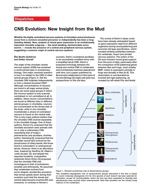
Bony vertebrates<br />
Sharks<br />
Agnathans<br />
Ascidians<br />
Cephalochordates<br />
Enteropneusts:<br />
Saccoglossus<br />
Pterobranchia<br />
Enteropneusts:<br />
Ptychodera<br />
Echinoderms<br />
Vertebrates<br />
Chordates<br />
Current Biology<br />
Figure 1. Revised evolutionary tree of deuterostome groups.<br />
This simplified phylogenetic tree of deuterostome groups discussed in the text is based<br />
on recent molecular phylogenies [12,13]. One species of each branch has been selected for<br />
illustration, except for hemichordates, where the tree has been expanded to illustrate that<br />
pterobranchs have evolved within enteropneusts. Note that ascidians are the closest living<br />
relatives of the vertebrates.<br />
Hemichordates
Dispatch<br />
R641<br />
expression in chordates as opposed to<br />
sea urchin and starfish larvae where<br />
these genes are expressed on the right<br />
side of the body [10,11]. Also, refined<br />
deuterostome phylogeny now<br />
indicates that ascidians are indeed<br />
secondarily simplified [12] and that<br />
the sessile pterobranchs stem from<br />
free-living acorn worms [13], safely<br />
ruling out that pterobranch- or<br />
ascidian-like organisms were among<br />
the chordate ancestors. These findings<br />
eliminated Romer’s influential theory<br />
from the list of plausible explanations<br />
for the origin of chordates.<br />
However, these findings were not<br />
enough to stop Dohrn’s views being<br />
disputed, as there is still a major<br />
objection regarding the common origin<br />
for the chordate, arthropod and annelid<br />
CNS. If they were indeed homologous,<br />
strands of centralised neurons that are<br />
similar to the chordate CNS should also<br />
exist in the more basal deuterostome<br />
groups, such as echinoderms or<br />
hemichordates (Figure 1). Clearly, the<br />
echinoderm CNS is very divergent<br />
and bears no resemblance to that of<br />
the chordates, but how about that of<br />
the hemichordates In juveniles of the<br />
enteropneust worm Saccoglossus<br />
kowalevski — a hemichordate<br />
species commonly known as ‘acorn<br />
worms’ — the scattered epithelial<br />
expression of neural markers and<br />
nervous system patterning genes<br />
seemed to suggest that these<br />
worm-shaped animals develop only a<br />
diffuse nervous system, with neurons<br />
distributed over the entire epidermis<br />
instead of being concentrated in a<br />
nerve cord [14]. This prompted the<br />
idea of ancient ‘skin brains’ [15], which<br />
proposes a non-centralised ancestral<br />
nervous system, where scattered<br />
neurons within the ectoderm would<br />
have evolved into a proper internal<br />
CNS independently in the more<br />
advanced protostome and<br />
deuterostome lineages.<br />
The new work by Brunet and<br />
collaborators [5] is now adding a fresh<br />
brushstroke to the picture by showing<br />
that adult acorn worms — unlike<br />
juveniles — actually possess a<br />
fully-formed CNS, which features what<br />
could be interpreted as a transition<br />
between the ventral protostome and the<br />
dorsal chordate CNS (Figure 2) [16]. By<br />
analysing the expression patterns<br />
of genes in the CNS, the authors<br />
demonstrate that in adult Ptychodera<br />
flava and S. kowalevski worms a ventral<br />
and a dorsal strand of centralised<br />
A<br />
B<br />
C<br />
np<br />
*<br />
Annelid<br />
cc<br />
Enteropneust<br />
Chordate<br />
neurons are present that merge<br />
anterior-dorsally at the level of the<br />
worm’s collar. Both species display<br />
atripartitebodythatconsistsofan<br />
anterior acorn-shaped proboscis, which<br />
they use for burrowing in the sand,<br />
followed by a short thick collar and<br />
a very long trunk. Further anteriorly, the<br />
collar cord extends into a neural platelike<br />
concentration of neurons in the<br />
proboscis stem, which harbors the<br />
thickest layers of neurons and<br />
underlying axons, and which extends<br />
ventrally to fully encircle the proboscis<br />
stem just behind the proboscis<br />
(Figure 2). In other body regions,<br />
neurons show a much lower density and<br />
ct<br />
ds<br />
dc<br />
vc<br />
D<br />
V<br />
D<br />
V<br />
V<br />
D<br />
*<br />
cc<br />
cc<br />
ct<br />
are interpreted as a peripheral nervous<br />
system (PNS). In line with a clear<br />
separation into a CNS and a PNS, CNS<br />
markers such as hb9 and Drg,<br />
expressed by somatic motor and<br />
sensory neurons of the dorsal root<br />
ganglia, or VACht, expressed by<br />
cholinergic neurons, are only found<br />
within the cords. Otherwise, serotonin<br />
expressing cells are only found outside<br />
the cords. This centralisation of the<br />
nervous system is apparent from the<br />
earliest stages of metamorphosis<br />
between larva and adult. The authors<br />
conclude that the previously described<br />
‘diffuse’ nervous system present at<br />
earlier developmental stages in<br />
ct<br />
ds<br />
ds<br />
dc<br />
dc<br />
D<br />
V<br />
D<br />
D<br />
V<br />
D<br />
V<br />
D<br />
V<br />
Current Biology<br />
Figure 2. Comparative anatomy of the CNS.<br />
Schematic comparison of centralised nerve cords in an (A) annelid, (B) enteropneust [5] and<br />
(C) chordate (note that this schematic is dorsoventrally inverted). Superficial and internalised<br />
portions of the CNS are depicted in bright and dark yellow, respectively. Left panels: Lateral<br />
views. Right panels: Animals cut open along the midline (black dashed line; dorsal in the<br />
annelid and enteropneust, ventral in the chordate) and flattened. Red dashed line indicates<br />
the midline on the opposite body side, internalised to form the floorplate of the neural tube<br />
in the chordate. A grey dotted line demarcates possibly homologous CNS strands, as initially<br />
put forward by Nübler-Jung and <strong>Arendt</strong> [16]. In the chordate, the asterisks indicate the position<br />
of the ancient (now dorsal) mouth at the bottom of the brain and the arrow depicts where<br />
a modified gill slit will form the new mouth. Note that the ventral neurogenic strand along<br />
the ventral midline will disintegrate (yellow dashed line). cc: collar cord; ct: circumesophageal<br />
tract; dc: dorsal cord; np: neural plate; vc: ventral cord; vs: ventral strand.
Current Biology Vol 19 No 15<br />
R642<br />
Saccoglossus is a transitory feature that<br />
may correspond to the larval nervous<br />
system of other enteropneusts.<br />
This work opens up new avenues of<br />
comparative CNS research. Clearly,<br />
these data, together with the inverted<br />
BMP patterning in acorn worms [17],<br />
are consistent with the view that the<br />
neural plate of the proboscis stem, the<br />
collar cord, the circumesophageal tract<br />
and ventral cord together correspond<br />
to the chordate CNS as a whole and to<br />
the CNS of other invertebrates where<br />
inversion has not occurred, as<br />
proposed earlier [16]. Yet, a more<br />
detailed comparative picture still<br />
remains to be drawn. So far, knowledge<br />
of neuron types in enteropneusts and<br />
of their differential distribution is rather<br />
scarce and will require a much closer<br />
inspection of a larger number of<br />
neuronal markers. Also, a link with the<br />
detailed orthologous gene expression<br />
data in vertebrates, similar to that<br />
described for Saccoglossus [14], will<br />
have to be established. Only then will it<br />
be possible to firmly homologise any<br />
portion of the enteropneust CNS with<br />
that of chordates or even annelids<br />
or arthropods. As a start, the<br />
concentration of GABAergic neurons in<br />
the proboscis stem, apparently located<br />
at the interface between the six3 and otx<br />
territory [14], may correspond to<br />
GABAergic populations in the vertebrate<br />
[18] and in the annelid forebrain (R.<br />
Tomer and D.A., unpublished results).<br />
If indeed the CNS represents ancient<br />
bilaterian heritage and vertebrates<br />
inverted their DV axis, one prominent<br />
problem still remains, as discussed by<br />
Brunet and colleagues [5] (Figure 2):<br />
The dorsal portions of the enteropneust<br />
CNS are located exactly where the<br />
chordates would have evolved their<br />
(new) mouth — on their new ventral<br />
(formerly dorsal) body side now facing<br />
the substrate. How can we reconcile<br />
this Dohrn [4] had suggested that the<br />
new chordate mouth evolved from the<br />
ventral relocation of gill slits (Figure 2),<br />
as is suggested by the amphioxus<br />
mouth, which is thought to represent<br />
a ventrally shifted gill slit [19] — hence<br />
the name Branchiostoma, meaning ‘gill<br />
slit mouth’. Interestingly, a strand of<br />
neurogenic tissue has recently been<br />
discovered along the amphioxus<br />
ventral midline giving rise to scattered<br />
neuronal precursors that further<br />
migrate dorsally [20] before the mouth<br />
takes its place. Future molecular<br />
comparisons of the neuronal cell types<br />
involved will reveal whether this<br />
transitory neurogenic ventral strand<br />
in amphioxus might be related to the<br />
dorsal strand of neurons in acorn<br />
worms or rather represents an<br />
independent acquisition that either<br />
could be an apomorphy or could be<br />
related to a second wave of<br />
centralisation: namely the dorsal<br />
reunion of a primitive neuronal<br />
population with placode-neural crest<br />
characteristics. With these new insights<br />
derived from mud- and sand-living<br />
acorn worms, comparative research on<br />
chordate nervous system evolution<br />
appears more exciting than ever.<br />
References<br />
1. Romer, A.S. (1972). The vertebrate as a dual<br />
animal - somatic and visceral. Evol. Biol. 6,<br />
121–156.<br />
2. Garstang, W. (1894). Preliminary note on a new<br />
theory of the ancestry of the Chordata. Zool.<br />
Anz. 17, 122–125.<br />
3. Geoffroy St.-Hilaire, E. (1822). Considérations<br />
générales sur la vertèbre. Mém. Mus. Hist. Nat.<br />
9, 89–119.<br />
4. Dohrn, A. (1875). Der Ursprung der Wirbelthiere<br />
und das Princip des Functionswechsels<br />
(Leipzig: Verlag von Wilhelm Engelmann).<br />
5. Nomaksteinsky, M., Röttinger, E., Dufour, H.D.,<br />
Chettouh, Z., Lowe, C.J., Martindale, M.Q., and<br />
Brunet, J.-F. (<strong>2009</strong>). Centralization of the<br />
deuterostome nervous system predates<br />
chordates. Curr. Biol. 19, 1264–1269.<br />
6. <strong>Arendt</strong>, D., and Nübler-Jung, K. (1994).<br />
Inversion of dorsoventral axis Nature 371, 26.<br />
7. <strong>Arendt</strong>, D., and Nübler-Jung, K. (1999).<br />
Comparison of early nerve cord development<br />
in insects and vertebrates. Development<br />
126, 2309–2325.<br />
8. Denes, A.S., Jekely, G., Steinmetz, P.R.,<br />
Raible, F., Snyman, H., Prud’homme, B.,<br />
Ferrier, D.E., Balavoine, G., and <strong>Arendt</strong>, D.<br />
(2007). Molecular architecture of annelid nerve<br />
cord supports common origin of nervous system<br />
centralization in bilateria. Cell 129, 277–288.<br />
9. Holley, S.A., Jackson, P.D., Sasai, Y., Lu, B.,<br />
De Robertis, E.M., Hoffmann, F.M., and<br />
Ferguson, E.L. (1995). A conserved system for<br />
dorsal-ventral patterning in insects and<br />
vertebrates involving sog and chordin. Nature<br />
376, 249–253.<br />
10. Duboc, V., Rottinger, E., Lapraz, F.,<br />
Besnardeau, L., and Lepage, T. (2005).<br />
Left-right asymmetry in the sea urchin embryo<br />
is regulated by nodal signaling on the right side.<br />
Dev. Cell 9, 147–158.<br />
11. Hibino, T., Nishino, A., and Amemiya, S. (2006).<br />
Phylogenetic correspondence of the body axes<br />
in bilaterians is revealed by the right-sided<br />
expression of Pitx genes in echinoderm larvae.<br />
Dev. Growth Differ. 48, 587–595.<br />
12. Delsuc, F., Brinkmann, H., Chourrout, D., and<br />
Philippe, H. (2006). Tunicates and not<br />
cephalochordates are the closest living<br />
relatives of vertebrates. Nature 439, 965–968.<br />
13. Cannon, J.T., Rychel, A.L., Eccleston, H.,<br />
Halanych, K.M., and Swalla, B.J. (<strong>2009</strong>).<br />
Molecular phylogeny of hemichordata, with<br />
updated status of deep-sea enteropneusts.<br />
Mol. Phylogenet. Evol. 52, 17–24.<br />
14. Lowe, C.J., Wu, M., Salic, A., Evans, L., Lander, E.,<br />
Stange-Thomann, N., Gruber, C.E., Gerhart, J.,<br />
and Kirschner, M. (2003). Anteroposterior<br />
patterning in Hemichordates and the origins of<br />
the chordate nervous system. Cell 113,853–865.<br />
15. Holland, N.D. (2003). Early central nervous<br />
system evolution: an era of skin brains Nat.<br />
Rev. Neurosci. 4, 1–11.<br />
16. Nübler-Jung, K., and <strong>Arendt</strong>, D. (1996).<br />
Enteropneusts and chordate evolution.<br />
Curr. Biol. 6, 352–353.<br />
17. Lowe, C.J., Terasaki, M., Wu, M.,<br />
Freeman, R.M., Jr., Runft, L., Kwan, K.,<br />
Haigo, S., Aronowicz, J., Lander, E., Gruber, C.,<br />
et al. (2006). Dorsoventral patterning in<br />
hemichordates: insights into early chordate<br />
evolution. PLoS Biol. 4, e291.<br />
18. Sugino, K., Hempel, C.M., Miller, M.N.,<br />
Hattox, A.M., Shapiro, P., Wu, C., Huang, Z.J.,<br />
and Nelson, S.B. (2006). Molecular taxonomy of<br />
major neuronal classes in the adult mouse<br />
forebrain. Nat. Neurosci. 9, 99–107.<br />
19. Conklin, E.G. (1932). The embryology of<br />
amphioxus. J. Morph. 54, 69–118.<br />
20. <strong>Benito</strong>-<strong>Gutierrez</strong>, E., Nake, C., Llovera, M.,<br />
Comella, J.X., and Garcia-Fernandez, J. (2005).<br />
The single AmphiTrk receptor highlights<br />
increased complexity of neurotrophin signalling<br />
in vertebrates and suggests an early role in<br />
developing sensory neuroepidermal cells.<br />
Development 132, 2191–2202.<br />
Developmental Biology Unit, European<br />
Molecular Biology Laboratory, D-69117<br />
Heidelberg, Germany.<br />
*E-mail: arendt@embl.de<br />
DOI: 10.1016/j.cub.<strong>2009</strong>.06.020<br />
Cancer: CINful Centrosomes<br />
The regulation of centrosome number is lost in many tumors and the presence<br />
of extra centrosomes correlates with chromosomal instability. Recent work<br />
now reveals how extra centrosomes cause chromosome mis-segregation<br />
in tumor cells.<br />
Samuel F. Bakhoum<br />
and Duane A. Compton*<br />
Centrosomes are pivotal organizers<br />
of the microtubule cytoskeleton and<br />
their duplication and inheritance is<br />
strictly controlled during the cell cycle<br />
in a manner that parallels genome<br />
duplication [1]. This control is lost<br />
in many cancer cells, making the<br />
presence of extra centrosomes<br />
a discernible feature of many tumors<br />
[2]. This defect has long been<br />
associated with aneuploidy in cancer<br />
and it is postulated that additional<br />
centrosomes induce chromosome<br />
mis-segregation, which then<br />
contributes to tumorigenesis [3–6].