Benito-Gutierrez & Arendt 2009
Benito-Gutierrez & Arendt 2009
Benito-Gutierrez & Arendt 2009

You also want an ePaper? Increase the reach of your titles
YUMPU automatically turns print PDFs into web optimized ePapers that Google loves.
Dispatch<br />
R641<br />
expression in chordates as opposed to<br />
sea urchin and starfish larvae where<br />
these genes are expressed on the right<br />
side of the body [10,11]. Also, refined<br />
deuterostome phylogeny now<br />
indicates that ascidians are indeed<br />
secondarily simplified [12] and that<br />
the sessile pterobranchs stem from<br />
free-living acorn worms [13], safely<br />
ruling out that pterobranch- or<br />
ascidian-like organisms were among<br />
the chordate ancestors. These findings<br />
eliminated Romer’s influential theory<br />
from the list of plausible explanations<br />
for the origin of chordates.<br />
However, these findings were not<br />
enough to stop Dohrn’s views being<br />
disputed, as there is still a major<br />
objection regarding the common origin<br />
for the chordate, arthropod and annelid<br />
CNS. If they were indeed homologous,<br />
strands of centralised neurons that are<br />
similar to the chordate CNS should also<br />
exist in the more basal deuterostome<br />
groups, such as echinoderms or<br />
hemichordates (Figure 1). Clearly, the<br />
echinoderm CNS is very divergent<br />
and bears no resemblance to that of<br />
the chordates, but how about that of<br />
the hemichordates In juveniles of the<br />
enteropneust worm Saccoglossus<br />
kowalevski — a hemichordate<br />
species commonly known as ‘acorn<br />
worms’ — the scattered epithelial<br />
expression of neural markers and<br />
nervous system patterning genes<br />
seemed to suggest that these<br />
worm-shaped animals develop only a<br />
diffuse nervous system, with neurons<br />
distributed over the entire epidermis<br />
instead of being concentrated in a<br />
nerve cord [14]. This prompted the<br />
idea of ancient ‘skin brains’ [15], which<br />
proposes a non-centralised ancestral<br />
nervous system, where scattered<br />
neurons within the ectoderm would<br />
have evolved into a proper internal<br />
CNS independently in the more<br />
advanced protostome and<br />
deuterostome lineages.<br />
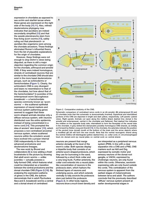
The new work by Brunet and<br />
collaborators [5] is now adding a fresh<br />
brushstroke to the picture by showing<br />
that adult acorn worms — unlike<br />
juveniles — actually possess a<br />
fully-formed CNS, which features what<br />
could be interpreted as a transition<br />
between the ventral protostome and the<br />
dorsal chordate CNS (Figure 2) [16]. By<br />
analysing the expression patterns<br />
of genes in the CNS, the authors<br />
demonstrate that in adult Ptychodera<br />
flava and S. kowalevski worms a ventral<br />
and a dorsal strand of centralised<br />
A<br />
B<br />
C<br />
np<br />
*<br />
Annelid<br />
cc<br />
Enteropneust<br />
Chordate<br />
neurons are present that merge<br />
anterior-dorsally at the level of the<br />
worm’s collar. Both species display<br />
atripartitebodythatconsistsofan<br />
anterior acorn-shaped proboscis, which<br />
they use for burrowing in the sand,<br />
followed by a short thick collar and<br />
a very long trunk. Further anteriorly, the<br />
collar cord extends into a neural platelike<br />
concentration of neurons in the<br />
proboscis stem, which harbors the<br />
thickest layers of neurons and<br />
underlying axons, and which extends<br />
ventrally to fully encircle the proboscis<br />
stem just behind the proboscis<br />
(Figure 2). In other body regions,<br />
neurons show a much lower density and<br />
ct<br />
ds<br />
dc<br />
vc<br />
D<br />
V<br />
D<br />
V<br />
V<br />
D<br />
*<br />
cc<br />
cc<br />
ct<br />
are interpreted as a peripheral nervous<br />
system (PNS). In line with a clear<br />
separation into a CNS and a PNS, CNS<br />
markers such as hb9 and Drg,<br />
expressed by somatic motor and<br />
sensory neurons of the dorsal root<br />
ganglia, or VACht, expressed by<br />
cholinergic neurons, are only found<br />
within the cords. Otherwise, serotonin<br />
expressing cells are only found outside<br />
the cords. This centralisation of the<br />
nervous system is apparent from the<br />
earliest stages of metamorphosis<br />
between larva and adult. The authors<br />
conclude that the previously described<br />
‘diffuse’ nervous system present at<br />
earlier developmental stages in<br />
ct<br />
ds<br />
ds<br />
dc<br />
dc<br />
D<br />
V<br />
D<br />
D<br />
V<br />
D<br />
V<br />
D<br />
V<br />
Current Biology<br />
Figure 2. Comparative anatomy of the CNS.<br />
Schematic comparison of centralised nerve cords in an (A) annelid, (B) enteropneust [5] and<br />
(C) chordate (note that this schematic is dorsoventrally inverted). Superficial and internalised<br />
portions of the CNS are depicted in bright and dark yellow, respectively. Left panels: Lateral<br />
views. Right panels: Animals cut open along the midline (black dashed line; dorsal in the<br />
annelid and enteropneust, ventral in the chordate) and flattened. Red dashed line indicates<br />
the midline on the opposite body side, internalised to form the floorplate of the neural tube<br />
in the chordate. A grey dotted line demarcates possibly homologous CNS strands, as initially<br />
put forward by Nübler-Jung and <strong>Arendt</strong> [16]. In the chordate, the asterisks indicate the position<br />
of the ancient (now dorsal) mouth at the bottom of the brain and the arrow depicts where<br />
a modified gill slit will form the new mouth. Note that the ventral neurogenic strand along<br />
the ventral midline will disintegrate (yellow dashed line). cc: collar cord; ct: circumesophageal<br />
tract; dc: dorsal cord; np: neural plate; vc: ventral cord; vs: ventral strand.