Benito-Gutierrez & Arendt 2009
Benito-Gutierrez & Arendt 2009
Benito-Gutierrez & Arendt 2009

Create successful ePaper yourself
Turn your PDF publications into a flip-book with our unique Google optimized e-Paper software.
Current Biology Vol 19 No 15<br />
R640<br />
Dispatches<br />
CNS Evolution: New Insight from the Mud<br />
Whether the highly centralised nervous systems of chordates and protostomes<br />
arose from a common ancestral precursor or independently has been a longstanding<br />
debate. Now, analysis of neural gene expression in an evolutionarily<br />
important chordate outgroup — the sand-dwelling, hemichordate acorn<br />
worms — reveals the presence of a central and peripheral nervous system,<br />
suggesting a common origin of central nervous systems.<br />
Èlia <strong>Benito</strong>-Gutiérrez<br />
and Detlev <strong>Arendt</strong>*<br />
The origin of the chordate central<br />
nervous system (CNS) has remained<br />
a controversial topic in evolutionary<br />
biology. One major unsolved question<br />
is how it is related to the CNS of other<br />
animal groups (Figure 1). Did the<br />
chordate CNS originate independently<br />
or from a shared ancestral CNS<br />
Although concentrations of neurons<br />
are found in all major animal phyla,<br />
there are some basal groups in which<br />
the nervous system is only scarcely<br />
centralised or not centralised at all. In<br />
addition, these neuron concentrations<br />
are found at different sites in different<br />
animal groups: in chordates, neurons<br />
are concentrated at the dorsal side of<br />
the body, while in non-chordate<br />
invertebrates a strand of centralised<br />
neurons is found on the ventral side.<br />
This is why many authors believe that<br />
the chordate CNS evolved separately<br />
in the chordate lineage. One of these<br />
authors, Romer [1], proposed that the<br />
chordate ancestors were animals with<br />
no or only a rudimentary CNS<br />
resembling that of today’s<br />
pterobranchs and ascidians. Another<br />
author, Garstang [2], proposed that the<br />
chordate neural tube evolved by the<br />
dorsal fusion of ciliary bands, like those<br />
found in echinoderm or enteropneust<br />
larvae. However, there is an alternative<br />
view. Inspired by Geoffroy St. Hilaire’s<br />
‘unite´ de plan’ [3], and despite the<br />
opposite locations, comparative<br />
anatomist Anton Dohrn [4] proposed<br />
that the chordate CNS was<br />
homologous to that of protostome<br />
annelids and arthropods. He assumed<br />
that vertebrates evolved from<br />
worm-shaped, annelid-like ancestors<br />
that turned upside-down during their<br />
evolution such that the dorsal and<br />
ventral sides became inverted (DV-axis<br />
inversion). In contrast to Romer’s<br />
scenario, Dohrn considered ascidians<br />
to be secondarily modified forms with<br />
a simplified larval CNS. Dohrn’s<br />
proposed homology between the<br />
dorsal and ventral CNS of vertebrates<br />
and invertebrates had been abandoned<br />
with time, but a paper published by<br />
Brunet and collaborators in this issue of<br />
Current Biology [5] might well yield new<br />
perspectives on this old idea.<br />
The revival of Dohrn’s ideas could<br />
have been already anticipated based<br />
on gene expression data from different<br />
organisms during neural patterning and<br />
neuronal cell-type specification, which<br />
revealed striking similarities between<br />
the vertebrate, insect and annelid<br />
nervous systems. The notion of the<br />
DV-axis inversion found great support<br />
from this pool of data, particularly after<br />
the comparison of DV patterning genes<br />
between flies and frogs, most notably<br />
BMP, which is expressed ventrally in<br />
frogs and dorsally in flies [6–9]. This<br />
observation is corroborated by<br />
inverted left-right patterning, as<br />
revealed by left-sided Pitx and Nodal<br />
Bony vertebrates<br />
Sharks<br />
Agnathans<br />
Ascidians<br />
Cephalochordates<br />
Enteropneusts:<br />
Saccoglossus<br />
Pterobranchia<br />
Enteropneusts:<br />
Ptychodera<br />
Echinoderms<br />
Vertebrates<br />
Chordates<br />
Current Biology<br />
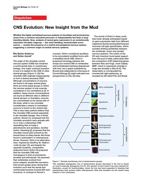
Figure 1. Revised evolutionary tree of deuterostome groups.<br />
This simplified phylogenetic tree of deuterostome groups discussed in the text is based<br />
on recent molecular phylogenies [12,13]. One species of each branch has been selected for<br />
illustration, except for hemichordates, where the tree has been expanded to illustrate that<br />
pterobranchs have evolved within enteropneusts. Note that ascidians are the closest living<br />
relatives of the vertebrates.<br />
Hemichordates
Dispatch<br />
R641<br />
expression in chordates as opposed to<br />
sea urchin and starfish larvae where<br />
these genes are expressed on the right<br />
side of the body [10,11]. Also, refined<br />
deuterostome phylogeny now<br />
indicates that ascidians are indeed<br />
secondarily simplified [12] and that<br />
the sessile pterobranchs stem from<br />
free-living acorn worms [13], safely<br />
ruling out that pterobranch- or<br />
ascidian-like organisms were among<br />
the chordate ancestors. These findings<br />
eliminated Romer’s influential theory<br />
from the list of plausible explanations<br />
for the origin of chordates.<br />
However, these findings were not<br />
enough to stop Dohrn’s views being<br />
disputed, as there is still a major<br />
objection regarding the common origin<br />
for the chordate, arthropod and annelid<br />
CNS. If they were indeed homologous,<br />
strands of centralised neurons that are<br />
similar to the chordate CNS should also<br />
exist in the more basal deuterostome<br />
groups, such as echinoderms or<br />
hemichordates (Figure 1). Clearly, the<br />
echinoderm CNS is very divergent<br />
and bears no resemblance to that of<br />
the chordates, but how about that of<br />
the hemichordates In juveniles of the<br />
enteropneust worm Saccoglossus<br />
kowalevski — a hemichordate<br />
species commonly known as ‘acorn<br />
worms’ — the scattered epithelial<br />
expression of neural markers and<br />
nervous system patterning genes<br />
seemed to suggest that these<br />
worm-shaped animals develop only a<br />
diffuse nervous system, with neurons<br />
distributed over the entire epidermis<br />
instead of being concentrated in a<br />
nerve cord [14]. This prompted the<br />
idea of ancient ‘skin brains’ [15], which<br />
proposes a non-centralised ancestral<br />
nervous system, where scattered<br />
neurons within the ectoderm would<br />
have evolved into a proper internal<br />
CNS independently in the more<br />
advanced protostome and<br />
deuterostome lineages.<br />
The new work by Brunet and<br />
collaborators [5] is now adding a fresh<br />
brushstroke to the picture by showing<br />
that adult acorn worms — unlike<br />
juveniles — actually possess a<br />
fully-formed CNS, which features what<br />
could be interpreted as a transition<br />
between the ventral protostome and the<br />
dorsal chordate CNS (Figure 2) [16]. By<br />
analysing the expression patterns<br />
of genes in the CNS, the authors<br />
demonstrate that in adult Ptychodera<br />
flava and S. kowalevski worms a ventral<br />
and a dorsal strand of centralised<br />
A<br />
B<br />
C<br />
np<br />
*<br />
Annelid<br />
cc<br />
Enteropneust<br />
Chordate<br />
neurons are present that merge<br />
anterior-dorsally at the level of the<br />
worm’s collar. Both species display<br />
atripartitebodythatconsistsofan<br />
anterior acorn-shaped proboscis, which<br />
they use for burrowing in the sand,<br />
followed by a short thick collar and<br />
a very long trunk. Further anteriorly, the<br />
collar cord extends into a neural platelike<br />
concentration of neurons in the<br />
proboscis stem, which harbors the<br />
thickest layers of neurons and<br />
underlying axons, and which extends<br />
ventrally to fully encircle the proboscis<br />
stem just behind the proboscis<br />
(Figure 2). In other body regions,<br />
neurons show a much lower density and<br />
ct<br />
ds<br />
dc<br />
vc<br />
D<br />
V<br />
D<br />
V<br />
V<br />
D<br />
*<br />
cc<br />
cc<br />
ct<br />
are interpreted as a peripheral nervous<br />
system (PNS). In line with a clear<br />
separation into a CNS and a PNS, CNS<br />
markers such as hb9 and Drg,<br />
expressed by somatic motor and<br />
sensory neurons of the dorsal root<br />
ganglia, or VACht, expressed by<br />
cholinergic neurons, are only found<br />
within the cords. Otherwise, serotonin<br />
expressing cells are only found outside<br />
the cords. This centralisation of the<br />
nervous system is apparent from the<br />
earliest stages of metamorphosis<br />
between larva and adult. The authors<br />
conclude that the previously described<br />
‘diffuse’ nervous system present at<br />
earlier developmental stages in<br />
ct<br />
ds<br />
ds<br />
dc<br />
dc<br />
D<br />
V<br />
D<br />
D<br />
V<br />
D<br />
V<br />
D<br />
V<br />
Current Biology<br />
Figure 2. Comparative anatomy of the CNS.<br />
Schematic comparison of centralised nerve cords in an (A) annelid, (B) enteropneust [5] and<br />
(C) chordate (note that this schematic is dorsoventrally inverted). Superficial and internalised<br />
portions of the CNS are depicted in bright and dark yellow, respectively. Left panels: Lateral<br />
views. Right panels: Animals cut open along the midline (black dashed line; dorsal in the<br />
annelid and enteropneust, ventral in the chordate) and flattened. Red dashed line indicates<br />
the midline on the opposite body side, internalised to form the floorplate of the neural tube<br />
in the chordate. A grey dotted line demarcates possibly homologous CNS strands, as initially<br />
put forward by Nübler-Jung and <strong>Arendt</strong> [16]. In the chordate, the asterisks indicate the position<br />
of the ancient (now dorsal) mouth at the bottom of the brain and the arrow depicts where<br />
a modified gill slit will form the new mouth. Note that the ventral neurogenic strand along<br />
the ventral midline will disintegrate (yellow dashed line). cc: collar cord; ct: circumesophageal<br />
tract; dc: dorsal cord; np: neural plate; vc: ventral cord; vs: ventral strand.
Current Biology Vol 19 No 15<br />
R642<br />
Saccoglossus is a transitory feature that<br />
may correspond to the larval nervous<br />
system of other enteropneusts.<br />
This work opens up new avenues of<br />
comparative CNS research. Clearly,<br />
these data, together with the inverted<br />
BMP patterning in acorn worms [17],<br />
are consistent with the view that the<br />
neural plate of the proboscis stem, the<br />
collar cord, the circumesophageal tract<br />
and ventral cord together correspond<br />
to the chordate CNS as a whole and to<br />
the CNS of other invertebrates where<br />
inversion has not occurred, as<br />
proposed earlier [16]. Yet, a more<br />
detailed comparative picture still<br />
remains to be drawn. So far, knowledge<br />
of neuron types in enteropneusts and<br />
of their differential distribution is rather<br />
scarce and will require a much closer<br />
inspection of a larger number of<br />
neuronal markers. Also, a link with the<br />
detailed orthologous gene expression<br />
data in vertebrates, similar to that<br />
described for Saccoglossus [14], will<br />
have to be established. Only then will it<br />
be possible to firmly homologise any<br />
portion of the enteropneust CNS with<br />
that of chordates or even annelids<br />
or arthropods. As a start, the<br />
concentration of GABAergic neurons in<br />
the proboscis stem, apparently located<br />
at the interface between the six3 and otx<br />
territory [14], may correspond to<br />
GABAergic populations in the vertebrate<br />
[18] and in the annelid forebrain (R.<br />
Tomer and D.A., unpublished results).<br />
If indeed the CNS represents ancient<br />
bilaterian heritage and vertebrates<br />
inverted their DV axis, one prominent<br />
problem still remains, as discussed by<br />
Brunet and colleagues [5] (Figure 2):<br />
The dorsal portions of the enteropneust<br />
CNS are located exactly where the<br />
chordates would have evolved their<br />
(new) mouth — on their new ventral<br />
(formerly dorsal) body side now facing<br />
the substrate. How can we reconcile<br />
this Dohrn [4] had suggested that the<br />
new chordate mouth evolved from the<br />
ventral relocation of gill slits (Figure 2),<br />
as is suggested by the amphioxus<br />
mouth, which is thought to represent<br />
a ventrally shifted gill slit [19] — hence<br />
the name Branchiostoma, meaning ‘gill<br />
slit mouth’. Interestingly, a strand of<br />
neurogenic tissue has recently been<br />
discovered along the amphioxus<br />
ventral midline giving rise to scattered<br />
neuronal precursors that further<br />
migrate dorsally [20] before the mouth<br />
takes its place. Future molecular<br />
comparisons of the neuronal cell types<br />
involved will reveal whether this<br />
transitory neurogenic ventral strand<br />
in amphioxus might be related to the<br />
dorsal strand of neurons in acorn<br />
worms or rather represents an<br />
independent acquisition that either<br />
could be an apomorphy or could be<br />
related to a second wave of<br />
centralisation: namely the dorsal<br />
reunion of a primitive neuronal<br />
population with placode-neural crest<br />
characteristics. With these new insights<br />
derived from mud- and sand-living<br />
acorn worms, comparative research on<br />
chordate nervous system evolution<br />
appears more exciting than ever.<br />
References<br />
1. Romer, A.S. (1972). The vertebrate as a dual<br />
animal - somatic and visceral. Evol. Biol. 6,<br />
121–156.<br />
2. Garstang, W. (1894). Preliminary note on a new<br />
theory of the ancestry of the Chordata. Zool.<br />
Anz. 17, 122–125.<br />
3. Geoffroy St.-Hilaire, E. (1822). Considérations<br />
générales sur la vertèbre. Mém. Mus. Hist. Nat.<br />
9, 89–119.<br />
4. Dohrn, A. (1875). Der Ursprung der Wirbelthiere<br />
und das Princip des Functionswechsels<br />
(Leipzig: Verlag von Wilhelm Engelmann).<br />
5. Nomaksteinsky, M., Röttinger, E., Dufour, H.D.,<br />
Chettouh, Z., Lowe, C.J., Martindale, M.Q., and<br />
Brunet, J.-F. (<strong>2009</strong>). Centralization of the<br />
deuterostome nervous system predates<br />
chordates. Curr. Biol. 19, 1264–1269.<br />
6. <strong>Arendt</strong>, D., and Nübler-Jung, K. (1994).<br />
Inversion of dorsoventral axis Nature 371, 26.<br />
7. <strong>Arendt</strong>, D., and Nübler-Jung, K. (1999).<br />
Comparison of early nerve cord development<br />
in insects and vertebrates. Development<br />
126, 2309–2325.<br />
8. Denes, A.S., Jekely, G., Steinmetz, P.R.,<br />
Raible, F., Snyman, H., Prud’homme, B.,<br />
Ferrier, D.E., Balavoine, G., and <strong>Arendt</strong>, D.<br />
(2007). Molecular architecture of annelid nerve<br />
cord supports common origin of nervous system<br />
centralization in bilateria. Cell 129, 277–288.<br />
9. Holley, S.A., Jackson, P.D., Sasai, Y., Lu, B.,<br />
De Robertis, E.M., Hoffmann, F.M., and<br />
Ferguson, E.L. (1995). A conserved system for<br />
dorsal-ventral patterning in insects and<br />
vertebrates involving sog and chordin. Nature<br />
376, 249–253.<br />
10. Duboc, V., Rottinger, E., Lapraz, F.,<br />
Besnardeau, L., and Lepage, T. (2005).<br />
Left-right asymmetry in the sea urchin embryo<br />
is regulated by nodal signaling on the right side.<br />
Dev. Cell 9, 147–158.<br />
11. Hibino, T., Nishino, A., and Amemiya, S. (2006).<br />
Phylogenetic correspondence of the body axes<br />
in bilaterians is revealed by the right-sided<br />
expression of Pitx genes in echinoderm larvae.<br />
Dev. Growth Differ. 48, 587–595.<br />
12. Delsuc, F., Brinkmann, H., Chourrout, D., and<br />
Philippe, H. (2006). Tunicates and not<br />
cephalochordates are the closest living<br />
relatives of vertebrates. Nature 439, 965–968.<br />
13. Cannon, J.T., Rychel, A.L., Eccleston, H.,<br />
Halanych, K.M., and Swalla, B.J. (<strong>2009</strong>).<br />
Molecular phylogeny of hemichordata, with<br />
updated status of deep-sea enteropneusts.<br />
Mol. Phylogenet. Evol. 52, 17–24.<br />
14. Lowe, C.J., Wu, M., Salic, A., Evans, L., Lander, E.,<br />
Stange-Thomann, N., Gruber, C.E., Gerhart, J.,<br />
and Kirschner, M. (2003). Anteroposterior<br />
patterning in Hemichordates and the origins of<br />
the chordate nervous system. Cell 113,853–865.<br />
15. Holland, N.D. (2003). Early central nervous<br />
system evolution: an era of skin brains Nat.<br />
Rev. Neurosci. 4, 1–11.<br />
16. Nübler-Jung, K., and <strong>Arendt</strong>, D. (1996).<br />
Enteropneusts and chordate evolution.<br />
Curr. Biol. 6, 352–353.<br />
17. Lowe, C.J., Terasaki, M., Wu, M.,<br />
Freeman, R.M., Jr., Runft, L., Kwan, K.,<br />
Haigo, S., Aronowicz, J., Lander, E., Gruber, C.,<br />
et al. (2006). Dorsoventral patterning in<br />
hemichordates: insights into early chordate<br />
evolution. PLoS Biol. 4, e291.<br />
18. Sugino, K., Hempel, C.M., Miller, M.N.,<br />
Hattox, A.M., Shapiro, P., Wu, C., Huang, Z.J.,<br />
and Nelson, S.B. (2006). Molecular taxonomy of<br />
major neuronal classes in the adult mouse<br />
forebrain. Nat. Neurosci. 9, 99–107.<br />
19. Conklin, E.G. (1932). The embryology of<br />
amphioxus. J. Morph. 54, 69–118.<br />
20. <strong>Benito</strong>-<strong>Gutierrez</strong>, E., Nake, C., Llovera, M.,<br />
Comella, J.X., and Garcia-Fernandez, J. (2005).<br />
The single AmphiTrk receptor highlights<br />
increased complexity of neurotrophin signalling<br />
in vertebrates and suggests an early role in<br />
developing sensory neuroepidermal cells.<br />
Development 132, 2191–2202.<br />
Developmental Biology Unit, European<br />
Molecular Biology Laboratory, D-69117<br />
Heidelberg, Germany.<br />
*E-mail: arendt@embl.de<br />
DOI: 10.1016/j.cub.<strong>2009</strong>.06.020<br />
Cancer: CINful Centrosomes<br />
The regulation of centrosome number is lost in many tumors and the presence<br />
of extra centrosomes correlates with chromosomal instability. Recent work<br />
now reveals how extra centrosomes cause chromosome mis-segregation<br />
in tumor cells.<br />
Samuel F. Bakhoum<br />
and Duane A. Compton*<br />
Centrosomes are pivotal organizers<br />
of the microtubule cytoskeleton and<br />
their duplication and inheritance is<br />
strictly controlled during the cell cycle<br />
in a manner that parallels genome<br />
duplication [1]. This control is lost<br />
in many cancer cells, making the<br />
presence of extra centrosomes<br />
a discernible feature of many tumors<br />
[2]. This defect has long been<br />
associated with aneuploidy in cancer<br />
and it is postulated that additional<br />
centrosomes induce chromosome<br />
mis-segregation, which then<br />
contributes to tumorigenesis [3–6].