Meditation experience is associated with increased cortical thickness
Meditation experience is associated with increased cortical thickness
Meditation experience is associated with increased cortical thickness
- No tags were found...

You also want an ePaper? Increase the reach of your titles
YUMPU automatically turns print PDFs into web optimized ePapers that Google loves.
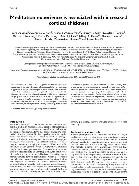
AGEINGNEUROREPORT<strong>Meditation</strong> <strong>experience</strong> <strong>is</strong> <strong>associated</strong> <strong>with</strong> <strong>increased</strong><strong>cortical</strong> <strong>thickness</strong>Sara W. Lazar a ,CatherineE.Kerr b , Rachel H. Wasserman a,b , Jeremy R. Gray c ,DouglasN.Greve d ,Michael T. Treadway a ,MettaMcGarvey e ,BrianT.Quinn d ,Je¡eryA.Dusek f,g ,HerbertBenson f,g ,Scott L. Rauch a ,Chr<strong>is</strong>topherI.Moore h,i and Bruce F<strong>is</strong>chl d,ja Psychiatric Neuroimaging Research Program, Massachusetts General Hospital, b Osher Institute, Harvard Medical School, Boston, Massachusettsc Department of Psychology,Yale University, New Haven,Connecticut, d Athinoula A. Martinos Center for Biomedical Imaging, MassachusettsGeneral Hospital, Boston, e Graduate School of Education, Harvard University,Cambridge, f Mind/Body Medical Institute, Chestnut Hill,g Department of Medicine, Beth Israel Deaconess Medical Center, Harvard Medical School, Boston, h Department of Brain and Cognitive Sciences,Massachusetts Institute of Technology, i McGovern Institute for Brain Research and j Computer Science and AI Lab (CSAIL),Massachusetts Institute of Technology,Cambridge, Massachusetts,USA.Correspondence and requests for reprints to Sara W. Lazar, PhD, Room 2609,14913th St.Charlestown, MA 02129,USATel: + 1617 724 7108; fax: + 1617 726 4078; e-mail: lazar@nmr.mgh.harvard.eduSponsorship:Th<strong>is</strong> work was supported by NIH/NCCAM K01AT00694-01, NCRR (P41RR14075), the MIND Institute, and CDC Grants H75/CCH119124 andH75/CCH123424.C.K. was supported by Grant R21AT002860-02.Received 26 August 2005; rev<strong>is</strong>ed16 September 2005; accepted19 September 2005Previous research indicates that long-term meditation practice <strong>is</strong><strong>associated</strong> <strong>with</strong> altered resting electroencephalogram patterns,suggestive of long lasting changes in brain activity. We hypothesizedthat meditation practice might also be <strong>associated</strong> <strong>with</strong>changes in the brain’s physical structure. Magnetic resonanceimaging was used to assess <strong>cortical</strong> <strong>thickness</strong> in 20 participants<strong>with</strong> extensive Insight meditation <strong>experience</strong>, which involvesfocused attention to internal <strong>experience</strong>s. Brain regions <strong>associated</strong><strong>with</strong> attention, interoception and sensory processing were thickerin meditation participants than matched controls, including theprefrontal cortex and right anterior insula. Between-group di¡erencesin prefrontal <strong>cortical</strong> <strong>thickness</strong> were most pronouncedin older participants, suggesting that meditation might o¡setage-related <strong>cortical</strong> thinning. Finally, the <strong>thickness</strong> of two regionscorrelated <strong>with</strong> meditation <strong>experience</strong>. These data provide the¢rst structural evidence for <strong>experience</strong>-dependent<strong>cortical</strong> plasticity<strong>associated</strong> <strong>with</strong> meditation practice. NeuroReport 16:1893^1897c 2005 Lippincott Williams & Wilkins.Keywords: insula, meditation, plasticity, prefrontal cortexIntroduction<strong>Meditation</strong> <strong>is</strong> a form of mental exerc<strong>is</strong>e that has become apopular US health practice. Regular practice of meditation <strong>is</strong>reported to produce changes in mental state and restingelectroencephalogram patterns that pers<strong>is</strong>t beyond the timeperiodof active practice [1]. We hypothesized that regularmeditation practice should also result in significant changesin the <strong>cortical</strong> structure in regions that are routinely engagedduring th<strong>is</strong> mental exerc<strong>is</strong>e. To test th<strong>is</strong> hypothes<strong>is</strong>, we usedmagnetic resonance imaging to v<strong>is</strong>ualize differences in the<strong>thickness</strong> of the cerebral cortex of <strong>experience</strong>d Buddh<strong>is</strong>tInsight meditation practitioners. Th<strong>is</strong> form of meditationdoes not utilize mantra or chanting. Rather, the main focusof Insight meditation <strong>is</strong> the cultivation of attention and amental capacity termed ‘mindfulness’, which <strong>is</strong> a specificnonjudgmental awareness of present-moment stimuli <strong>with</strong>outcognitive elaboration [2]. Formal practice involvessustained mindful attention to internal and external sensorystimuli. Thus, we tested the hypothes<strong>is</strong> that between-groupand <strong>experience</strong>-dependent differences in <strong>cortical</strong> <strong>thickness</strong>would be found in brain regions involved in attention andsensory processing, thereby showing evidence of <strong>cortical</strong>plasticity.Participants and methodsTwenty participants <strong>with</strong> extensive training in Insightmeditation were recruited from local meditation communities.These participants were not monks, but rather typicalWestern meditation practitioners who incorporate theirpractice into a daily routine involving career, family, friendsand outside interests. Two participants were full-timemeditation teachers, three were part-time yoga or meditationteachers and the rest meditated an average of once aday for 40 min, while pursuing traditional careers in fieldssuch as healthcare and law. On average, participants had9.177.1 years of meditation <strong>experience</strong> and practiced6.274.0 h per week. Participants were required to haveparticipated in at least 1 week-long Insight meditationretreat, which entails approximately 10 h of meditation perday. Fifteen control participants <strong>with</strong> no meditation or yoga<strong>experience</strong> were also recruited. The meditation and control0959-4965 c Lippincott Williams & Wilkins Vol 16 No 17 28 November 2005 1893Copyright © Lippincott Williams & Wilkins. Unauthorized reproduction of th<strong>is</strong> article <strong>is</strong> prohibited.
NEUROREPORTLAZAR ETAL.participants were matched for sex (meditators 65% male,controls 67%), age (meditators 38.2 years old, controls 36.8years old), race (both groups 100% Caucasian) and years ofeducation (meditators 17.3 years, controls 17.4 years). Allparticipants were physically and psychologically healthy.Two meditation participants were left-handed; exclusion ofthe left-handed participants did not significantly alterresults. All participants provided written, informed consentand the study was approved by the Institutional ReviewBoard at the Massachusetts General Hospital.The present methods utilized a well-validated computationalapproach to measure the <strong>thickness</strong> of the cerebralcortex [3,4]. Cortical <strong>thickness</strong> was estimated from twomagnetization prepared rapid gradient echo (MPRAGE)structural images collected from each participant that werethen motion-corrected and averaged together to form asingle high-resolution image [3–5]. An initial estimate of thegray/white matter boundary was constructed by classifyingall white matter voxels in a magnetic resonance imagingvolume using a combination of geometric and intensitybasedinformation. A surface-deformation procedure wasthen used to obtain subvoxel resolution in the gray/whiteboundary and in the pial surface using a combination ofsmoothness constraints and intensity terms. The resulting<strong>cortical</strong> surface models for all participants were aligned toan atlas of <strong>cortical</strong> folding patterns using a high-dimensionalnonlinear reg<strong>is</strong>tration technique.(a)(b)341 2ResultsThe mean <strong>thickness</strong> across the entire cortex did not differsignificantly between the groups for either hem<strong>is</strong>phere(P40.10), indicating that it was not the case that the cortexof meditators <strong>is</strong> nonspecifically thicker everywhere. Stat<strong>is</strong>tical<strong>thickness</strong>-difference maps constructed using theKolmogorov–Smirnoff stat<strong>is</strong>tics (one-tailed, a-levelP¼0.05), however, indicated that significant differences inthe ‘d<strong>is</strong>tribution’ of <strong>thickness</strong> ex<strong>is</strong>ted between groups acrossboth hem<strong>is</strong>pheres (k¼3.89, P¼0.0001), and in each hem<strong>is</strong>phereseparately (k¼3.02, P¼0.0025 for left hem<strong>is</strong>phere;k¼2.49, P¼0.013 for right hem<strong>is</strong>phere). Th<strong>is</strong> finding indicatesthat the pattern of relative <strong>thickness</strong> across eachhem<strong>is</strong>phere was different between groups. Protected bysignificant unidirectional results for the omnibus test foreach hem<strong>is</strong>phere, an unpaired t-test was performed to testfor specific loci of significant between-group differences inregional <strong>cortical</strong> <strong>thickness</strong>. Specifically, we tested the a priorihypotheses that differences would be observed <strong>with</strong>inprefrontal, interoceptive and unimodal sensory <strong>cortical</strong>regions. A false d<strong>is</strong>covery rate of 0.05 corresponding to anuncorrected P¼3.5 10 4 was used to correct for multiplecompar<strong>is</strong>ons [6].Within the search territory, a large region of right anteriorinsula (P¼1.2 10 5 ) and right middle and superior frontalsulci corresponding approximately to Brodmann areas (BA)9 and 10 (P¼1.8 10 5 ) were significantly thicker inmeditators than in controls (Fig. 1). The left superiortemporal gyrus (auditory cortex, P¼3.7 10 4 ) and a smallregion in the fundus of the central sulcus, (BA 3a,somatosensory cortex, P¼6.0 10 4 ) showed trends towardsa significantly thicker cortex in meditation participants thanin controls. Analys<strong>is</strong> of the right frontal BA 9/10 subregionresulted in a significant age by group interaction,F(1,31)¼10.85, P¼0.002, <strong>with</strong> typical age-related decreases(c)Thickness (mm)2.52.32.11.91.72025 30 35 40 45 50 55Age(d)Thicknessobserved in the control group [r(13)¼ 0.76, P¼0.001] butnot in the meditation group [r(18)¼ 0.05, P¼0.83]. Significantinteractions were not observed in any other brainregion.As a further confirmation that meditation can influence<strong>experience</strong>-dependent plasticity, we tested whether objectivemeasures of meditation <strong>experience</strong> correlated <strong>with</strong><strong>cortical</strong> <strong>thickness</strong>. As frequency of daily practice varies2.2P
MEDITATION AND CORTICALTHICKNESSNEUROREPORT(a)(b) 2.3Thickness (mm)2.11.91.71.5−10−50 5Change in breathing rate (bpm)Fig. 2 V<strong>is</strong>ual area correlated <strong>with</strong> meditation <strong>experience</strong>. (a) Stat<strong>is</strong>ticalmap depicting <strong>cortical</strong> <strong>thickness</strong> correlated <strong>with</strong> change in respirationrate. (b) Scatter plot of mean <strong>cortical</strong> <strong>thickness</strong> of each participant fromthe circled region <strong>with</strong>in the inferior occipitotemporal lobe plottedversus change in respiration rate. Note: negative change in breathing rate(left side) <strong>is</strong> <strong>associated</strong> <strong>with</strong> more hours of meditation <strong>experience</strong> and athicker cortex.between meditation practitioners, using the total number ofyears of practice <strong>is</strong> not a sensitive metric of <strong>experience</strong>. Oneeffect of regular meditation practice <strong>is</strong> a significant drop inrespiration rate during formal practice [7,8]. We thereforetested whether changes in respiration rate between rest andmeditation could serve as an objective measure of meditation<strong>experience</strong>. The change in mean respiration rate from a6-min baseline period to the first 6 min of the meditationperiod was calculated for each participant and thencorrelated <strong>with</strong> the self-reported total number of hours offormal sitting meditation over the participant’s lifetime(r¼ 0.75, Po0.001). The correlation between respirationrate and total number of years the participant had beenpracticing was also significant (r¼ 0.57, P¼0.009); however,the correlation <strong>with</strong> total hours of formal sitting practiceresulted in a higher coefficient.To directly test for cumulative effects of meditation<strong>experience</strong> on brain structure, a correlation was performedbetween <strong>cortical</strong> <strong>thickness</strong> and change in respiration rate.After correcting for multiple compar<strong>is</strong>ons using a falsed<strong>is</strong>covery rate <strong>associated</strong> <strong>with</strong> a P¼0.05, the analys<strong>is</strong>revealed one significant region <strong>with</strong>in the inferior occipitotemporalv<strong>is</strong>ual cortex (Fig. 2). Among the meditationgroup, the zero-order correlation between <strong>thickness</strong> in th<strong>is</strong>region and change in respiration rate was r(18)¼0.72,Po0.001, which was effectively unchanged when controllingfor individual right-hem<strong>is</strong>phere mean <strong>thickness</strong> (as ameasure of nonspecific effects on <strong>cortical</strong> <strong>thickness</strong>), partialr(17)¼0.73, Po0.001, and still further when controlling forage, partial r(16)¼0.75, Po0.001. When controlling for ageand individual right-hem<strong>is</strong>phere average <strong>thickness</strong>, a partialcorrelation between <strong>thickness</strong> in th<strong>is</strong> region and years of<strong>experience</strong> remained significant [partial r(15)¼0.627,P¼0.007]. These findings are cons<strong>is</strong>tent <strong>with</strong> the hypothes<strong>is</strong>that meditation practice promoted thickening in th<strong>is</strong> region.The most <strong>experience</strong>d participants were also among theoldest. As age-related decreases in <strong>cortical</strong> <strong>thickness</strong> aregreatest in frontal regions [5], it <strong>is</strong> possible that the effect ofage may obscure the modest effects of meditation practice inthese areas. The Pearson correlations between respirationrate and <strong>cortical</strong> <strong>thickness</strong> in the insula and BA 9/10 werenot significant (r¼ 0.36, P¼0.12 and r¼ 0.23, P¼0.33,respectively), although they became so in the insula aftercontrolling for age [partial r(17)¼0.48, P¼0.04]. The correlationbetween these parameters for the BA 9/10 region wasessentially unchanged [partial r(17)¼ 0.25, P¼0.30].D<strong>is</strong>cussionOur data indicate that regular practice of meditation <strong>is</strong><strong>associated</strong> <strong>with</strong> <strong>increased</strong> <strong>thickness</strong> in a subset of <strong>cortical</strong>regions related to somatosensory, auditory, v<strong>is</strong>ual andinteroceptive processing. Further, regular meditation practicemay slow age-related thinning of the frontal cortex.Previous studies of <strong>cortical</strong> plasticity in animals andhumans have shown that when a task requires that attentionbe cons<strong>is</strong>tently directed towards a behaviorally relevantsensory stimulus (e.g. a somatosensory [9] or auditorystimulus [10]) over repeated practice sessions [11], robustchanges in sensory <strong>cortical</strong> maps result ([12] and Kerr CE,Wasserman RH and Moore CI. Cortical plasticity as atherapeutic mechan<strong>is</strong>m for touch healing, under reveiw).Additional studies suggest that relaxation facilitates thelearning-based process that underlies such <strong>cortical</strong> plasticity[13]. It may be useful to conceptualize meditation practice asengaging in an analogous set of <strong>cortical</strong> remodelingprocesses: namely, directing attention towards behaviorallyrelevant sensory stimuli <strong>with</strong>in a relaxing setting overrepeated practice sessions [2,7]. Increased <strong>cortical</strong> <strong>thickness</strong>could be due to greater arborization per neuron, <strong>increased</strong>glial volume or <strong>increased</strong> regional vasculature. The methodsemployed do not d<strong>is</strong>tingu<strong>is</strong>h between these possibilities;however, each of these mechan<strong>is</strong>ms <strong>is</strong> supportive of<strong>increased</strong> neural function.We hypothesized that meditation practice should promoteneural plasticity in regions that are routinely engagedduring formal practice. Many factors including age, sex,genetics, neuropathology and psychopathology [4,5,14,15],however, influence the <strong>thickness</strong> of cortex nonspecifically,confounding these analyses. Perhaps the largest of theseconfounds <strong>is</strong> the effect of age. The rate of age-dependentthinning <strong>is</strong> highly variable across the <strong>cortical</strong> surface [5].<strong>Meditation</strong>-related effects on <strong>thickness</strong> may have beencounterbalanced by the effects of age on <strong>cortical</strong> thinning,thereby minimizing our ability to detect significant correlations.Thinning <strong>is</strong> most pronounced in the frontal lobe, andindeed there were many regions in the parietal, temporaland occipital lobe where there was little if any difference inthe average <strong>thickness</strong> in our older and younger participants(data not shown). Such age-related effects may account forVol 16 No17 28 November 2005 1895Copyright © Lippincott Williams & Wilkins. Unauthorized reproduction of th<strong>is</strong> article <strong>is</strong> prohibited.
NEUROREPORTLAZAR ETAL.the fact that the strongest correlation <strong>with</strong> <strong>experience</strong> wasfound in the occipitotemporal region, while other regions ofinterest, which all lie in frontal regions, had only lowcorrelation <strong>with</strong> <strong>experience</strong>. Interestingly, despite the effectsof aging on the prefrontal cortex, in one focal region of BA9/10 the average <strong>cortical</strong> <strong>thickness</strong> of the 40–50-year-oldmeditation participants was similar to the average <strong>thickness</strong>of the 20–30-year-old meditators and controls, suggestingthat regular practice of meditation may slow the rate ofneural degeneration at th<strong>is</strong> specific locus. Future longitudinalstudies will be required to verify th<strong>is</strong> finding.Another factor possibly confounding our ability to detectcorrelations between <strong>thickness</strong> and <strong>experience</strong> <strong>is</strong> heterogeneityin the specific mental exerc<strong>is</strong>es that Insight practitionersengage in over time. Beginners are taught tomaintain focused awareness on interoceptive stimuli andthen are gradually taught to expand their awareness tofocus on thoughts, emotions and external stimul<strong>is</strong>uch as sounds, although there <strong>is</strong> no prescribed scheduleor order in which these practices are taught. Correspondingly,the insula, an area <strong>associated</strong> <strong>with</strong> the interoceptiveprocesses and breath awareness techniques common tobeginning and <strong>experience</strong>d meditators, had the largest andmost significant between-group difference, while unimodalsensory areas, which may be <strong>associated</strong> <strong>with</strong> moreadvanced and heterogeneous practices, had less significantdifferences.As a result of the cross-sectional nature of the study, thefindings are necessarily correlational, and a causal relationshipbetween <strong>cortical</strong> <strong>thickness</strong> and meditation cannot beinferred. For example, it <strong>is</strong> possible that people <strong>with</strong> thickersensory cortex are for some reason drawn to meditation.Several factors, however, suggest that these findings relateto the meditative practice itself. First, although there weresignificant ‘regional’ differences in <strong>thickness</strong> betweengroups, there was no between-group difference in ‘global’mean <strong>cortical</strong> <strong>thickness</strong>, indicating that these findings areunlikely to be due to spurious between-group differencesthat might impact <strong>cortical</strong> structure nonspecifically. Second,the regions of <strong>cortical</strong> thickening correspond well to thespecific activities that practitioners of Insight repeatedlyengage in over time – paying attention to breathingsensations and sensory stimuli. It <strong>is</strong> unlikely that nonspecificlifestyle effects such as diet would be <strong>associated</strong> <strong>with</strong>the specific pattern of differences found. The most plausibleexplanation for the specific pattern observed <strong>is</strong> <strong>experience</strong>dependent<strong>cortical</strong> plasticity.Finally, both years of practice and change in respirationrate (a physiological measure of cumulative meditation<strong>experience</strong>) were correlated <strong>with</strong> <strong>cortical</strong> <strong>thickness</strong> in tworegions, the inferior occipitotemporal v<strong>is</strong>ual cortex and rightanterior insula. These findings are cons<strong>is</strong>tent <strong>with</strong> othercross-sectional reports of <strong>experience</strong>-dependent differencesin neural volume [16,17]. In addition, a longitudinal study[18] has demonstrated that learning to juggle <strong>is</strong> <strong>associated</strong><strong>with</strong> increases in v<strong>is</strong>ual motion <strong>cortical</strong> areas. Our finding ofa correlation between the <strong>thickness</strong> in two regions andamount of <strong>experience</strong> lends support to the hypothes<strong>is</strong> thatthe observed differences are acquired through extensivepractice of meditation, and are not simply due to preex<strong>is</strong>tingor incidental between-group differences.Most of the regions identified in th<strong>is</strong> study were found inthe right hem<strong>is</strong>phere. The right hem<strong>is</strong>phere <strong>is</strong> essential forsustaining attention [19], which <strong>is</strong> a central practice ofInsight meditation. The largest between-group differencewas in the <strong>thickness</strong> of right anterior insula. Functionalimaging and electrophysiological studies in humans andmonkeys have implicated the right anterior insula in tasksrelated to bodily attention and <strong>increased</strong> v<strong>is</strong>ceral awareness[20,21]. Structural measures of gray matter volume of theright anterior insula predict accuracy of objective measuresof interoceptive performance, as well as subjective ratings ofglobal v<strong>is</strong>ceral awareness [21]. The differential <strong>thickness</strong>between groups in th<strong>is</strong> region <strong>is</strong> cons<strong>is</strong>tent <strong>with</strong> <strong>increased</strong>capacity for awareness of internal states by meditators,particularly awareness of breathing sensations. Right BA9/10 has been shown to be involved in the integration ofemotion and cognition [22]. It has been hypothesized that bybecoming increasingly more aware of sensory stimuliduring formal practice, the meditation practitioner <strong>is</strong>gradually able to use th<strong>is</strong> self-awareness to more successfullynavigate through potentially stressful encounters thatar<strong>is</strong>e throughout the day [2,23]. Th<strong>is</strong> eastern philosophy ofemotion dovetails <strong>with</strong> Damasio’s theory that connectionsbetween sensory cortices and emotion cortices play a crucialrole in processing of emotionally salient material andadaptive dec<strong>is</strong>ion making [24].Other forms of yoga and meditation will likely have asimilar impact on <strong>cortical</strong> structure, although each traditionwould be expected to have a slightly different pattern of<strong>cortical</strong> thickening based on the specific mental exerc<strong>is</strong>esinvolved [7,8,25]. Although numerous studies haveshown that indices of <strong>cortical</strong> size can decrease as a resultof aging and pathology (e.g. [4,5]), there are limited dataindicating mechan<strong>is</strong>ms that promote <strong>cortical</strong> thickening[16–18]. Our findings suggest that <strong>cortical</strong> plasticity canoccur, in adults, in areas important for cognitive andemotional processing.ConclusionOur initial results suggest that meditation may be <strong>associated</strong><strong>with</strong> structural changes in areas of the brain that areimportant for sensory, cognitive and emotional processing.The data further suggest that meditation may impact agerelateddeclines in <strong>cortical</strong> structure.AcknowledgementsWe thank R. Gollub, D. Salat, M. Bar, G. Kuperberg andS. Stufflebeam for helpful d<strong>is</strong>cussions. We also thankI. Rosman for technical ass<strong>is</strong>tance, J. Zaki for manuscriptediting, and D. Salat and D. Rosas for access to data.References1. Lutz A, Gre<strong>is</strong>char LL, Rawlings NB, Ricard M, Davidson RJ. Long-termmeditators self-induce high-amplitude gamma synchrony during mentalpractice. Proc Natl Acad Sci USA 2004; 101:16369–16373.2. Goldstein J, Kornfield J. Seeking the heart of w<strong>is</strong>dom: The path of Insight<strong>Meditation</strong>. Boston: Shambhala Publications; 1987.3. F<strong>is</strong>chl B, Dale AM. Measuring the <strong>thickness</strong> of the human cerebral cortexfrom magnetic resonance images. Proc Natl Acad Sci USA 2000; 97:11050–11055.4. Rosas HD, Liu AK, Hersch S, Glessner M, Ferrante RJ, Salat DH, et al.Regional and progressive thinning of the <strong>cortical</strong> ribbon in Huntington’sd<strong>is</strong>ease. Neurology 2002; 58:695–701.5. Salat DH, Buckner RL, Snyder AZ, Greve DN, Desikan RSR, Busa E et al.Thinning of the cerebral cortex in aging. Cereb Cortex 2004; 14:721–730.1896 Vol16No1728November2005Copyright © Lippincott Williams & Wilkins. Unauthorized reproduction of th<strong>is</strong> article <strong>is</strong> prohibited.
MEDITATION AND CORTICALTHICKNESSNEUROREPORT6. Genovese CR, Lazar NA, Nichols TE. Thresholding of stat<strong>is</strong>tical maps infunctional neuroimaging using the false d<strong>is</strong>covery rate. Neuroimage 2002;15:870–878.7. Wallace RK, Benson H, Wilson AF. A wakeful hypometabolicphysiological state. Am J Physiol 1971; 221:795–799.8. Lehrer P, Sasaki Y, Sauti Y. Zazen and cardiac variability. Psychosom Med1999; 61:812–821.9. Recanzone GH, Merzenich MM, Jenkins WM, Grajski KA, Dinse HR.Topographic reorganization of the hand representation in <strong>cortical</strong> area 3bowl monkeys trained in a frequency-d<strong>is</strong>crimination task. J Neurophys1992; 67:1031–1056.10. Bao S, Chang EF, Woods J, Merzenich MM. Temporal plasticity in theprimary auditory cortex induced by operant perceptual learning. NatNeurosci 2004; 7:974–981.11. Bangert M, Altenmuller EO. Mapping perception to action in pianopractice: a longitudinal DC-EEG study. BMC Neurosci 2003; 4:26.12. Merzenich MM, DeCharms RC. Neural representations, <strong>experience</strong> andchange. Boston: MIT Press; 1996.13. Gottselig JM, Hofer-Tinguely G, Borbely AA, Regel SJ, Landolt HP, ReteyJV, Achermann P. Sleep and rest facilitate auditory learning. Neurosci2004; 127:557–561.14. Kuperberg GR, Broome MR, McGuire PK, David AS, Eddy M, Ozawa F,et al. Regionally localized thinning of the cerebral cortex in schizophrenia.Arch Gen Psychiatry 2003; 60:878–888.15. Rauch SL, Wright CI, Mart<strong>is</strong> B, Busa E, McMullin KG, Shin LM, et al.A magnetic resonance imaging study of <strong>cortical</strong> <strong>thickness</strong> in animalphobia. Biol Psychiatry 2004; 55:946–952.16. Maguire EA, Gadian DG, Johnsrude IS, Good CD, Ashburner J,Frackowiak RSJ, Frith CD. Navigation-related structural change in thehippocampi of taxi drivers. Proc Natl Acad Sci USA 2000; 97:4398–4403.17. Mechelli A, Crinion JT, Noppeney U, O’Doherty J, Ashburner J,Frackowiak RS, Price CJ. Neurolingu<strong>is</strong>tics: structural plasticity in thebilingual brain. Proficiency in a second language and age at acqu<strong>is</strong>itionaffect grey-matter density. Nature 2004; 431.18. Draganski B, Gaser C, Busch V, Schuierer G, Bogdahn U, May A. Changesin grey matter induced by training. Nature 2004; 427:311–312.19. Posner MI, Peterson SE. The attention system of the human brain. AnnuRev Neurosci 1990; 13:25–42.20. Craig AD. Interoception: the sense of the physiological condition of thebody. Curr Opin Neurobiol 2003; 13:500–505.21. Critchley HG, Wiens S, Rotshtein P, Ohman A, Dolan RJ. Neural systemssupporting interoceptive awareness. Nat Neurosci 2004; 7:189–195.22. Gray JR, Braver TS, Raichle ME. Integration of emotion and cognition inthe lateral prefrontal cortex. Proc Natl Acad Sci USA 2002; 99:4115–4120.23. Segal ZV, Williams JMG, Teasdale JD. Mindfulness-based cognitive therapyfor depression: a new approach to preventing relapse. New York: GuilfordPress; 2002.24. Damasio AR. The somatic marker hypothes<strong>is</strong> and the possible functionsof the prefrontal cortex. Philos Trans R Soc Lond B Biol Sci 1996; 351:1413–1420.25. Lazar SW, Bush G, Gollub RL, Fricchione GL, Khalsa G, Benson H.Functional brain mapping of the relaxation response and meditation.Neuroreport 2000; 11:1581–1585.Vol 16 No17 28 November 2005 1897Copyright © Lippincott Williams & Wilkins. Unauthorized reproduction of th<strong>is</strong> article <strong>is</strong> prohibited.