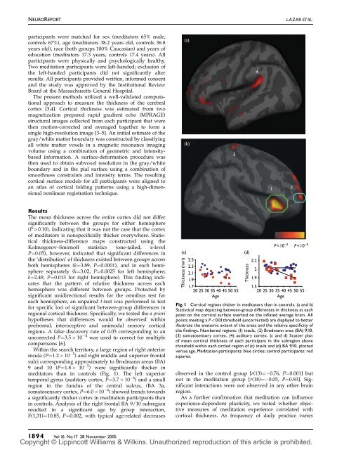
NEUROREPORTLAZAR ETAL.participants were matched for sex (meditators 65% male,controls 67%), age (meditators 38.2 years old, controls 36.8years old), race (both groups 100% Caucasian) and years ofeducation (meditators 17.3 years, controls 17.4 years). Allparticipants were physically and psychologically healthy.Two meditation participants were left-handed; exclusion ofthe left-handed participants did not significantly alterresults. All participants provided written, informed consentand the study was approved by the Institutional ReviewBoard at the Massachusetts General Hospital.The present methods utilized a well-validated computationalapproach to measure the <strong>thickness</strong> of the cerebralcortex [3,4]. Cortical <strong>thickness</strong> was estimated from twomagnetization prepared rapid gradient echo (MPRAGE)structural images collected from each participant that werethen motion-corrected and averaged together to form asingle high-resolution image [3–5]. An initial estimate of thegray/white matter boundary was constructed by classifyingall white matter voxels in a magnetic resonance imagingvolume using a combination of geometric and intensitybasedinformation. A surface-deformation procedure wasthen used to obtain subvoxel resolution in the gray/whiteboundary and in the pial surface using a combination ofsmoothness constraints and intensity terms. The resulting<strong>cortical</strong> surface models for all participants were aligned toan atlas of <strong>cortical</strong> folding patterns using a high-dimensionalnonlinear reg<strong>is</strong>tration technique.(a)(b)341 2ResultsThe mean <strong>thickness</strong> across the entire cortex did not differsignificantly between the groups for either hem<strong>is</strong>phere(P40.10), indicating that it was not the case that the cortexof meditators <strong>is</strong> nonspecifically thicker everywhere. Stat<strong>is</strong>tical<strong>thickness</strong>-difference maps constructed using theKolmogorov–Smirnoff stat<strong>is</strong>tics (one-tailed, a-levelP¼0.05), however, indicated that significant differences inthe ‘d<strong>is</strong>tribution’ of <strong>thickness</strong> ex<strong>is</strong>ted between groups acrossboth hem<strong>is</strong>pheres (k¼3.89, P¼0.0001), and in each hem<strong>is</strong>phereseparately (k¼3.02, P¼0.0025 for left hem<strong>is</strong>phere;k¼2.49, P¼0.013 for right hem<strong>is</strong>phere). Th<strong>is</strong> finding indicatesthat the pattern of relative <strong>thickness</strong> across eachhem<strong>is</strong>phere was different between groups. Protected bysignificant unidirectional results for the omnibus test foreach hem<strong>is</strong>phere, an unpaired t-test was performed to testfor specific loci of significant between-group differences inregional <strong>cortical</strong> <strong>thickness</strong>. Specifically, we tested the a priorihypotheses that differences would be observed <strong>with</strong>inprefrontal, interoceptive and unimodal sensory <strong>cortical</strong>regions. A false d<strong>is</strong>covery rate of 0.05 corresponding to anuncorrected P¼3.5 10 4 was used to correct for multiplecompar<strong>is</strong>ons [6].Within the search territory, a large region of right anteriorinsula (P¼1.2 10 5 ) and right middle and superior frontalsulci corresponding approximately to Brodmann areas (BA)9 and 10 (P¼1.8 10 5 ) were significantly thicker inmeditators than in controls (Fig. 1). The left superiortemporal gyrus (auditory cortex, P¼3.7 10 4 ) and a smallregion in the fundus of the central sulcus, (BA 3a,somatosensory cortex, P¼6.0 10 4 ) showed trends towardsa significantly thicker cortex in meditation participants thanin controls. Analys<strong>is</strong> of the right frontal BA 9/10 subregionresulted in a significant age by group interaction,F(1,31)¼10.85, P¼0.002, <strong>with</strong> typical age-related decreases(c)Thickness (mm)2.52.32.11.91.72025 30 35 40 45 50 55Age(d)Thicknessobserved in the control group [r(13)¼ 0.76, P¼0.001] butnot in the meditation group [r(18)¼ 0.05, P¼0.83]. Significantinteractions were not observed in any other brainregion.As a further confirmation that meditation can influence<strong>experience</strong>-dependent plasticity, we tested whether objectivemeasures of meditation <strong>experience</strong> correlated <strong>with</strong><strong>cortical</strong> <strong>thickness</strong>. As frequency of daily practice varies2.2P
MEDITATION AND CORTICALTHICKNESSNEUROREPORT(a)(b) 2.3Thickness (mm)2.11.91.71.5−10−50 5Change in breathing rate (bpm)Fig. 2 V<strong>is</strong>ual area correlated <strong>with</strong> meditation <strong>experience</strong>. (a) Stat<strong>is</strong>ticalmap depicting <strong>cortical</strong> <strong>thickness</strong> correlated <strong>with</strong> change in respirationrate. (b) Scatter plot of mean <strong>cortical</strong> <strong>thickness</strong> of each participant fromthe circled region <strong>with</strong>in the inferior occipitotemporal lobe plottedversus change in respiration rate. Note: negative change in breathing rate(left side) <strong>is</strong> <strong>associated</strong> <strong>with</strong> more hours of meditation <strong>experience</strong> and athicker cortex.between meditation practitioners, using the total number ofyears of practice <strong>is</strong> not a sensitive metric of <strong>experience</strong>. Oneeffect of regular meditation practice <strong>is</strong> a significant drop inrespiration rate during formal practice [7,8]. We thereforetested whether changes in respiration rate between rest andmeditation could serve as an objective measure of meditation<strong>experience</strong>. The change in mean respiration rate from a6-min baseline period to the first 6 min of the meditationperiod was calculated for each participant and thencorrelated <strong>with</strong> the self-reported total number of hours offormal sitting meditation over the participant’s lifetime(r¼ 0.75, Po0.001). The correlation between respirationrate and total number of years the participant had beenpracticing was also significant (r¼ 0.57, P¼0.009); however,the correlation <strong>with</strong> total hours of formal sitting practiceresulted in a higher coefficient.To directly test for cumulative effects of meditation<strong>experience</strong> on brain structure, a correlation was performedbetween <strong>cortical</strong> <strong>thickness</strong> and change in respiration rate.After correcting for multiple compar<strong>is</strong>ons using a falsed<strong>is</strong>covery rate <strong>associated</strong> <strong>with</strong> a P¼0.05, the analys<strong>is</strong>revealed one significant region <strong>with</strong>in the inferior occipitotemporalv<strong>is</strong>ual cortex (Fig. 2). Among the meditationgroup, the zero-order correlation between <strong>thickness</strong> in th<strong>is</strong>region and change in respiration rate was r(18)¼0.72,Po0.001, which was effectively unchanged when controllingfor individual right-hem<strong>is</strong>phere mean <strong>thickness</strong> (as ameasure of nonspecific effects on <strong>cortical</strong> <strong>thickness</strong>), partialr(17)¼0.73, Po0.001, and still further when controlling forage, partial r(16)¼0.75, Po0.001. When controlling for ageand individual right-hem<strong>is</strong>phere average <strong>thickness</strong>, a partialcorrelation between <strong>thickness</strong> in th<strong>is</strong> region and years of<strong>experience</strong> remained significant [partial r(15)¼0.627,P¼0.007]. These findings are cons<strong>is</strong>tent <strong>with</strong> the hypothes<strong>is</strong>that meditation practice promoted thickening in th<strong>is</strong> region.The most <strong>experience</strong>d participants were also among theoldest. As age-related decreases in <strong>cortical</strong> <strong>thickness</strong> aregreatest in frontal regions [5], it <strong>is</strong> possible that the effect ofage may obscure the modest effects of meditation practice inthese areas. The Pearson correlations between respirationrate and <strong>cortical</strong> <strong>thickness</strong> in the insula and BA 9/10 werenot significant (r¼ 0.36, P¼0.12 and r¼ 0.23, P¼0.33,respectively), although they became so in the insula aftercontrolling for age [partial r(17)¼0.48, P¼0.04]. The correlationbetween these parameters for the BA 9/10 region wasessentially unchanged [partial r(17)¼ 0.25, P¼0.30].D<strong>is</strong>cussionOur data indicate that regular practice of meditation <strong>is</strong><strong>associated</strong> <strong>with</strong> <strong>increased</strong> <strong>thickness</strong> in a subset of <strong>cortical</strong>regions related to somatosensory, auditory, v<strong>is</strong>ual andinteroceptive processing. Further, regular meditation practicemay slow age-related thinning of the frontal cortex.Previous studies of <strong>cortical</strong> plasticity in animals andhumans have shown that when a task requires that attentionbe cons<strong>is</strong>tently directed towards a behaviorally relevantsensory stimulus (e.g. a somatosensory [9] or auditorystimulus [10]) over repeated practice sessions [11], robustchanges in sensory <strong>cortical</strong> maps result ([12] and Kerr CE,Wasserman RH and Moore CI. Cortical plasticity as atherapeutic mechan<strong>is</strong>m for touch healing, under reveiw).Additional studies suggest that relaxation facilitates thelearning-based process that underlies such <strong>cortical</strong> plasticity[13]. It may be useful to conceptualize meditation practice asengaging in an analogous set of <strong>cortical</strong> remodelingprocesses: namely, directing attention towards behaviorallyrelevant sensory stimuli <strong>with</strong>in a relaxing setting overrepeated practice sessions [2,7]. Increased <strong>cortical</strong> <strong>thickness</strong>could be due to greater arborization per neuron, <strong>increased</strong>glial volume or <strong>increased</strong> regional vasculature. The methodsemployed do not d<strong>is</strong>tingu<strong>is</strong>h between these possibilities;however, each of these mechan<strong>is</strong>ms <strong>is</strong> supportive of<strong>increased</strong> neural function.We hypothesized that meditation practice should promoteneural plasticity in regions that are routinely engagedduring formal practice. Many factors including age, sex,genetics, neuropathology and psychopathology [4,5,14,15],however, influence the <strong>thickness</strong> of cortex nonspecifically,confounding these analyses. Perhaps the largest of theseconfounds <strong>is</strong> the effect of age. The rate of age-dependentthinning <strong>is</strong> highly variable across the <strong>cortical</strong> surface [5].<strong>Meditation</strong>-related effects on <strong>thickness</strong> may have beencounterbalanced by the effects of age on <strong>cortical</strong> thinning,thereby minimizing our ability to detect significant correlations.Thinning <strong>is</strong> most pronounced in the frontal lobe, andindeed there were many regions in the parietal, temporaland occipital lobe where there was little if any difference inthe average <strong>thickness</strong> in our older and younger participants(data not shown). Such age-related effects may account forVol 16 No17 28 November 2005 1895Copyright © Lippincott Williams & Wilkins. Unauthorized reproduction of th<strong>is</strong> article <strong>is</strong> prohibited.