Meditation experience is associated with increased cortical thickness
Meditation experience is associated with increased cortical thickness
Meditation experience is associated with increased cortical thickness
- No tags were found...

You also want an ePaper? Increase the reach of your titles
YUMPU automatically turns print PDFs into web optimized ePapers that Google loves.
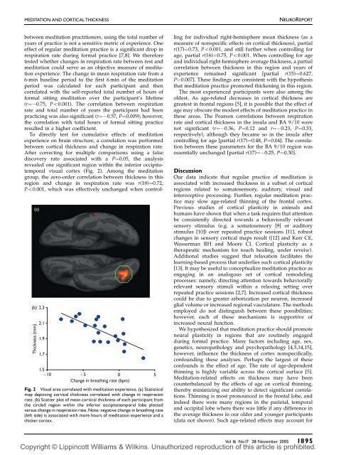
MEDITATION AND CORTICALTHICKNESSNEUROREPORT(a)(b) 2.3Thickness (mm)2.11.91.71.5−10−50 5Change in breathing rate (bpm)Fig. 2 V<strong>is</strong>ual area correlated <strong>with</strong> meditation <strong>experience</strong>. (a) Stat<strong>is</strong>ticalmap depicting <strong>cortical</strong> <strong>thickness</strong> correlated <strong>with</strong> change in respirationrate. (b) Scatter plot of mean <strong>cortical</strong> <strong>thickness</strong> of each participant fromthe circled region <strong>with</strong>in the inferior occipitotemporal lobe plottedversus change in respiration rate. Note: negative change in breathing rate(left side) <strong>is</strong> <strong>associated</strong> <strong>with</strong> more hours of meditation <strong>experience</strong> and athicker cortex.between meditation practitioners, using the total number ofyears of practice <strong>is</strong> not a sensitive metric of <strong>experience</strong>. Oneeffect of regular meditation practice <strong>is</strong> a significant drop inrespiration rate during formal practice [7,8]. We thereforetested whether changes in respiration rate between rest andmeditation could serve as an objective measure of meditation<strong>experience</strong>. The change in mean respiration rate from a6-min baseline period to the first 6 min of the meditationperiod was calculated for each participant and thencorrelated <strong>with</strong> the self-reported total number of hours offormal sitting meditation over the participant’s lifetime(r¼ 0.75, Po0.001). The correlation between respirationrate and total number of years the participant had beenpracticing was also significant (r¼ 0.57, P¼0.009); however,the correlation <strong>with</strong> total hours of formal sitting practiceresulted in a higher coefficient.To directly test for cumulative effects of meditation<strong>experience</strong> on brain structure, a correlation was performedbetween <strong>cortical</strong> <strong>thickness</strong> and change in respiration rate.After correcting for multiple compar<strong>is</strong>ons using a falsed<strong>is</strong>covery rate <strong>associated</strong> <strong>with</strong> a P¼0.05, the analys<strong>is</strong>revealed one significant region <strong>with</strong>in the inferior occipitotemporalv<strong>is</strong>ual cortex (Fig. 2). Among the meditationgroup, the zero-order correlation between <strong>thickness</strong> in th<strong>is</strong>region and change in respiration rate was r(18)¼0.72,Po0.001, which was effectively unchanged when controllingfor individual right-hem<strong>is</strong>phere mean <strong>thickness</strong> (as ameasure of nonspecific effects on <strong>cortical</strong> <strong>thickness</strong>), partialr(17)¼0.73, Po0.001, and still further when controlling forage, partial r(16)¼0.75, Po0.001. When controlling for ageand individual right-hem<strong>is</strong>phere average <strong>thickness</strong>, a partialcorrelation between <strong>thickness</strong> in th<strong>is</strong> region and years of<strong>experience</strong> remained significant [partial r(15)¼0.627,P¼0.007]. These findings are cons<strong>is</strong>tent <strong>with</strong> the hypothes<strong>is</strong>that meditation practice promoted thickening in th<strong>is</strong> region.The most <strong>experience</strong>d participants were also among theoldest. As age-related decreases in <strong>cortical</strong> <strong>thickness</strong> aregreatest in frontal regions [5], it <strong>is</strong> possible that the effect ofage may obscure the modest effects of meditation practice inthese areas. The Pearson correlations between respirationrate and <strong>cortical</strong> <strong>thickness</strong> in the insula and BA 9/10 werenot significant (r¼ 0.36, P¼0.12 and r¼ 0.23, P¼0.33,respectively), although they became so in the insula aftercontrolling for age [partial r(17)¼0.48, P¼0.04]. The correlationbetween these parameters for the BA 9/10 region wasessentially unchanged [partial r(17)¼ 0.25, P¼0.30].D<strong>is</strong>cussionOur data indicate that regular practice of meditation <strong>is</strong><strong>associated</strong> <strong>with</strong> <strong>increased</strong> <strong>thickness</strong> in a subset of <strong>cortical</strong>regions related to somatosensory, auditory, v<strong>is</strong>ual andinteroceptive processing. Further, regular meditation practicemay slow age-related thinning of the frontal cortex.Previous studies of <strong>cortical</strong> plasticity in animals andhumans have shown that when a task requires that attentionbe cons<strong>is</strong>tently directed towards a behaviorally relevantsensory stimulus (e.g. a somatosensory [9] or auditorystimulus [10]) over repeated practice sessions [11], robustchanges in sensory <strong>cortical</strong> maps result ([12] and Kerr CE,Wasserman RH and Moore CI. Cortical plasticity as atherapeutic mechan<strong>is</strong>m for touch healing, under reveiw).Additional studies suggest that relaxation facilitates thelearning-based process that underlies such <strong>cortical</strong> plasticity[13]. It may be useful to conceptualize meditation practice asengaging in an analogous set of <strong>cortical</strong> remodelingprocesses: namely, directing attention towards behaviorallyrelevant sensory stimuli <strong>with</strong>in a relaxing setting overrepeated practice sessions [2,7]. Increased <strong>cortical</strong> <strong>thickness</strong>could be due to greater arborization per neuron, <strong>increased</strong>glial volume or <strong>increased</strong> regional vasculature. The methodsemployed do not d<strong>is</strong>tingu<strong>is</strong>h between these possibilities;however, each of these mechan<strong>is</strong>ms <strong>is</strong> supportive of<strong>increased</strong> neural function.We hypothesized that meditation practice should promoteneural plasticity in regions that are routinely engagedduring formal practice. Many factors including age, sex,genetics, neuropathology and psychopathology [4,5,14,15],however, influence the <strong>thickness</strong> of cortex nonspecifically,confounding these analyses. Perhaps the largest of theseconfounds <strong>is</strong> the effect of age. The rate of age-dependentthinning <strong>is</strong> highly variable across the <strong>cortical</strong> surface [5].<strong>Meditation</strong>-related effects on <strong>thickness</strong> may have beencounterbalanced by the effects of age on <strong>cortical</strong> thinning,thereby minimizing our ability to detect significant correlations.Thinning <strong>is</strong> most pronounced in the frontal lobe, andindeed there were many regions in the parietal, temporaland occipital lobe where there was little if any difference inthe average <strong>thickness</strong> in our older and younger participants(data not shown). Such age-related effects may account forVol 16 No17 28 November 2005 1895Copyright © Lippincott Williams & Wilkins. Unauthorized reproduction of th<strong>is</strong> article <strong>is</strong> prohibited.