Sponges of the New Caledonian lagoon - IRD
Sponges of the New Caledonian lagoon - IRD
Sponges of the New Caledonian lagoon - IRD

Create successful ePaper yourself
Turn your PDF publications into a flip-book with our unique Google optimized e-Paper software.
212<br />
<strong>Sponges</strong><br />
<strong>of</strong> <strong>the</strong> <strong>New</strong><br />
Caledonion<br />
Lagoon<br />
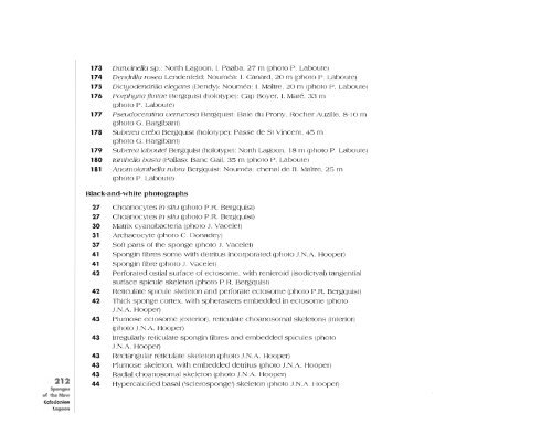
173 DarwineJla sp.: North Lagoon, I. Paaba, 27 m (photo P. Laboute)<br />
174 Dendrilla rosea Lendenfeld: Noumea: I. Canard, 20 m (photo P. Laboute)<br />
175 Dictyodendrilla elegans (Dendy): Noumea: I. Maltre, 20 m (photo P. Laboute)<br />
176 Porphyria jlinrae Bergquist (holotype): Cap Boyer, I. Mare, 33 m<br />
(photo P. Laboute)<br />
177 Pseudoceratina uerrucosa Bergquist: Baie du Prony, Rocher Auzille, 8-10 m<br />
(photo G. Bargibant)<br />
178 Suberea creba Bergquist (holotype): Passe de St Vincent, 45 m<br />
(photo G. Bargibant)<br />
179 Suberea laboutei Bergquist (holotype): North Lagoon, 18 m (photo P. Laboute)<br />
180 Ian<strong>the</strong>lla basta (pallas): Banc Gail, 35 m (photo P. Laboute)<br />
181 Anomoian<strong>the</strong>Jla rubra Bergquist: Noumea: chenal de 1'1. Maltre, 25 m<br />
(photo P. Laboute)<br />
Black-and-white photographs<br />
27<br />
27<br />
30<br />
31<br />
37<br />
41<br />
41<br />
42<br />
42<br />
42<br />
43<br />
43<br />
43<br />
43<br />
43<br />
44<br />
Choanocytes in situ (photo P.R. Bergquist)<br />
Choanocytes in situ (photo P.R. Bergquist)<br />
Matrix cyanobacteria (photo J. Vacelet)<br />
Archaeocyte (photo C Donadey)<br />
S<strong>of</strong>t parts <strong>of</strong> <strong>the</strong> sponge (photo J. Vacelet)<br />
Spongin fibres some with detritus incorporated (photo J.N.A. Hooper)<br />
Spongin fibre (photo J. vacelet)<br />
Perforated ostial surface <strong>of</strong> ectosome, with renieroid (isodictyal) tangential<br />
surface spicule skeleton (photo P.R. Bergquist)<br />
Reticulate spicule skeleton and perforate ectosome (photo P.R. Bergquist)<br />
Thick sponge cortex, with spherasters embedded in ectosome (photo<br />
J.N.A. Hooper)<br />
Plumose ectosome(exterior), reticulate choanosomal skeletons (interior)<br />
(photo J.N.A. Hooper)<br />
Irregularly reticulate spongin fibres and embedded spicules (photo<br />
J.N.A. Hooper)<br />
Rectangular reticulate skeleton (photo J.N.A. Hooper)<br />
Plumose skeleton, with embedded detritus (photo J.N.A. Hooper)<br />
Radial choanosomal skeleton (photo J.N.A. Hooper)<br />
Hypercalcified basal ('sclerosponge') skeleton (photo J.N.A. Hooper)