DEVELOPMENT OF MESODERM,
DEVELOPMENT OF MESODERM,
DEVELOPMENT OF MESODERM,

You also want an ePaper? Increase the reach of your titles
YUMPU automatically turns print PDFs into web optimized ePapers that Google loves.
<strong>DEVELOPMENT</strong> <strong>OF</strong> <strong>MESODERM</strong>,<br />
PARAXIAL <strong>MESODERM</strong> AND<br />
SCLEROMYOTOME AND FORMATION <strong>OF</strong> CARTILAGES<br />
LEARNING OBJECTIVES<br />
At the end of lecture students should be able to know,<br />
What is gastrulation.<br />
Development of mesoderm.<br />
Somitogenesis.<br />
Formation of cartilage.<br />
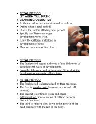
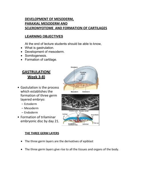
GASTRULATION(<br />
Week 3-8)<br />
• Gastulation is the process<br />
which establishes the<br />
formation of three germ<br />
layered embryo:<br />
– Ectoderm<br />
– Mesoderm<br />
– Endoderm<br />
• Formation of trilaminar<br />
embryonic disc by day 21.<br />
THE THREE GERM LAYERS<br />
The three germ layers are the derivatives of epiblast<br />
The three germ layers give rise to all the tissues and organs of the body.
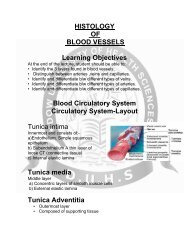
<strong>DEVELOPMENT</strong> <strong>OF</strong> <strong>MESODERM</strong><br />
Paraxial mesoderm<br />
Intermediate mesoderm<br />
Lateral mesoderm
PARAXIAL <strong>MESODERM</strong><br />
Thick plate of mesoderm located on each side of midline<br />
Becomes organized into segments called somatomeres in cranio-caudal<br />
sequence.<br />
INTERMEDIATE <strong>MESODERM</strong>
Longitudinal dorsal ridge of mesoderm located between paraxial and lateral<br />
mesoderm<br />
This ridge forms the Urogenital ridge<br />
Urogenital ridge forms the future kidneys and gonads<br />
LATERAL <strong>MESODERM</strong><br />
Thin plate of mesoderm located along the lateral side of embryo.<br />
Large spaces develop in the lateral plate mesoderm and coalesce to form<br />
intraembryonic coelom<br />
Intraembryonic coelom divides lateral mesoderm into:<br />
Intraembryonic somatic mesoderm<br />
Intraembryonic visceral mesoderm<br />
SOMITOGENESIS<br />
Process of segmentation development of<br />
axial system vertebrae, muscles and
innervations.<br />
Somites form from paraxial mesoderm in<br />
anterior-posterior gradient, begins at<br />
neurulation.<br />
Two parallel columns of mesodermal cells<br />
form along the longitudinal axis, on each<br />
side of the notochord and neural tube.<br />
Transverse fissures form in the<br />
columns forming somitomeres in cranio caudal direction.<br />
SOMITOMERES
Somitomere 1-7 do not form somites, but contribute mesoderm to<br />
pharyngeal arches<br />
The remaining somitomeres condense in cranio-caudal sequence forming<br />
41-42 pairs of somites.<br />
SOMITES
Somites differentialte into:<br />
Sclerotome<br />
Forms cartilage and bone component of veebral column<br />
Myotome<br />
Forms epimeric and hypomeric muscles<br />
Dermatome<br />
Forms dermis and subcutaneous area of skin<br />
SUMMARY <strong>OF</strong> <strong>MESODERM</strong>AL <strong>DEVELOPMENT</strong><br />
FORMATION <strong>OF</strong> CARTILAGE<br />
Cartilage is derived from mesenchyme<br />
Avascular mesenchyme contains stem cells embedded in a delicate collagen<br />
matrix
CHONDROBLAST<br />
Mesenchyme cells begin to differentiate into protochondral cells<br />
Protochondral cells divide to produce chondroblasts<br />
CARTILAGE <strong>DEVELOPMENT</strong>
The center fills with chondroblasts as the top and bottom edges continue to<br />
differentiate.<br />
Stem cells remain on the edges<br />
Chondroblasts remain in the center and produce cartilage matrix.<br />
Stem cells at the edges produce fibroblasts and make the fibrous<br />
perichondrium<br />
Chondroblasts in the center continue to divide
THANK YOU