HISTO Histology of blood vessels
HISTO Histology of blood vessels
HISTO Histology of blood vessels

You also want an ePaper? Increase the reach of your titles
YUMPU automatically turns print PDFs into web optimized ePapers that Google loves.
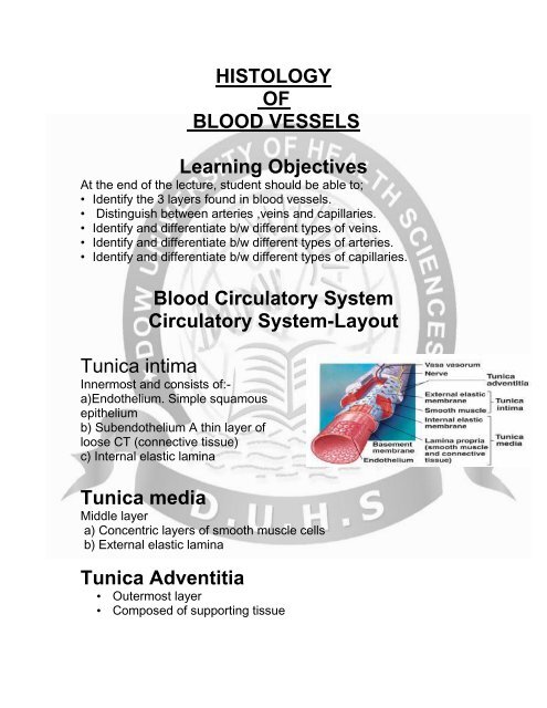
<strong>HISTO</strong>LOGY<br />
OF<br />
BLOOD VESSELS<br />
Learning Objectives<br />
At the end <strong>of</strong> the lecture, student should be able to;<br />
• Identify the 3 layers found in <strong>blood</strong> <strong>vessels</strong>.<br />
• Distinguish between arteries ,veins and capillaries.<br />
• Identify and differentiate b/w different types <strong>of</strong> veins.<br />
• Identify and differentiate b/w different types <strong>of</strong> arteries.<br />
• Identify and differentiate b/w different types <strong>of</strong> capillaries.<br />
Blood Circulatory System<br />
Circulatory System-Layout<br />
Tunica intima<br />
Innermost and consists <strong>of</strong>:-<br />
a)Endothelium. Simple squamous<br />
epithelium<br />
b) Subendothelium A thin layer <strong>of</strong><br />
loose CT (connective tissue)<br />
c) Internal elastic lamina<br />
Tunica media<br />
Middle layer<br />
a) Concentric layers <strong>of</strong> smooth muscle cells<br />
b) External elastic lamina<br />
Tunica Adventitia<br />
• Outermost layer<br />
• Composed <strong>of</strong> supporting tissue
Basic structure <strong>of</strong> the vessel<br />
CAPILLARIES<br />
• Endothelial tubes.<br />
• Diameter – average 7 to 9 μm<br />
• Length – usually 0.25 to 1mm.<br />
• Usually arranged in network<br />
form.<br />
• Endothelial cells- polygonal in shape & arranged as<br />
a mosaic.<br />
• Cell borders are wavy or serrated, and held<br />
togather by tight junctions.<br />
• Centrally located nucleus bulge into the lumen <strong>of</strong><br />
capillary.
CAPILLARIES<br />
Endothelial cells usually rest on basal lamina. Outer to<br />
basal lamina, there is thin layer <strong>of</strong> CT consists <strong>of</strong> delicate<br />
collagenous & reticular<br />
fibers, and pericytes.<br />
Classification <strong>of</strong> Capillaries<br />
1. Continuous Capillaries<br />
2. Fenestrated Capillaries<br />
3. Sinusoidal Capillaries<br />
Continuous Capillaries<br />
• No pores, gapes and discontinuities in their walls.<br />
• Usually numerous pinocytotic vesicles in the cytoplasm <strong>of</strong> cells<br />
for transport <strong>of</strong> fluid across the capillary wall<br />
Fenestrated Capillaries<br />
• Numerous circular pores or fenestrae (60-80 nm) in their wall.<br />
• Pores are closed by diaphragm (except glomerular capillaries).<br />
• A continuous basal lamina is present.
Sinusoidal Capillaries<br />
• Enlarged diameter(30-40 um). Irregular & tortuous path.<br />
• Wide gaps b/w cells.<br />
• Multiple pores (no diaphragm) in cell wall<br />
• Incomplete basal lamina<br />
ARTERIES<br />
According to size, 4 types:-<br />
1. Arterioles<br />
2. Small Arteries<br />
3. Medium size Arteries<br />
4. Large Arteries<br />
ARTERIOLES<br />
Less than 0.5 mm diameter (narrow lumen &thick wall).<br />
T. Intima : Only Endothelium<br />
T. media: 1 or 2 layers<br />
T. Adventitia: Thin, but Relatively thick.
SMALL ARERTIES<br />
T. intima: Endothelium<br />
Very thin Sub – endothelium<br />
T. media: up to 8 layers<br />
T. Adventitia: Thin<br />
MEDIUM SIZE ARTERIES<br />
(Muscular artery OR Distributing Artery)<br />
Examples: Named Arteries.<br />
T. intima:<br />
Endothelium:<br />
Sub-endothelium: Somewhat thicker, few smooth muscle fibers<br />
may be present.<br />
Internal elastic lamina: Very prominent, Forms a thick fenestrated<br />
band <strong>of</strong> closely interwoven elastic fibers.<br />
T. media:<br />
Several layers (up to 40), Small quantity <strong>of</strong> C.T. composed mainly<br />
<strong>of</strong> collagenous fibers.<br />
External elastic lamina: May be seen<br />
T. Adventitia:<br />
Sufficiently thick (may be as thick as Tunica media<br />
Muscular Artery;<br />
diagrammatic representation
LARGE ARTERIES<br />
Elastic Artery (yellow) OR Conducting artery.<br />
Examples: Aorta & Pulmonary trunk<br />
LARGE ARTERIES<br />
T. intima:<br />
Endothelium:<br />
Sub-endothelium: Thicker, In addition to C & E fibers,<br />
fibroblasts and few S.M. fibers also<br />
Internal elastic lamina: Not separately Identified<br />
T. media: A series <strong>of</strong> concentrically arranged fenestrated<br />
elastic membranes. Number increased with age (40 in new<br />
born to 70 in adult).<br />
External elastic lamina: Absent.<br />
T. Adventitia: Thin.<br />
Muscular artery (left) & Elastic artery (right)<br />
The tunica media <strong>of</strong> muscular artery contains predominantly<br />
smooth muscle, whereas the tunica media <strong>of</strong> an elastic artery<br />
is formed by layers <strong>of</strong> smooth muscle intercalated by elastic<br />
laminas. The adventitia and the outer part <strong>of</strong> media have<br />
small <strong>blood</strong> <strong>vessels</strong> (vasa vasorum) and elastic& collagenous<br />
fibers.
VEINS<br />
“Capacitance <strong>vessels</strong>” because more than70%<strong>of</strong> total <strong>blood</strong> volume<br />
is in this portion <strong>of</strong> CVS at any time.<br />
Structural difference B/W veins & arteries.<br />
1. Caliber (Int.diameter) <strong>of</strong> vein is larger than same size artery,<br />
because veins have thinner walls due to scarcity <strong>of</strong> muscular<br />
and elastic components<br />
1. Unlike arteries,small& medium sized veins have valves.<br />
valves :-Consists <strong>of</strong> 2 semi lunar folds <strong>of</strong> tunica intima that project<br />
into the lumen.They composed <strong>of</strong> elastic CT and are lined on both<br />
sides by endothelium.<br />
They are specially numerous in veins <strong>of</strong> lower limbs.<br />
Comparison <strong>of</strong> artery and vein<br />
VEINS<br />
4 types, according to size.<br />
1. Venule.<br />
2. Small veins.<br />
3. Medium sized veins.<br />
4. Large veins.<br />
Small veins :- They have similar structure <strong>of</strong><br />
venules, except, diameter (1 – 2 mm), few smooth
layers in the tunica media and presence <strong>of</strong> internal valves.<br />
VEINS Cont.<br />
VENULE<br />
Diameter from 0.2 to 1mm.<br />
T. intima: Only Endothelium<br />
T. media: None to few smooth muscle fibers.<br />
T. adventitia: Relatively Thickest, Consists <strong>of</strong> longitudinally oriented<br />
collagenous fibers.<br />
VEINS Cont.<br />
MEDIUM SIZED VEINS<br />
Diameter from 2 to 9 mm. Examples – Named veins.<br />
T. intima:<br />
Endothelium<br />
Sub-endothelium: Thin<br />
Internal elastic lamina: May be distinguish<br />
T. media: Small bundles <strong>of</strong> smooth muscle fibers<br />
intermixed with reticular & delicate network <strong>of</strong><br />
elastic fibers.<br />
T. adventitia: Well developed. Forms bulk <strong>of</strong> vein’s wall, composed <strong>of</strong><br />
thick bundles <strong>of</strong> collagenous fibers, running in longitudinal direction.<br />
VEINS Cont.<br />
LARGE VEINS<br />
Examples – superior & inferior vena cava & portal vein.<br />
T. INTIMA<br />
Endothelium<br />
Sub-endothelium: Thin<br />
Internal elastic lamina: May be distinguish<br />
T. MEDIA Thin & poorly developed.<br />
Mainly consists <strong>of</strong> collagenous fibers.<br />
T. ADVENTITIA EXTREMELY THICKEST & has 3 ZONES<br />
Inner most: Dense fibroelastic C.T. with coarse
collagenous fibers, running spirally.<br />
Middle: Longitudinally running smooth muscle fibers.<br />
Outer: A network <strong>of</strong> coarse collagenous & elastic fibers.<br />
Cross section <strong>of</strong> two venules and four small arterioles<br />
The walls <strong>of</strong> the arteries are thicker than the walls <strong>of</strong> the<br />
veins.<br />
Note the cross sections <strong>of</strong> smooth muscle cells and the<br />
field <strong>of</strong> loose connective tissue that surrounds the <strong>vessels</strong>.<br />
COMPARISON 0F MEDIUM SIZE ARTERY<br />
& VEIN<br />
Diagram comparing the structure <strong>of</strong> a muscular artery (left)<br />
and accompanying vein (right).<br />
Note that the tunica intima and tunica media are highly developed in the<br />
artery but not in the vein
Artery, vein, nerve, elastin stain<br />
Comparison <strong>of</strong> artery, vein and capillary<br />
REFERENCE<br />
• MEDICAL <strong>HISTO</strong>LOGY BY LAIQUE HUSSAIN<br />
5TH EDITION