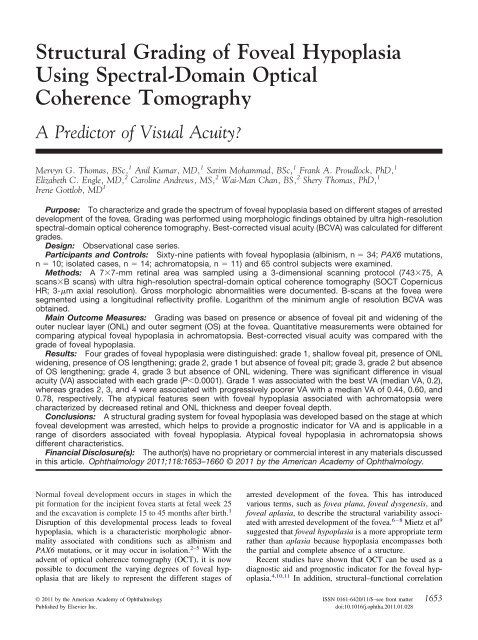
Structural Grading of Foveal Hypoplasia Using Spectral-Domain ...
Structural Grading of Foveal Hypoplasia Using Spectral-Domain ...
Structural Grading of Foveal Hypoplasia Using Spectral-Domain ...

Create successful ePaper yourself
Turn your PDF publications into a flip-book with our unique Google optimized e-Paper software.
<strong>Structural</strong> <strong>Grading</strong> <strong>of</strong> <strong>Foveal</strong> <strong>Hypoplasia</strong><br />
<strong>Using</strong> <strong>Spectral</strong>-<strong>Domain</strong> Optical<br />
Coherence Tomography<br />
A Predictor <strong>of</strong> Visual Acuity?<br />
Mervyn G. Thomas, BSc, 1 Anil Kumar, MD, 1 Sarim Mohammad, BSc, 1 Frank A. Proudlock, PhD, 1<br />
Elizabeth C. Engle, MD, 2 Caroline Andrews, MS, 2 Wai-Man Chan, BS, 2 Shery Thomas, PhD, 1<br />
Irene Gottlob, MD 1<br />
Purpose: To characterize and grade the spectrum <strong>of</strong> foveal hypoplasia based on different stages <strong>of</strong> arrested<br />
development <strong>of</strong> the fovea. <strong>Grading</strong> was performed using morphologic findings obtained by ultra high-resolution<br />
spectral-domain optical coherence tomography. Best-corrected visual acuity (BCVA) was calculated for different<br />
grades.<br />
Design: Observational case series.<br />
Participants and Controls: Sixty-nine patients with foveal hypoplasia (albinism, n 34; PAX6 mutations,<br />
n 10; isolated cases, n 14; achromatopsia, n 11) and 65 control subjects were examined.<br />
Methods: A 77-mm retinal area was sampled using a 3-dimensional scanning protocol (74375, A<br />
scansB scans) with ultra high-resolution spectral-domain optical coherence tomography (SOCT Copernicus<br />
HR; 3-m axial resolution). Gross morphologic abnormalities were documented. B-scans at the fovea were<br />
segmented using a longitudinal reflectivity pr<strong>of</strong>ile. Logarithm <strong>of</strong> the minimum angle <strong>of</strong> resolution BCVA was<br />
obtained.<br />
Main Outcome Measures: <strong>Grading</strong> was based on presence or absence <strong>of</strong> foveal pit and widening <strong>of</strong> the<br />
outer nuclear layer (ONL) and outer segment (OS) at the fovea. Quantitative measurements were obtained for<br />
comparing atypical foveal hypoplasia in achromatopsia. Best-corrected visual acuity was compared with the<br />
grade <strong>of</strong> foveal hypoplasia.<br />
Results: Four grades <strong>of</strong> foveal hypoplasia were distinguished: grade 1, shallow foveal pit, presence <strong>of</strong> ONL<br />
widening, presence <strong>of</strong> OS lengthening; grade 2, grade 1 but absence <strong>of</strong> foveal pit; grade 3, grade 2 but absence<br />
<strong>of</strong> OS lengthening; grade 4, grade 3 but absence <strong>of</strong> ONL widening. There was significant difference in visual<br />
acuity (VA) associated with each grade (P0.0001). Grade 1 was associated with the best VA (median VA, 0.2),<br />
whereas grades 2, 3, and 4 were associated with progressively poorer VA with a median VA <strong>of</strong> 0.44, 0.60, and<br />
0.78, respectively. The atypical features seen with foveal hypoplasia associated with achromatopsia were<br />
characterized by decreased retinal and ONL thickness and deeper foveal depth.<br />
Conclusions: A structural grading system for foveal hypoplasia was developed based on the stage at which<br />
foveal development was arrested, which helps to provide a prognostic indicator for VA and is applicable in a<br />
range <strong>of</strong> disorders associated with foveal hypoplasia. Atypical foveal hypoplasia in achromatopsia shows<br />
different characteristics.<br />
Financial Disclosure(s): The author(s) have no proprietary or commercial interest in any materials discussed<br />
in this article. Ophthalmology 2011;118:1653–1660 © 2011 by the American Academy <strong>of</strong> Ophthalmology.<br />
Normal foveal development occurs in stages in which the<br />
pit formation for the incipient fovea starts at fetal week 25<br />
and the excavation is complete 15 to 45 months after birth. 1<br />
Disruption <strong>of</strong> this developmental process leads to foveal<br />
hypoplasia, which is a characteristic morphologic abnormality<br />
associated with conditions such as albinism and<br />
PAX6 mutations, or it may occur in isolation. 2–5 With the<br />
advent <strong>of</strong> optical coherence tomography (OCT), it is now<br />
possible to document the varying degrees <strong>of</strong> foveal hypoplasia<br />
that are likely to represent the different stages <strong>of</strong><br />
arrested development <strong>of</strong> the fovea. This has introduced<br />
various terms, such as fovea plana, foveal dysgenesis, and<br />
foveal aplasia, to describe the structural variability associated<br />
with arrested development <strong>of</strong> the fovea. 6–8 Mietz et al 9<br />
suggested that foveal hypoplasia is a more appropriate term<br />
rather than aplasia because hypoplasia encompasses both<br />
the partial and complete absence <strong>of</strong> a structure.<br />
Recent studies have shown that OCT can be used as a<br />
diagnostic aid and prognostic indicator for the foveal hypoplasia.<br />
4,10,11 In addition, structural–functional correlation<br />
© 2011 by the American Academy <strong>of</strong> Ophthalmology ISSN 0161-6420/11/$–see front matter<br />
Published by Elsevier Inc. doi:10.1016/j.ophtha.2011.01.028<br />
1653
studies have been performed to document OCT findings in<br />
foveal hypoplasia, although most <strong>of</strong> them were disease<br />
specific for albinism. Harvey et al 12 showed a weak but<br />
significant correlation between macular thickness and visual<br />
acuity. In contrast, Holmstrom et al 10 did not find significant<br />
correlation between central macular thickness and visual<br />
acuity (VA). Seo et al 11 proposed a grading system for<br />
foveal hypoplasia in albinism based on: (1) foveal hyporeflectivity,<br />
(2) choroidal transillumination, (3) tram tract<br />
sign, and (4) foveal depression. They showed in 13 patients<br />
that VA correlates well with degree <strong>of</strong> foveal hypoplasia,<br />
although subsequently they suggested that the role <strong>of</strong> foveal<br />
depression as prognostic indicator remains unclear because<br />
<strong>of</strong> the small sample size. 13 Chong et al 14 also characterized<br />
the foveal morphologic abnormalities in 2 patients with<br />
ocular albinism and in 5 patients with suspected ocular<br />
albinism and reported that there were no foveal hyporeflectivity<br />
or tram tract signs in their study population. Marmor<br />
et al 7 noted a spectrum <strong>of</strong> different visual acuities in 4<br />
patients who all lacked a foveal pit. Furthermore, they<br />
showed that foveal cone specialization, represented on OCT<br />
by the lengthening <strong>of</strong> the outer segment (OS) at the fovea,<br />
can be preserved even in the absence <strong>of</strong> a foveal pit.<br />
However, it remains unclear how the morphologic variability<br />
associated with foveal hypoplasia relates to visual prognosis<br />
and what specific features at the fovea may be more<br />
important in determining visual performance.<br />
This study sought to characterize the spectrum <strong>of</strong> foveal<br />
hypoplasia and to develop a structural grading system for<br />
foveal hypoplasia based on foveal development that can be<br />
applied to various disorders associated with foveal hypoplasia.<br />
The purpose for establishing a grading system was<br />
3-fold: (1) to document the stage at which foveal development<br />
was arrested, (2) to provide a prognostic indicator for<br />
visual acuity, and (3) to ensure applicability in a range <strong>of</strong><br />
disorders associated with foveal hypoplasia. A high-speed<br />
ultra high-resolution spectral-domain OCT (axial resolution,<br />
3 m) was used to image the foveal region in patients<br />
with foveal hypoplasia associated with different diseases<br />
and controls.<br />
Patients and Methods<br />
Patients<br />
The study population consisted <strong>of</strong> 69 patients with foveal hypoplasia<br />
(defined as incursion or continuation <strong>of</strong> inner retinal layers) and<br />
65 controls with a mean age <strong>of</strong> 28.5 years (standard deviation, 15.7<br />
years; range, 5–64 years) and 33.5 years (standard deviation, 14.3<br />
years; range, 8–62 years), respectively. The patient cohort consisted<br />
<strong>of</strong> 58 white persons (84%) and 11 Asians (16%); similarly,<br />
the control cohort consisted <strong>of</strong> 48 white persons (74%) and 17<br />
Asians (26%). In the patient group, there were 38 males (55%) and<br />
31 females (45%); in the control group, there were 31 males (48%)<br />
and 34 females (52%). All patients underwent an ophthalmologic<br />
examination that included slit-lamp examination, fundus examination,<br />
and measurement <strong>of</strong> binocular best-corrected visual acuity<br />
(BCVA) using standardized logarithm <strong>of</strong> the minimum angle <strong>of</strong><br />
resolution (logMAR) charts. The BCVA with both eyes open was<br />
used for comparison with OCT grading because it represents the<br />
best achievable VA and, for example, would reduce other causes<br />
1654<br />
Ophthalmology Volume 118, Number 8, August 2011<br />
<strong>of</strong> decreased VA such as amblyopia or latent nystagmus. Full-field<br />
electroretinography response and visual evoked potentials were<br />
recorded based on International Society for Clinical Electrophysiology<br />
<strong>of</strong> Vision standards. The patients were diagnosed with<br />
albinism (n 34), PAX6 mutations (n 10), isolated foveal<br />
hypoplasia (n 14), and achromatopsia (n 11). The diagnosis<br />
<strong>of</strong> albinism and achromatopsia were based on the clinical findings<br />
and on electrodiagnostic tests. All patients diagnosed with albinism<br />
had asymmetric visual evoked potentials and transillumination<br />
defects <strong>of</strong> the iris. Patients with achromatopsia had extinguished<br />
photopic electroretinography results. All other patients<br />
with normal visual evoked potential and electroretinography results<br />
underwent bidirectional sequence analysis for PAX6 mutations.<br />
PAX6 mutations were detected in 10 patients, <strong>of</strong> whom 3 had<br />
aniridia. One patient with a PAX6 mutation had to be excluded<br />
because <strong>of</strong> poor scan quality resulting from corneal opacity. All<br />
patients in the study population had nystagmus. There was no<br />
history <strong>of</strong> premature birth among the patients evaluated. All other<br />
patients and controls chosen for this study had clear media. None<br />
<strong>of</strong> the controls had nystagmus or a history <strong>of</strong> premature birth.<br />
Informed consent was obtained from all volunteers participating in<br />
this study. The study adhered to the tenets <strong>of</strong> the Declaration <strong>of</strong><br />
Helsinki and was approved by the local ethics committee.<br />
Optical Coherence Tomography Image Acquisition<br />
An ultra high-resolution spectral-domain OCT (SOCT Copernicus<br />
HR; OPTOPOL Technology S.A., Zawiercie, Poland) was used<br />
to acquire tomograms from the patient and control cohorts. A<br />
3-dimensional scan program (74375; A scansB scans) was<br />
used to obtain a 77-mm retinal area centered at the fovea. The<br />
effective axial and transverse resolution for this machine was<br />
approximately 3 and 12 m, respectively. Because this OCT<br />
machine achieves quite a high scanning speed (52 000 A-scans/<br />
second), reproducible, quantitative OCT measurements could be<br />
obtained with no associated motion artifact. The OCT images were<br />
obtained from both eyes in the study population. The tomograms<br />
were viewed using SOCT s<strong>of</strong>tware (version 4.10) and subsequently<br />
were graded. Intraretinal thickness measurements were<br />
obtained using reflectivity pr<strong>of</strong>iles as described elsewhere. 15 The<br />
thickness measurements derived from both eyes were averaged<br />
and analyzed subsequently, similar to a 1-eye study design as<br />
described by Ray and O’Day. 16<br />
<strong>Grading</strong> <strong>Foveal</strong> <strong>Hypoplasia</strong><br />
The rationale behind the grading system that was adopted was<br />
based on the unique developmental processes occurring at the<br />
fovea (Fig 1). During development <strong>of</strong> the fovea, there is (1)<br />
centrifugal displacement <strong>of</strong> cells <strong>of</strong> the inner retina toward the<br />
periphery, (2) centripetal migration <strong>of</strong> cone photoreceptors toward<br />
the location <strong>of</strong> the incipient fovea, and (3) cone specialization<br />
<strong>of</strong> the foveolar cones. 1,17 Because <strong>of</strong> the centrifugal displacement<br />
<strong>of</strong> the inner retinal cells, the foveal depression continues to deepen<br />
until 15 months after birth, and this is seen as complete extrusion<br />
<strong>of</strong> the inner nuclear and plexiform layers posterior to the foveola<br />
(see example <strong>of</strong> normal OCT scan in Fig 2). 18 The centripetal<br />
migration <strong>of</strong> the cone photoreceptors is represented by the outer<br />
nuclear layer (ONL) widening. The cone outer segment undergoes<br />
both a decrease in diameter and increase an in length (i.e., cone<br />
specialization); this allows an increase in foveolar cone packing<br />
density. 17 The change in cone diameter continues up to 45 months<br />
after birth. The cone specialization is represented on OCT by the<br />
OS lengthening. 7 The grading system used takes into account each<br />
<strong>of</strong> these developmental steps.
Figure 1. Chart showing the 3 developmental processes involved in<br />
formation <strong>of</strong> a structural and functional fovea. In grade 1 foveal hypoplasia,<br />
all processes occur to a certain extent. However, in grade 4 foveal<br />
hypoplasia, none <strong>of</strong> these processes occur; thus, the retina resembles that<br />
<strong>of</strong> the parafovea. In grade 2 and 3 foveal hypoplasia, there is outer nuclear<br />
layer widening, but no foveal pit. The difference between grade 2 and 3<br />
foveal hypoplasia is occurrence <strong>of</strong> cone photoreceptor specialization. Identifying<br />
these specific features on optical coherence tomography (OCT)<br />
enables us to understand whether the respective developmental process<br />
has occurred.<br />
Statistical Methods<br />
Normality <strong>of</strong> the VA and intraretinal thickness measurements data<br />
were tested using the Shapiro-Wilk test. Nonparametric tests<br />
(Kruskal-Wallis) were used (because <strong>of</strong> nonnormality) to test the<br />
difference in (1) logMAR VA between the different grades <strong>of</strong><br />
foveal hypoplasia, (2) retinal thickness, (3) ONL thickness, and (4)<br />
foveal depth at the fovea between controls and patients with<br />
typical and atypical forms <strong>of</strong> foveal hypoplasia. Multiple comparisons<br />
were performed with Bonferroni correction. The Mann–<br />
Whitney U test was used to test the differences in VA between<br />
typical and atypical forms <strong>of</strong> foveal hypoplasia.<br />
Results<br />
Gross Morphologic Features Associated with<br />
<strong>Foveal</strong> <strong>Hypoplasia</strong><br />
Examples <strong>of</strong> the gross features <strong>of</strong> foveal hypoplasia detectable on<br />
ultra high-resolution spectral-domain OCT are shown in Figure 2.<br />
The hallmark <strong>of</strong> foveal hypoplasia detectable on OCT is the<br />
incursion <strong>of</strong> the inner retinal layers posterior to the foveola. In<br />
addition to this, other features seen on OCT included: shallower or<br />
absent foveal pit, diminished ONL widening, decreased OS lengthening,<br />
and overall thickening <strong>of</strong> the retina. However, there is<br />
considerable phenotypic variability associated with foveal hypoplasia.<br />
Figure 2 shows examples <strong>of</strong> foveal hypoplasia and the<br />
degree <strong>of</strong> variability seen.<br />
<strong>Grading</strong> and Functional Implications <strong>of</strong> <strong>Foveal</strong><br />
<strong>Hypoplasia</strong><br />
To derive a structural grading system for foveal hypoplasia, the<br />
foveal region was subdivided according to foveal development<br />
into the structural elements that represent the unique features <strong>of</strong> the<br />
fovea detectable using OCT (see Fig 3A). Progressive loss <strong>of</strong> the<br />
foveal elements is represented as increasing grades.<br />
Incursion <strong>of</strong> the plexiform layers was present in all types <strong>of</strong><br />
foveal hypoplasia because it was the criteria used to diagnose<br />
foveal hypoplasia. Subsequently, the grading system gives most<br />
importance to the integrity <strong>of</strong> the outer segment (OS; i.e., whether<br />
this region disrupted to rule out atypical forms <strong>of</strong> foveal hypopla-<br />
Thomas et al <strong>Structural</strong> <strong>Grading</strong> <strong>of</strong> <strong>Foveal</strong> <strong>Hypoplasia</strong><br />
sia). Then, importance is given to the development <strong>of</strong> this region<br />
(detected on OCT by the lengthening <strong>of</strong> the OS; this feature is<br />
present in grades 1 and 2 but not in grades 3 and 4). Subsequently,<br />
the features <strong>of</strong> foveal development anterior to the inner segment<br />
(IS) and OS were assessed. Widening <strong>of</strong> the ONL differentiated<br />
between grades 3 and 4, and foveal pit formation differentiated<br />
between grades 1 and 2. The features <strong>of</strong> each grade are illustrated<br />
in Figure 3B. An algorithm was devised to grade foveal hypoplasia<br />
structurally (Fig 4).<br />
Whether the grade <strong>of</strong> foveal hypoplasia significantly predicted<br />
the BCVA was assessed. There was a significant difference in<br />
BCVA between the grades <strong>of</strong> foveal hypoplasia (P0.0001; see<br />
Fig 5). Grade 1 foveal hypoplasia was associated with the best VA<br />
(median BCVA, 0.2 logMAR), whereas grades 2, 3, and 4 were<br />
associated with progressively poorer VA with a median BCVA <strong>of</strong><br />
0.44, 0.60, and 0.78 logMAR, respectively (Fig 5B). The results <strong>of</strong><br />
the multiple comparisons <strong>of</strong> VA between the grades are shown in<br />
Figure 5B. A significant effect <strong>of</strong> gender, age, or ethnicity on the<br />
grade <strong>of</strong> foveal hypoplasia was not found.<br />
Comparison <strong>of</strong> Typical and Atypical <strong>Foveal</strong><br />
<strong>Hypoplasia</strong><br />
The number <strong>of</strong> patients within each grade and their diagnosis<br />
is shown in Figure 5A. Most patients with albinism had grade<br />
3 foveal hypoplasia, whereas most isolated cases and PAX6 patients<br />
had grade 1 foveal hypoplasia. Overall, patients with albinism<br />
were associated with the worst BCVA (median BCVA, 0.6<br />
logMAR; interquartile range, 0.30), followed by patients with<br />
PAX6 mutations (median BCVA, 0.4 logMAR; interquartile range,<br />
0.35), and then isolated cases (median BCVA, 0.2 logMAR; standard<br />
deviation, 0.30). However, if the grade <strong>of</strong> foveal hypoplasia<br />
was considered, there was no significant difference in BCVA<br />
between the 3 disorders for grade 1 foveal hypoplasia (P 0.83).<br />
(Only grade 1 had sufficient sample numbers within each diagnosis<br />
to make multiple comparisons.)<br />
As shown in Figure 2, achromatopsia is associated with disruption<br />
<strong>of</strong> the IS/OS junction; however, it also can be associated<br />
with a shallower pit and incursion <strong>of</strong> the plexiform layers posterior<br />
to the foveola. This results in an atypical foveal hypoplasia because<br />
it is associated with photoreceptor degeneration. The other<br />
atypical features associated with achromatopsia that do not follow<br />
the pattern seen with the typical forms <strong>of</strong> foveal hypoplasia include:<br />
a significantly decreased retinal thickness in comparison<br />
with both controls (P0.0001) and the patients with typical foveal<br />
hypoplasia (P0.0001; grades 1 through 4; Fig 5D). Similarly,<br />
there is a significantly thinner outer nuclear layer in comparison<br />
with both controls (P0.0001) and patients with typical foveal<br />
hypoplasia (P0.0001; Fig 5E). Although achromatopsia is associated<br />
with significantly shallower pit in comparison with the<br />
controls (P0.0001), the foveal pit in achromatopsia is significantly<br />
deeper in comparison with the patients with grade 1 foveal<br />
hypoplasia (P 0.005; only grade 1 foveal hypoplasia was used<br />
for this comparison because only these patients have a rudimentary<br />
foveal pit; see Fig 5F).<br />
Discussion<br />
This study proposed a structural grading system for foveal<br />
hypoplasia based on loss <strong>of</strong> unique elements that form the<br />
normal fovea that likely have been arrested during early<br />
development. The proposed grading system has 3 advantages:<br />
(1) it gives insight into the degree <strong>of</strong> development <strong>of</strong><br />
the fovea, (2) it provides a prognostic indicator from a<br />
1655
Figure 2. A, Optical coherence tomography scan showing a normal fovea with description <strong>of</strong> the normal foveal elements. Optical coherence tomography<br />
scans showing the spectrum <strong>of</strong> foveal hypoplasia seen in various conditions, including: (B, C) albinism, (D, E) associated with PAX6 mutations, (F, G)<br />
isolated cases, and (H, I) an atypical form <strong>of</strong> foveal hypoplasia seen in achromatopsia. A hyporeflective zone (cavitation) is also seen (I) that is a sign<br />
<strong>of</strong> cone photoreceptor degeneration. Both foveal hypoplasia and fovea plana were seen in all disorders except the achromatopsia, which results in an<br />
atypical form <strong>of</strong> foveal hypoplasia with a shallower pit, incursion <strong>of</strong> the plexiform layers, and disruption <strong>of</strong> the inner segment (IS)/outer segment (OS)<br />
junction. INL inner nuclear layer; NFL nerve fiber layer; ONL outer nuclear layer.<br />
morphologic OCT scan, and (3) it can be applied to most<br />
disorders associated with foveal hypoplasia. The study also<br />
showed that achromatopsia can be associated with foveal<br />
hypoplasia, although it is associated with atypical features<br />
such as IS/OS disruption and ONL thinning, which are signs<br />
<strong>of</strong> photoreceptor degeneration, reduced retinal thickness<br />
(RT), and a deeper foveal pit in comparison with the typical<br />
forms <strong>of</strong> foveal hypoplasia.<br />
From a developmental perspective, each grade suggests<br />
developmental arrest <strong>of</strong> the 3 key events to varying degrees,<br />
as shown in Figure 1. All patients with foveal hypoplasia in<br />
this study had incursion <strong>of</strong> the plexiform layer posterior to<br />
1656<br />
Ophthalmology Volume 118, Number 8, August 2011<br />
the foveola, suggesting that pit formation was incomplete in<br />
all patients. A partial displacement <strong>of</strong> the inner retinal layers<br />
results in a rudimentary pit, as encountered with grade 1<br />
foveal hypoplasia and atypical foveal hypoplasia. This process<br />
has failed to occur in grades 2, 3, and 4 <strong>of</strong> foveal<br />
hypoplasia. Lengthening <strong>of</strong> the OS is a sign <strong>of</strong> cone photoreceptor<br />
specialization, and this occurs to some extent in<br />
grades 1 and 2, but not grades 3 and 4. Lengthening <strong>of</strong> the<br />
OS can occur in the absence <strong>of</strong> a foveal pit, as seen in grade<br />
2 foveal hypoplasia, and also as suggested by Marmor et al. 7<br />
However, no cases where a foveal pit was present with no<br />
OS lengthening (i.e., all patients with a foveal pit had OS
Thomas et al <strong>Structural</strong> <strong>Grading</strong> <strong>of</strong> <strong>Foveal</strong> <strong>Hypoplasia</strong><br />
Figure 3. A, Illustration showing the unique features <strong>of</strong> a normal fovea detectable on optical coherence tomography. B, Illustration <strong>of</strong> typical and atypical grades<br />
<strong>of</strong> foveal (this is a CE) hypoplasia. All grades <strong>of</strong> foveal hypoplasia had incursion <strong>of</strong> inner retinal layers. Atypical foveal hypoplasia also had incursion <strong>of</strong> the inner<br />
retinal layers. Grade 1 foveal hypoplasia is associated with a shallow foveal pit, outer nuclear layer (ONL) widening, and outer segment (OS) lengthening relative<br />
to the parafoveal ONL and OS length, respectively. In Grade 2 foveal hypoplasia, all features <strong>of</strong> grade 1 are present except the presence <strong>of</strong> a foveal pit. Grade<br />
3 foveal hypoplasia consists <strong>of</strong> all features <strong>of</strong> grade 2 foveal hypoplasia except the widening <strong>of</strong> the cone outer segment. Grade 4 foveal hypoplasia represents all<br />
the features seen in grade 3 except there is no widening <strong>of</strong> the ONL at the fovea. Finally, an atypical form <strong>of</strong> foveal hypoplasia also is described in which there<br />
is a shallower pit with disruption <strong>of</strong> the inner segment/outer segment (IS/OS) junction, possibly a sign <strong>of</strong> photoreceptor degeneration. The atypical form <strong>of</strong> foveal<br />
hypoplasia is seen with achromatopsia, whereas grades 1 through 4 are seen with albinism, PAX-6 mutations, and isolated cases. ELM external limiting<br />
membrane; GCL ganglion cell layer; INL inner nuclear layer; IPL inner plexiform layer; OPL outer plexiform layer; RNFL retinal nerve fibre layer;<br />
RPE retinal pigment epithelium.<br />
1657
Figure 4. Algorithm used for grading foveal hypoplasia based on optical coherence tomography findings. The hallmark <strong>of</strong> foveal hypoplasia is incursion<br />
<strong>of</strong> the inner retinal layers. Based on disruption <strong>of</strong> the inner segment/outer segment (IS/OS) junction <strong>of</strong> the photoreceptor, the foveal hypoplasia is classified<br />
into either typical or atypical foveal hypoplasia. The grade <strong>of</strong> foveal hypoplasia can be determined based on whether the following features are present<br />
or absent: outer segment (OS) lengthening, foveal pit, and outer nuclear layer (ONL) widening.<br />
lengthening) were encountered, suggesting that pit formation<br />
may be dependent partly on OS lengthening. Successful<br />
centripetal migration <strong>of</strong> cone photoreceptors is represented<br />
by ONL widening (seen in grades 1, 2, and 3). However, in<br />
grade 4 foveal hypoplasia, this developmental process is<br />
completely arrested and has failed to occur.<br />
This grading system provides a prognostic visual function<br />
indicator by showing that the grade <strong>of</strong> foveal hypoplasia<br />
is significantly related to VA. It can be used easily in a<br />
clinical setting and does not require thickness measurements<br />
to predict and provide a likely visual prognosis for the<br />
patient with foveal hypoplasia. The grading system proposed<br />
accounts for the retinal structural basis for the decreased<br />
VA, but not for additional causes reducing VA,<br />
such as nystagmus, amblyopia, anterior segment disorder,<br />
or refractive error. Hence, morphologic grading <strong>of</strong> foveal<br />
hypoplasia can help in deciding whether further investigation<br />
or treatment are necessary to improve the patient’s VA.<br />
For instance, if a patient has grade 1 foveal hypoplasia and<br />
a VA <strong>of</strong> 0.7 logMAR, factors other than the foveal hypoplasia<br />
are likely to be contributing to the poor VA. Similarly,<br />
if a patient has a VA <strong>of</strong> 0.6 logMAR and has a grade<br />
3 foveal hypoplasia, one would not expect the vision to<br />
improve past 0.5 logMAR because the structural development<br />
<strong>of</strong> the fovea is the limiting factor. An algorithm was<br />
also developed that is easy to follow and that can be used to<br />
derive the structural grade for foveal hypoplasia. Although<br />
4 representative disorders associated with foveal hypoplasia<br />
were included, the grading system was not applied in patients<br />
with other forms <strong>of</strong> foveal hypoplasia, such as nanophthalmos<br />
19 and retinopathy <strong>of</strong> prematurity. 20 In this study<br />
1658<br />
Ophthalmology Volume 118, Number 8, August 2011<br />
population, VA correlated well with the grade <strong>of</strong> foveal<br />
hypoplasia; however, validation <strong>of</strong> the grading scheme in<br />
larger cohorts is necessary.<br />
The previous study by Seo et al 11 used OCT signs in<br />
albinism, which are dependent on reflectivity <strong>of</strong> structures<br />
in the tomogram such as foveal hyporeflectivity, choroidal<br />
transillumination, and tram tract sign. Presence or absence<br />
<strong>of</strong> foveal depression also was used to grade the foveal<br />
hypoplasia. Determining reflectivity features can be subjective,<br />
especially when using spectral-domain OCT, because<br />
they are associated with a sensitivity roll <strong>of</strong>f, that is, the<br />
decrease in reflectivity values as the image moves away<br />
from the zero delay line. Hence, foveal hyporeflectivity may<br />
be difficult to interpret between an image that is close to the<br />
0 delay line (which would be associated with higher reflectivity)<br />
and one that is further away (which would be less<br />
reflective). 21 This study avoided using reflectivity values<br />
because <strong>of</strong> the inherent difficulty in standardizing these<br />
values with spectral-domain OCT and because these signs<br />
may be specific for albinism. This study also showed that<br />
atypical foveal hypoplasia (seen in achromatopsia) is associated<br />
with worse visual prognosis in comparison with the<br />
typical disorders associated with foveal hypoplasia; this is<br />
because <strong>of</strong> the photoreceptor degeneration, visualized on<br />
OCT as IS/OS disruption. 22,23 Interestingly, in achromatopsia<br />
there is also a reduction in RT, whereas in typical foveal<br />
hypoplasia, there is an increase in RT compared with controls.<br />
This paradoxical reduction in RT in achromats is<br />
because <strong>of</strong> the ONL thinning resulting from photoreceptor<br />
degeneration. 22 However in achromatopsia, there is still a<br />
significantly shallower foveal pit compared with that in
controls and incursion <strong>of</strong> the inner retinal layers. This suggests<br />
that the centrifugal displacement <strong>of</strong> the inner retinal<br />
layers is not complete in some patients with achromatopsia.<br />
In summary, a structural grading system for foveal hypoplasia<br />
that relates to developmental stages and visual prognosis<br />
and that can be applied to most disorders associated<br />
with foveal hypoplasia was developed. Atypical forms <strong>of</strong><br />
Thomas et al <strong>Structural</strong> <strong>Grading</strong> <strong>of</strong> <strong>Foveal</strong> <strong>Hypoplasia</strong><br />
Figure 5. A, Bar graph showing the number <strong>of</strong> patients within each grade <strong>of</strong> foveal hypoplasia; the proportion <strong>of</strong> different disorders within each grade<br />
are shown with different shades. B, Box plots <strong>of</strong> visual acuity (VA) for each grade <strong>of</strong> foveal hypoplasia. The results <strong>of</strong> multiple comparisons <strong>of</strong> how grade<br />
<strong>of</strong> foveal hypoplasia affects VA are shown with the respective P values and median difference (d) in VA measured in logarithm <strong>of</strong> the minimum angle<br />
<strong>of</strong> resolution (logMAR) units. C, Box plot showing that similarly, there was a significant difference in visual acuity between the typical forms <strong>of</strong> foveal<br />
hypoplasia and atypical foveal hypoplasia. The other features that were significantly different between the controls and typical and atypical forms <strong>of</strong> foveal<br />
hypoplasia were: (D) retinal thickness at the fovea, (E) outer nuclear layer thickness, and (F) foveal depth. For all box plots, the whiskers represents the<br />
maximum and minimum range <strong>of</strong> observations, whereas the box represents the interquartile range and the line dividing the box represents the median.<br />
All multiple comparisons are shown with the box plots with the significance values and median differences (d), units for which are logMAR (for B and<br />
C) or micrometers (for D, E, and F).<br />
foveal hypoplasia seen with achromatopsia that are associated<br />
with worse visual prognosis resulting from photoreceptor<br />
degeneration also were investigated. This grading is<br />
especially suitable for clinical use because it is qualitative<br />
and fast, does not need measurements or specialized analytical<br />
s<strong>of</strong>tware, and hence can be used just by visualizing<br />
the OCT.<br />
1659
References<br />
1. Hendrickson AE, Yuodelis C. The morphological development<br />
<strong>of</strong> the human fovea. Ophthalmology 1984;91:603–12.<br />
2. Hingorani M, Williamson KA, Moore AT, van Heyningen<br />
V. Detailed ophthalmologic evaluation <strong>of</strong> 43 individuals<br />
with PAX6 mutations. Invest Ophthalmol Vis Sci 2009;<br />
50:2581–90.<br />
3. McAllister JT, Dubis AM, Tait DM, et al. Arrested<br />
development: high-resolution imaging <strong>of</strong> foveal morphology<br />
in albinism. Vision Res 2010;50:810–7.<br />
4. Cronin TH, Hertle RW, Ishikawa H, Schuman JS. <strong>Spectral</strong><br />
domain optical coherence tomography for detection <strong>of</strong> foveal<br />
morphology in patients with nystagmus. J AAPOS 2009;13:<br />
563–6.<br />
5. Querques G, Bux AV, Iaculli C, Delle Noci N. Isolated foveal<br />
hypoplasia. Retina 2008;28:1552–3.<br />
6. Recchia FM, Carvalho-Recchia CA, Trese MT. Optical coherence<br />
tomography in the diagnosis <strong>of</strong> foveal hypoplasia.<br />
Arch Ophthalmol 2002;120:1587–8.<br />
7. Marmor MF, Choi SS, Zawadzki RJ, Werner JS. Visual insignificance<br />
<strong>of</strong> the foveal pit: reassessment <strong>of</strong> foveal hypoplasia<br />
as fovea plana. Arch Ophthalmol 2008;126:907–13.<br />
8. McCulley TJ, Mayer K, Dahr SS, et al. Aniridia and optic<br />
nerve hypoplasia. Eye (Lond) 2005;19:762–4.<br />
9. Mietz H, Green WR, Wolff SM, Abundo GP. <strong>Foveal</strong> hypoplasia<br />
in complete oculocutaneous albinism: a histopathologic<br />
study. Retina 1992;12:254–60.<br />
10. Holmstrom G, Eriksson U, Hellgren K, Larsson E. Optical<br />
coherence tomography is helpful in the diagnosis <strong>of</strong> foveal<br />
hypoplasia. Acta Ophthalmol 2010;88:439–42.<br />
11. Seo JH, Yu YS, Kim JH, et al. Correlation <strong>of</strong> visual acuity<br />
with foveal hypoplasia grading by optical coherence tomography<br />
in albinism. Ophthalmology 2007;114:1547–51.<br />
12. Harvey PS, King RA, Summers CG. Spectrum <strong>of</strong> foveal<br />
development in albinism detected with optical coherence tomography.<br />
J AAPOS 2006;10:237–42.<br />
Footnotes and Financial Disclosures<br />
Originally received: July 26, 2010.<br />
Final revision: January 10, 2011.<br />
Accepted: January 10, 2011.<br />
Available online: April 29, 2011. Manuscript no. 2010-1032.<br />
1<br />
Ophthalmology Group, School <strong>of</strong> Medicine, University <strong>of</strong> Leicester,<br />
Leicester, United Kingdom.<br />
2 Departments <strong>of</strong> Neurology and Ophthalmology, The Manton Center for<br />
Orphan Disease Research, Children’s Hospital Boston, Boston, Massachusetts,<br />
and Howard Hughes Medical Institute, Chevy Chase, Maryland.<br />
1660<br />
Ophthalmology Volume 118, Number 8, August 2011<br />
13. Harvey PS, King RA, Summers CS. <strong>Foveal</strong> depression and<br />
albinism [letter]. Ophthalmology 2008;115:756; author reply<br />
756–7.<br />
14. Chong GT, Farsiu S, Freedman SF, et al. Abnormal foveal<br />
morphology in ocular albinism imaged with spectral-domain<br />
optical coherence tomography. Arch Ophthalmol 2009;127:<br />
37–44.<br />
15. Barthelmes D, Sutter FK, Kurz-Levin MM, et al. Quantitative<br />
analysis <strong>of</strong> OCT characteristics in patients with achromatopsia<br />
and blue-cone monochromatism. Invest Ophthalmol Vis Sci<br />
2006;47:1161–6.<br />
16. Ray WA, O’Day DM. Statistical analysis <strong>of</strong> multi-eye data in<br />
ophthalmic research. Invest Ophthalmol Vis Sci 1985;26:<br />
1186–8.<br />
17. Yuodelis C, Hendrickson A. A qualitative and quantitative<br />
analysis <strong>of</strong> the human fovea during development. Vision Res<br />
1986;26:847–55.<br />
18. Springer AD, Hendrickson AE. Development <strong>of</strong> the primate<br />
area <strong>of</strong> high acuity. 1. Use <strong>of</strong> finite element analysis models to<br />
identify mechanical variables affecting pit formation. Vis<br />
Neurosci 2004;21:53–62.<br />
19. Bijlsma WR, van Schooneveld MJ, Van der Lelij A. Optical<br />
coherence tomography findings for nanophthalmic eyes. Retina<br />
2008;28:1002–7.<br />
20. Hammer DX, Iftimia NV, Ferguson RD, et al. <strong>Foveal</strong> fine<br />
structure in retinopathy <strong>of</strong> prematurity: an adaptive optics<br />
Fourier domain optical coherence tomography study. Invest<br />
Ophthalmol Vis Sci 2008;49:2061–70.<br />
21. Ho J, Castro DP, Castro LC, et al. Clinical assessment <strong>of</strong><br />
mirror artifacts in spectral domain optical coherence tomography.<br />
Invest Ophthalmol Vis Sci 2010;51:3714–20.<br />
22. Thomas MG, Kumar A, Kohl S, et al. High-resolution in vivo<br />
imaging in achromatopsia. Ophthalmology 2011;118:882–7.<br />
23. Thiadens AA, Somervuo V, van den Born LI, et al. Progressive<br />
loss <strong>of</strong> cones in achromatopsia: an imaging study using<br />
spectral-domain optical coherence tomography. Invest Ophthalmol<br />
Vis Sci 2010;51:5952–7.<br />
Financial Disclosure(s):<br />
The author(s) have no proprietary or commercial interest in any materials<br />
discussed in this article.<br />
Supported by the National Eye Research Centre, Bristol Eye Hospital,<br />
Bristol, UK (grant nos.: RM61G0124 and RM61G0216); and Ulverscr<strong>of</strong>t<br />
Foundation, Anstey, Leicester, UK.<br />
Correspondence:<br />
Irene Gottlob, Ophthalmology Group, School <strong>of</strong> Medicine, University <strong>of</strong><br />
Leicester, RKCSB, P.O. Box 65, Leicester LE2 7LX, United Kingdom.<br />
E-mail: ig15@le.ac.uk.