Structural Grading of Foveal Hypoplasia Using Spectral-Domain ...
Structural Grading of Foveal Hypoplasia Using Spectral-Domain ...
Structural Grading of Foveal Hypoplasia Using Spectral-Domain ...

You also want an ePaper? Increase the reach of your titles
YUMPU automatically turns print PDFs into web optimized ePapers that Google loves.
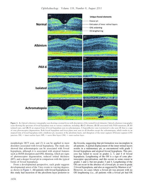
Figure 2. A, Optical coherence tomography scan showing a normal fovea with description <strong>of</strong> the normal foveal elements. Optical coherence tomography<br />
scans showing the spectrum <strong>of</strong> foveal hypoplasia seen in various conditions, including: (B, C) albinism, (D, E) associated with PAX6 mutations, (F, G)<br />
isolated cases, and (H, I) an atypical form <strong>of</strong> foveal hypoplasia seen in achromatopsia. A hyporeflective zone (cavitation) is also seen (I) that is a sign<br />
<strong>of</strong> cone photoreceptor degeneration. Both foveal hypoplasia and fovea plana were seen in all disorders except the achromatopsia, which results in an<br />
atypical form <strong>of</strong> foveal hypoplasia with a shallower pit, incursion <strong>of</strong> the plexiform layers, and disruption <strong>of</strong> the inner segment (IS)/outer segment (OS)<br />
junction. INL inner nuclear layer; NFL nerve fiber layer; ONL outer nuclear layer.<br />
morphologic OCT scan, and (3) it can be applied to most<br />
disorders associated with foveal hypoplasia. The study also<br />
showed that achromatopsia can be associated with foveal<br />
hypoplasia, although it is associated with atypical features<br />
such as IS/OS disruption and ONL thinning, which are signs<br />
<strong>of</strong> photoreceptor degeneration, reduced retinal thickness<br />
(RT), and a deeper foveal pit in comparison with the typical<br />
forms <strong>of</strong> foveal hypoplasia.<br />
From a developmental perspective, each grade suggests<br />
developmental arrest <strong>of</strong> the 3 key events to varying degrees,<br />
as shown in Figure 1. All patients with foveal hypoplasia in<br />
this study had incursion <strong>of</strong> the plexiform layer posterior to<br />
1656<br />
Ophthalmology Volume 118, Number 8, August 2011<br />
the foveola, suggesting that pit formation was incomplete in<br />
all patients. A partial displacement <strong>of</strong> the inner retinal layers<br />
results in a rudimentary pit, as encountered with grade 1<br />
foveal hypoplasia and atypical foveal hypoplasia. This process<br />
has failed to occur in grades 2, 3, and 4 <strong>of</strong> foveal<br />
hypoplasia. Lengthening <strong>of</strong> the OS is a sign <strong>of</strong> cone photoreceptor<br />
specialization, and this occurs to some extent in<br />
grades 1 and 2, but not grades 3 and 4. Lengthening <strong>of</strong> the<br />
OS can occur in the absence <strong>of</strong> a foveal pit, as seen in grade<br />
2 foveal hypoplasia, and also as suggested by Marmor et al. 7<br />
However, no cases where a foveal pit was present with no<br />
OS lengthening (i.e., all patients with a foveal pit had OS