Structural Grading of Foveal Hypoplasia Using Spectral-Domain ...
Structural Grading of Foveal Hypoplasia Using Spectral-Domain ...
Structural Grading of Foveal Hypoplasia Using Spectral-Domain ...

Create successful ePaper yourself
Turn your PDF publications into a flip-book with our unique Google optimized e-Paper software.
Thomas et al <strong>Structural</strong> <strong>Grading</strong> <strong>of</strong> <strong>Foveal</strong> <strong>Hypoplasia</strong><br />
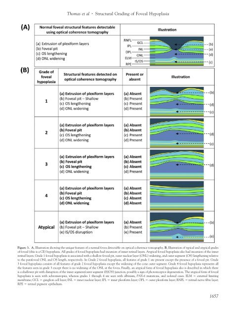
Figure 3. A, Illustration showing the unique features <strong>of</strong> a normal fovea detectable on optical coherence tomography. B, Illustration <strong>of</strong> typical and atypical grades<br />
<strong>of</strong> foveal (this is a CE) hypoplasia. All grades <strong>of</strong> foveal hypoplasia had incursion <strong>of</strong> inner retinal layers. Atypical foveal hypoplasia also had incursion <strong>of</strong> the inner<br />
retinal layers. Grade 1 foveal hypoplasia is associated with a shallow foveal pit, outer nuclear layer (ONL) widening, and outer segment (OS) lengthening relative<br />
to the parafoveal ONL and OS length, respectively. In Grade 2 foveal hypoplasia, all features <strong>of</strong> grade 1 are present except the presence <strong>of</strong> a foveal pit. Grade<br />
3 foveal hypoplasia consists <strong>of</strong> all features <strong>of</strong> grade 2 foveal hypoplasia except the widening <strong>of</strong> the cone outer segment. Grade 4 foveal hypoplasia represents all<br />
the features seen in grade 3 except there is no widening <strong>of</strong> the ONL at the fovea. Finally, an atypical form <strong>of</strong> foveal hypoplasia also is described in which there<br />
is a shallower pit with disruption <strong>of</strong> the inner segment/outer segment (IS/OS) junction, possibly a sign <strong>of</strong> photoreceptor degeneration. The atypical form <strong>of</strong> foveal<br />
hypoplasia is seen with achromatopsia, whereas grades 1 through 4 are seen with albinism, PAX-6 mutations, and isolated cases. ELM external limiting<br />
membrane; GCL ganglion cell layer; INL inner nuclear layer; IPL inner plexiform layer; OPL outer plexiform layer; RNFL retinal nerve fibre layer;<br />
RPE retinal pigment epithelium.<br />
1657