Cartilage tissue engineering: current scenario and challenges
Cartilage tissue engineering: current scenario and challenges
Cartilage tissue engineering: current scenario and challenges
You also want an ePaper? Increase the reach of your titles
YUMPU automatically turns print PDFs into web optimized ePapers that Google loves.
Review Article Adv. Mat. Lett. 2011, 2(2), 90-99 ADVANCED MATERIALS Letters<br />
factors, but till now it is difficult to recommend a single<br />
growth factor or a cocktail of different growth factors to<br />
promote cartilage repair either during in vitro or in vivo<br />
cartilage <strong>tissue</strong> <strong>engineering</strong>. This is because the actions of<br />
growth factors are not yet completely understood or<br />
sometimes even contradictory. Some studies have shown<br />
the synergistic effects of TGF- <strong>and</strong> FGF-2, in combination<br />
[48]. They interact to modulate their respective action,<br />
creating effector cascades <strong>and</strong> feedback loops of<br />
intercellular <strong>and</strong> intracellular events that control articular<br />
chondrocyte functions. However, some growth factors may<br />
elicit seemingly opposite effects under different<br />
experimental conditions [49]. Table 4 summarizes some of<br />
the important growth factors that enhance chondrogenesis.<br />
Table 4. Growth factors evaluated for their effects on chondrocyte growth<br />
<strong>and</strong> matrix production.<br />
Growth factors Chondrocyte growth Matrix production<br />
TGF- Promotes differentiation Proteoglycan synthesis<br />
FGF- Mitogenic differentiation Matrix synthesis<br />
IGF-1 Mitogenic differentiation Matrix synthesis<br />
PDGF(platelet<br />
factors)<br />
derived growth Mitogenic differentiation Matrix synthesis<br />
BMP-1 <strong>Cartilage</strong> proliferation Collagen synthesis<br />
BMP-2 Promotes cartilage formation by<br />
inducing<br />
production of cartilage matrix.<br />
Collagen synthesis<br />
BMP-4 Promotes cartilage formation by<br />
inducing<br />
MSCs to become chondroprogenitors<br />
<strong>and</strong><br />
chondrocyte maturation.<br />
Matrix synthesis<br />
BMP-5 Chondrocyte proliferation Matrix synthesis<br />
BMP-9 Potent anabolic factor for juvenile<br />
cartilage<br />
Matrix synthesis<br />
BMP-12 (GDF7)<br />
Modulates in vitro cartilage<br />
formation in<br />
a similar fashion as BMP-2 does<br />
Collagen synthesis<br />
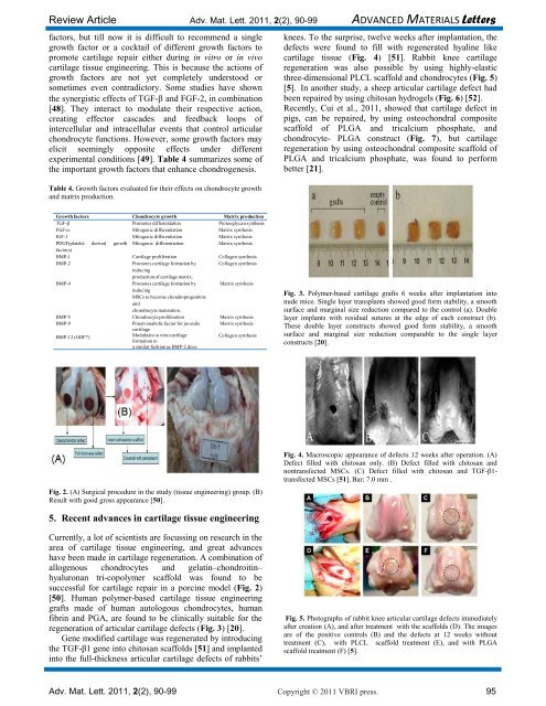
Fig. 2. (A) Surgical procedure in the study (<strong>tissue</strong> <strong>engineering</strong>) group. (B)<br />
Result with good gross appearance [50].<br />
5. Recent advances in cartilage <strong>tissue</strong> <strong>engineering</strong><br />
Currently, a lot of scientists are focussing on research in the<br />
area of cartilage <strong>tissue</strong> <strong>engineering</strong>, <strong>and</strong> great advances<br />
have been made in cartilage regeneration. A combination of<br />
allogenous chondrocytes <strong>and</strong> gelatin–chondroitin–<br />
hyaluronan tri-copolymer scaffold was found to be<br />
successful for cartilage repair in a porcine model (Fig. 2)<br />
[50]. Human polymer-based cartilage <strong>tissue</strong> <strong>engineering</strong><br />
grafts made of human autologous chondrocytes, human<br />
fibrin <strong>and</strong> PGA, are found to be clinically suitable for the<br />
regeneration of articular cartilage defects (Fig. 3) [20].<br />
Gene modified cartilage was regenerated by introducing<br />
the TGF-β1 gene into chitosan scaffolds [51] <strong>and</strong> implanted<br />
into the full-thickness articular cartilage defects of rabbits‟<br />
knees. To the surprise, twelve weeks after implantation, the<br />
defects were found to fill with regenerated hyaline like<br />
cartilage <strong>tissue</strong> (Fig. 4) [51]. Rabbit knee cartilage<br />
regeneration was also possible by using highly-elastic<br />
three-dimensional PLCL scaffold <strong>and</strong> chondrocytes (Fig. 5)<br />
[5]. In another study, a sheep articular cartilage defect had<br />
been repaired by using chitosan hydrogels (Fig. 6) [52].<br />
Recently, Cui et al., 2011, showed that cartilage defect in<br />
pigs, can be repaired, by using osteochondral composite<br />
scaffold of PLGA <strong>and</strong> tricalcium phosphate, <strong>and</strong><br />
chondrocyte- PLGA construct (Fig. 7), but cartilage<br />
regeneration by using osteochondral composite scaffold of<br />
PLGA <strong>and</strong> tricalcium phosphate, was found to perform<br />
better [21].<br />
Fig. 3. Polymer-based cartilage grafts 6 weeks after implantation into<br />
nude mice. Single layer transplants showed good form stability, a smooth<br />
surface <strong>and</strong> marginal size reduction compared to the control (a). Double<br />
layer implants with residual sutures at the edge of each construct (b).<br />
These double layer constructs showed good form stability, a smooth<br />
surface <strong>and</strong> marginal size reduction comparable to the single layer<br />
constructs [20].<br />
Fig. 4. Macroscopic appearance of defects 12 weeks after operation. (A)<br />
Defect filled with chitosan only. (B) Defect filled with chitosan <strong>and</strong><br />
nontransfected MSCs. (C) Defect filled with chitosan <strong>and</strong> TGF-β1transfected<br />
MSCs [51]. Bar: 7.0 mm .<br />
Fig. 5. Photographs of rabbit knee articular cartilage defects immediately<br />
after creation (A), <strong>and</strong> after treatment with the scaffolds (D). The images<br />
are of the positive controls (B) <strong>and</strong> the defects at 12 weeks without<br />
treatment (C), with PLCL scaffold treatment (E), <strong>and</strong> with PLGA<br />
scaffold treatment (F) [5].<br />
Adv. Mat. Lett. 2011, 2(2), 90-99 Copyright © 2011 VBRI press. 95


