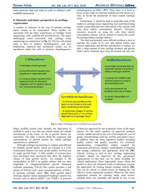
Review Article Adv. Mat. Lett. 2011, 2(2), 90-99 ADVANCED MATERIALS Letters more proteins that can stick to cells to enhance cellscaffold interaction. 8. Obstacles <strong>and</strong> future perspectives in cartilage regeneration A number of obstacles in the way of optimal cartilage repair remain to be surmounted. These hurdles are associated with the three cornerstones of cartilage <strong>tissue</strong> <strong>engineering</strong>: cells, scaffold <strong>and</strong> growth factors. The major challenging issues associated with cartilage <strong>tissue</strong> <strong>engineering</strong> are summarised in Fig. 11. The key cell related issue is how to enhance chondrogenesis, <strong>and</strong> how biophysical, chemical <strong>and</strong> mechanical stimuli can be introduced within the cells to promote chondrogenesis. Major scaffold related issue includes the fabrication of scaffold in such a way that can exactly mimic the natural environment of the <strong>tissue</strong>. As far as growth factors are concerned, very little is known about the sequences <strong>and</strong> concentrations of growth factors for which cartilage regeneration is optimized. Although cartilage regeneration is complex <strong>and</strong> affected by multiple growth factors which are released in a wellorchestrated manner, but most of the studies involved use of single growth factor. A little number of systems have been developed been developed that allow the biphasic release of dual growth factors. An example is the encapsulation of IGF-1 in gelatin spheres that are then suspended in oligo[poly(ethylene glycol) fumarate] scaffolds containing TGF-1.This system provides a fast release of TGF-1 <strong>and</strong> a more sustained delivery of IGF-1 to promote cartilage repair [56]. Dual growth factor releasing alginate based nanoparticle/hydrogel system was recently used to deliver BMP-7 <strong>and</strong> TGFβ-2 to promote Fig. 11. Major challenging issues in cartilage <strong>tissue</strong> <strong>engineering</strong>. chondrogenesis in MSCs [57]. Thus, there is a need to focus on the study of releasing multiple growth factors at a time, to favour the production of more natural cartilage <strong>tissue</strong>. Furthermore, it should be kept in mind that most of the studies in cartilage <strong>tissue</strong> <strong>engineering</strong> were performed using mostly young adult <strong>and</strong> even fetal animal cells, <strong>and</strong> not with cells from elderly osteoarthritis patients. Therefore, extensive research on using the cells from elderly osteoarthritis patients will be needed to extend the results for treating human cartilage defects. The final <strong>and</strong> probably most difficult problem is how to translate the results of in vitro <strong>and</strong> animal studies into clinical application <strong>and</strong> thereby introduction to market. To date, a large number of new cartilage products <strong>and</strong> growth factor carrier materials have been developed, but only few of them are approved for use in patients. Some of the major reasons for the small numbers of approved products include, hurdles posed by the cost of development, cost of goods, manufacturing scale-up, sterility <strong>and</strong> patent issues. In addition, there are many regulatory hurdles, including quality control <strong>and</strong> quality assurance for consistent manufacturing, comparability studies required for component <strong>and</strong> process changes, establishment of shipping <strong>and</strong> storage conditions, <strong>and</strong> appropriate shelf life [58]. Already existing as well as new cartilage <strong>engineering</strong> products should be approved by the government organizations so that they can be easily available for clinical applications. Most importantly, major efforts are needed to move the obtained results in laboratory towards the application for treating human cartilage defects [58]. This can be solved, to certain extent, by making the cost effective <strong>tissue</strong> engineered products. Moreover, the <strong>tissue</strong> engineered solution for cartilage repair must involve minimal donor site morbidity <strong>and</strong> should be free of any Adv. Mat. Lett. 2011, 2(2), 90-99 Copyright © 2011 VBRI press. 97
peri-operative complications. This will attract the patients suffering from cartilage defects towards <strong>tissue</strong> <strong>engineering</strong> solution rather than opting for other surgical interventions <strong>and</strong> prosthetics. Thus, it is clear that despite progress, further advances in cartilage <strong>tissue</strong> <strong>engineering</strong> are required to find optimal conditions for cartilage regeneration economically. 9. Conclusion <strong>Cartilage</strong> <strong>tissue</strong> <strong>engineering</strong> serves as a thriving area that has revolutionized the treatment of disease <strong>and</strong> damaged cartilage. It is an alternative to <strong>current</strong>ly used techniques that are full of limitations <strong>and</strong> do not serve as a permanent lifetime therapy. Although, there is progress in orthopaedic surgery, but the lack of efficient modalities in treatment of large chondral defects has prompted research on <strong>tissue</strong> <strong>engineering</strong>. Several points that still required more directed research includes: i) defining the best cell c<strong>and</strong>idates among chondrocytes <strong>and</strong> multipotent progenitor cells (e.g., multipotent mesenchymal stromal cells), in terms of readily available sources for isolation, expansion <strong>and</strong> repair potential; ii) <strong>engineering</strong> biocompatible <strong>and</strong> biodegradable scaffolds for enhancing growth <strong>and</strong> proliferation of cells; iii) identifying highly specific growth factors <strong>and</strong> the appropriate scheme of their application that will promote chondrogenesis <strong>and</strong> iv) there is a need to study more on simultaneous release of multiple growth factors to produce more natural cartilage <strong>tissue</strong>. Besides these, efforts must be made to avail the approved cartilage <strong>tissue</strong> <strong>engineering</strong> products from preclinical trials to the market. We hope that the development in this field will find more tremendous applications in our aging population by overcoming all the major roadblocks in cartilage regeneration in near future. 10. References 1. Tuli, R.; Li, J.;Tuan, R.S. Arthritis Res Ther. 2003, 5, 235. DOI: 10.1186/ar991 2. Jallali, N.; James, S.; Elmiyeh, B.; Searle, A.; Ghattaura, A.; Dwivedi, RC.; Kazi, R.; Harris, P. Indian J. Cancer. 2010, 47, 274. DOI: 10.4103/0019-509X.64722 3. Kinner, B.; Spector, M. J. Orthop. Res. 2001, 19, 233. DOI: 10.1016/S0736-0266(00)00081-4 4. Junying, Yu.; Thomsom, J. A. Regenerative medicine; Terese Winslow, 2006 ,pp 1-2 5. Chen, F.H., Rousche, K. T., Tuan, R.S. Nature clinical practice rheumatology. 2006, 2 (7), 373. DOI: 10.1038/ncprheum0216 6. Editorial, Joint Bone Spine. 2010, 77, 283. DOI: 10.1016/j.jbspin.2010.02.029 7. Puppi, D.; Chiellini, F.; Piras, A.M.; Chiellini, E. Progress in Polymer Science. 2010, 35,403. DOI: 10.1016/j.progpolymsci.2010.01.006 8. Stoop, R. Int. J. Care Injured. 2008, 39(1), 77. DOI: 10.1016/j.injury.2008.01.036 9. Vinatier, C.; Mrugala, D.; Jorgensen, C.; Guicheux, J., Noël, D. Trends Biotechnol. 2009, 27, 307. DOI: 10.1016/j.tibtech.2009.02.005 10. Pulkkinen, H., Tiitu, V., Lammentausta, E. et al. Biomed Mater Eng. 2006,16(4),S29. 11. Misch,C.E.; Suzuki,J.B. Tooth extraction, socket grafting, <strong>and</strong> barrier membrane bone regeneration. In: Contemporary Implant Dentistry. 3rd ed. St Louis, MO: Mosby. 2007, pp. 870-904 12. Fan, H.; Hu, Y.; Li, X. Int. J. Artif. Organs. 2006, 29(6), 602. PMID: 16841290 Sharma et al. 13. Wang, Y.; Kim, H.J.; Vunjak-Novakovic, G., et al. Biomaterials. 2006, 27(36), 6064. DOI: 10.3390/ijms10041514 14. Shi, C.; Zhu, Y.; Ran, X.; Wang, M.; Su, Y.; Cheng, T.; J Surg Res. 2006, 133(2), 185. DOI: 10.1016/j.jss.2005.12.013 15. Suh, J.K.F.; Matthew, H.W.T. Biomaterials, 2000, 21, 2589. DOI: 10.1016/S0142-9612(00)00126-5 16. Gutowska, A.; Jeong, B.; Jasionowski, M. Anat Rec. 2001, 263(4), 342. DOI: 10.1002/ar.1115 17. Salcedo, S.; Nieto, A.; Vallet-Reg, A. Chemical Engineering Journal. 2008, 137,62. DOI: 10.1016/j.cej.2007.09.011 18. Abbas, A.A.; Lee, S.Y.; Selvaratnam, L.; Yusof, N.; Kamarul, T. European Cells <strong>and</strong> Materials. 2008, 16(2), 50. DOI: 10.1002/jbm.a.33005 19. Gong, Y.; He, L.; Li, J.; Zhou, Q.; Ma, Z.; Gao, C.; Shen, J. J Biomed Mater Res B Appl Biomater. 2007, 82(1),192. DOI: 10.1002/jbm.b.30721 20. Endres, M.; Neumannb, K.; Schr¨oder, S.E.A. et al., Tissue <strong>and</strong> Cell 2007, 293, 39. DOI: 10.1016/j.tice.2007.05.002 21. Cui, W.; Wang, Q.; Chen, G, et al., Journal of Bioscience <strong>and</strong> Bio<strong>engineering</strong>, 2011, xx (xx), xxx. DOI: 10.1016/j.jbiosc.2010.11.023 22. Lee, C.R.; Grad, S.; Gorna, K. Tissue Eng. 2005, 11, 1562. DOI: 10.1089/ten.2005.11.1562 23. Larno, S.; Calvet, M.C.; Calvet, J., et al. Life Sci. 1985, 36(21), 2069. DOI: 10.1016/0024-3205(85)90458-8 24. Messner, K.; Gillquist, J. Biomaterials 1993, 14, 513. DOI: 10.1016/0142-9612(93)90240-3 25. Chen, J.P.; Cheng, T.H. Macromol. Biosci. 2006, 6(12), 1026. DOI: 10.1002/mabi.200600142 26. Holl<strong>and</strong>,T.A.; Tabata, Y.; Mikos, A.G. J. Control Release. 2005, 101(1−3), 111. DOI: 10.1016/j.jconrel.2004.07.004 27. Oliveira, J.M.; Rodrigues, M.T.; Silva, S.S. Biomaterials. 2006, 27(36), 6123. DOI: 10.1016/j.biomaterials.2006.07.034 28. Chou, C.H.; Cheng, W.T.; Kuo, T.F. J. Biomed. Mater. Res. A. 2007, 8(3), 757. DOI: 10.1002/jbm.a.31186 29. Garcia, A.; Collard, D.; Keselowsky, B. Biomimetic materials <strong>and</strong> designs, New York: Marcel Dekker, Inc. 2002, 29. 30. Harbers, G.; Barber, T.; <strong>and</strong> Stile, R. Biomimetic materials <strong>and</strong> design. New York: Marcel Dekker, Inc. 2002, 55. 31. Nuttelman, C.R.; Tripodi, M.C; Anseth, K.S. Matrix Biol. 2005, 24(3), 208. PMID: 15896949 32. Hwang, N.S.; Varghese, S.; Zhang, Z. Tissue Eng. 2006, 12(9), 2695. PMID: 16995803 33. Hsu, S.H.; Chang, S.H.; Yen, H.J. Artif. Organs. 2006, 30(1), 42. PMID: 16409397 34. Alsberg, E.; Anderson, K.W.; Albeiruti, A. Proc. Natl. Acad. Sci., U S A. 2002, 99(19), 12025. 35. Ibusuki, S.; Fujii, Y.; Iwamoto,Y.; Matsuda, T. Tissue Engineering. 2003, 9(2), 371. PMID: 12740100 36. Tan, H.; Chu, C.R.; Payne, K.A.; Marra, K.G. Biomaterials. 2009, 30(13), 2499. PMID: 19167750 37. S<strong>and</strong>ell, L.J.; Daniel, J.C. Tissue res.1988, 17, 11. PMID: 3383569 38. Vinatier, C.; Bouffi, C.; Merceron, C et al., Current Stem Cell Research & Therapy. 2009, 4, 318. DOI: 1574-888X/09 $55.00+.00 39. Martin, I.; vunjak-Novakovic, G.; Yang, J., Langer, R. <strong>and</strong> Freed, L. E. Exp. Cell Res. 1999, 253, 681. DOI: 10.1006/excr.1999.4708 40. Reddi, A.H.; Reddi, A. Cytokine & growth factor reviews. 2009, 20(5-6), 341. DOI: 10.1016/j.cytogfr.2009.10.015 41. Bleuming, S.A.; He, X.C.; Kodach, L.L.; Hardwick, J.C.; Koopman, F.A.; TenKate, F.J.; Van Deventer, S.J.; Hommes, D.W.; Adv. Mat. Lett. 2011, 2(2), 90-99 Copyright © 2011 VBRI press.