Cartilage tissue engineering: current scenario and challenges
Cartilage tissue engineering: current scenario and challenges
Cartilage tissue engineering: current scenario and challenges

You also want an ePaper? Increase the reach of your titles
YUMPU automatically turns print PDFs into web optimized ePapers that Google loves.
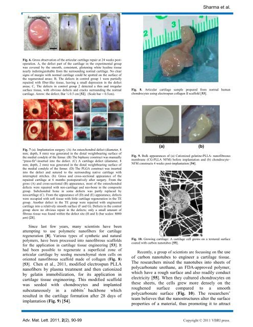
Fig. 6. Gross observation of the articular cartilage repair at 24 weeks postoperation.<br />
A, the defect part of the cartilage in the experimental group<br />
was covered by the smooth, consistent, glistening white hyaline <strong>tissue</strong><br />
nearly indistinguishable from the surrounding normal cartilage. No clear<br />
signs of margin with normal cartilage could be spotted on the surface of<br />
the regenerated areas; B, The defects in control group 1 were partially<br />
repaired with fiber-like <strong>tissue</strong>, leaving a small depression in the defect<br />
areas; C, The defects in control group 2 detected a thin <strong>and</strong> irregular<br />
surface <strong>tissue</strong>, with obvious defects <strong>and</strong> cracks surrounding the normal<br />
cartilage. Arrow: the defect; Bar ¼ 0.5 cm [52]. (Scale bar = 0.5cm).<br />
Fig. 7 (a). Implantation surgery. (A) An osteochondral defect (diameter, 8<br />
mm; depth, 8 mm) was generated in the distal weightbearing surface of<br />
the medial condyle of the femur. (B) The biphasic construct was manually<br />
“press-fit”-inserted into the defect. (C) A cartilage defect (diameter, 8<br />
mm; depth, 2 mm) was generated in the distal weightbearing surface of<br />
the medial condyle of the femur. (D) The PLGA construct was inserted<br />
into the defect <strong>and</strong> sutured to the surrounding native cartilage with<br />
interrupted stitches. (b): Gross <strong>and</strong> cross-sectional appearance of the<br />
repaired cartilage at 6 months postoperatively after surgery. From the<br />
gross (A) <strong>and</strong> cross-sectional (B) appearance, most of the osteochondral<br />
defects were repaired with neo-cartilage <strong>and</strong> neo-bone in the composite<br />
group. Subchondral bone in some defects was partly replaced by<br />
neocartilage (C). From the appearance of (D) <strong>and</strong> (E) appearance, defects<br />
were occupied with soft <strong>tissue</strong> with little cartilage regeneration in the TE<br />
group. Another defect in the TE group were repaired with engineered<br />
cartilage into a relatively smooth surface (F <strong>and</strong> G). Defects in the control<br />
group show no obvious repair in the defects; only a small amount of<br />
fibrous <strong>tissue</strong> was found within the defect site (H <strong>and</strong> I) (bar scales: 8000<br />
μm) [21].<br />
Since last few years, many scientists have been<br />
attempting to use polymeric nanofibers for cartilage<br />
regeneration [8]. Various types of synthetic <strong>and</strong> natural<br />
polymers, have been processed into nanofibrous scaffolds<br />
for the application in cartilage <strong>tissue</strong> <strong>engineering</strong> [53]. It<br />
had been possible to regenerate a superficial zone of<br />
articular cartilage by seedng mesenchymal stem cells on<br />
oriented nanofibrous scaffold made of collagen (Fig. 8)<br />
[53]. Chen et al., 2011, modified electrospun PLLA<br />
nanofibers by plasma treatment <strong>and</strong> then cationized<br />
by gelatin immobilization, for its application in<br />
cartilage <strong>tissue</strong> <strong>engineering</strong>. This modified scaffold<br />
was seeded with chondrocytes <strong>and</strong> implanted<br />
subcutaneously in a rabbits’backbone which<br />
resulted in the cartilage formation after 28 days of<br />
implantation (Fig. 9) [54].<br />
Sharma et al.<br />
Fig. 8. Articular cartilage sample prepared from normal human<br />
chondrocytes using electrospun collagen II scaffold [53].<br />
Fig. 9. Bulk appearances of (a) Cationized gelatine-PLLA- nanofibreous<br />
membrane (CG-PLLA NFM) before implantation <strong>and</strong> (b) chondrocyte–<br />
NFM constructs 4 weeks post-implantation [54].<br />
Fig. 10. Growing cartilage: A cartilage cell grows on a textured surface<br />
coated with carbon nanotubes [55].<br />
Recently, a group of scientists are focussing on the use<br />
of carbon nanotubes to engineer a cartilage <strong>tissue</strong>.<br />
The researchers mixed the nanotubes into sheets of<br />
polycarbonate urethane, an FDA-approved polymer,<br />
which have a rough surface <strong>and</strong> also readily conduct<br />
electricity [55]. When they cultured chondrocytes on<br />
these sheets, the cells grew more densely on the<br />
roughened surface compared to a smooth<br />
polycarbonate surface (Fig. 10). The researchers‟<br />
team believes that the nanostructures alter the surface<br />
properties of a material, thus promoting it to attract<br />
Adv. Mat. Lett. 2011, 2(2), 90-99 Copyright © 2011 VBRI press.