chapter 2 - Bentham Science
chapter 2 - Bentham Science
chapter 2 - Bentham Science
You also want an ePaper? Increase the reach of your titles
YUMPU automatically turns print PDFs into web optimized ePapers that Google loves.
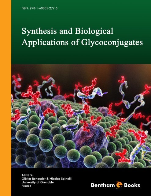
Synthesis and Biological Applications of<br />
Glycoconjugates<br />
Editors<br />
Olivier Renaudet and Nicolas Spinelli<br />
University of Grenoble<br />
France
eBooks End User License Agreement<br />
Please read this license agreement carefully before using this eBook. Your use of this eBook/<strong>chapter</strong> constitutes your agreement<br />
to the terms and conditions set forth in this License Agreement. <strong>Bentham</strong> <strong>Science</strong> Publishers agrees to grant the user of this<br />
eBook/<strong>chapter</strong>, a non-exclusive, nontransferable license to download and use this eBook/<strong>chapter</strong> under the following terms and<br />
conditions:<br />
1. This eBook/<strong>chapter</strong> may be downloaded and used by one user on one computer. The user may make one back-up copy of this<br />
publication to avoid losing it. The user may not give copies of this publication to others, or make it available for others to copy or<br />
download. For a multi-user license contact permission@bentham.org<br />
2. All rights reserved: All content in this publication is copyrighted and <strong>Bentham</strong> <strong>Science</strong> Publishers own the copyright. You may<br />
not copy, reproduce, modify, remove, delete, augment, add to, publish, transmit, sell, resell, create derivative works from, or in<br />
any way exploit any of this publication’s content, in any form by any means, in whole or in part, without the prior written<br />
permission from <strong>Bentham</strong> <strong>Science</strong> Publishers.<br />
3. The user may print one or more copies/pages of this eBook/<strong>chapter</strong> for their personal use. The user may not print pages from<br />
this eBook/<strong>chapter</strong> or the entire printed eBook/<strong>chapter</strong> for general distribution, for promotion, for creating new works, or for<br />
resale. Specific permission must be obtained from the publisher for such requirements. Requests must be sent to the permissions<br />
department at E-mail: permission@bentham.org<br />
4. The unauthorized use or distribution of copyrighted or other proprietary content is illegal and could subject the purchaser to<br />
substantial money damages. The purchaser will be liable for any damage resulting from misuse of this publication or any<br />
violation of this License Agreement, including any infringement of copyrights or proprietary rights.<br />
Warranty Disclaimer: The publisher does not guarantee that the information in this publication is error-free, or warrants that it<br />
will meet the users’ requirements or that the operation of the publication will be uninterrupted or error-free. This publication is<br />
provided "as is" without warranty of any kind, either express or implied or statutory, including, without limitation, implied<br />
warranties of merchantability and fitness for a particular purpose. The entire risk as to the results and performance of this<br />
publication is assumed by the user. In no event will the publisher be liable for any damages, including, without limitation,<br />
incidental and consequential damages and damages for lost data or profits arising out of the use or inability to use the publication.<br />
The entire liability of the publisher shall be limited to the amount actually paid by the user for the eBook or eBook license<br />
agreement.<br />
Limitation of Liability: Under no circumstances shall <strong>Bentham</strong> <strong>Science</strong> Publishers, its staff, editors and authors, be liable for<br />
any special or consequential damages that result from the use of, or the inability to use, the materials in this site.<br />
eBook Product Disclaimer: No responsibility is assumed by <strong>Bentham</strong> <strong>Science</strong> Publishers, its staff or members of the editorial<br />
board for any injury and/or damage to persons or property as a matter of products liability, negligence or otherwise, or from any<br />
use or operation of any methods, products instruction, advertisements or ideas contained in the publication purchased or read by<br />
the user(s). Any dispute will be governed exclusively by the laws of the U.A.E. and will be settled exclusively by the competent<br />
Court at the city of Dubai, U.A.E.<br />
You (the user) acknowledge that you have read this Agreement, and agree to be bound by its terms and conditions.<br />
Permission for Use of Material and Reproduction<br />
Photocopying Information for Users Outside the USA: <strong>Bentham</strong> <strong>Science</strong> Publishers Ltd. grants authorization for individuals<br />
to photocopy copyright material for private research use, on the sole basis that requests for such use are referred directly to the<br />
requestor's local Reproduction Rights Organization (RRO). The copyright fee is US $25.00 per copy per article exclusive of any<br />
charge or fee levied. In order to contact your local RRO, please contact the International Federation of Reproduction Rights<br />
Organisations (IFRRO), Rue du Prince Royal 87, B-I050 Brussels, Belgium; Tel: +32 2 551 08 99; Fax: +32 2 551 08 95; E-mail:<br />
secretariat@ifrro.org; url: www.ifrro.org This authorization does not extend to any other kind of copying by any means, in any<br />
form, and for any purpose other than private research use.<br />
Photocopying Information for Users in the USA: Authorization to photocopy items for internal or personal use, or the internal<br />
or personal use of specific clients, is granted by <strong>Bentham</strong> <strong>Science</strong> Publishers Ltd. for libraries and other users registered with the<br />
Copyright Clearance Center (CCC) Transactional Reporting Services, provided that the appropriate fee of US $25.00 per copy<br />
per <strong>chapter</strong> is paid directly to Copyright Clearance Center, 222 Rosewood Drive, Danvers MA 01923, USA. Refer also to<br />
www.copyright.com
CONTENTS<br />
Foreword i<br />
Preface ii<br />
List of Contributors iii<br />
CHAPTERS<br />
1. Bacterial Lectins and Adhesins: Structures, Ligands and Functions 3<br />
Anne Imberty<br />
2. Ligands for FimH 12<br />
Thisbe K. Lindhorst<br />
3. Multivalent Glycocalixarenes 36<br />
Francesco Sansone, Gabriele Rispoli, Alessandro Casnati and Rocco Ungaro<br />
4. Solving Promiscuous Protein Carbohydrate Recognition Domains with Multivalent<br />
Glycofullerenes 64<br />
Yoann M. Chabre and René Roy<br />
5. Monovalent and Multivalent Inhibitors of Bacterial Toxins 78<br />
Edward D. Hayes and W. Bruce Turnbull<br />
6. Monovalent and Multivalent Glycoconjugates as High Affinity Ligands for Galectins 92<br />
Sébastien Vidal<br />
7. Combinatorial Libraires of Dendritic Glycoclusters 116<br />
Jean-Louis Reymond and Tamis Darbre<br />
8. Cyclopeptide-Based Glycoclusters 129<br />
Olivier Renaudet, Didier Boturyn and Pascal Dumy<br />
9. Oligonucleotide-Carbohydrate Conjugates 145<br />
Nicolas Spinelli and Eric Defrancq<br />
10. Glycoliposomes and Metallic Glyconanoparticles in Glycoscience 164<br />
Marco Marradi, Fabrizio Chiodo, Isabel García and Soledad Penadés<br />
11. Chemoselective Glycosylation Techniques for the Synthesis of Bioactive Neoglycoconjugates,<br />
Glyconanoparticles and Glycoarrays 203<br />
Francesco Peri<br />
12. Glycosidases in Synthesis of Glycomimetics and Unnatural Carbohydrates 226<br />
Vladimír Křen<br />
13. Diels-Alder Based Synthesis of Glycomimetics 240<br />
Cristina Nativi, Elisa Dragoni, Barbara Richichi and Stefano Roelens
14. Analytical Tools for Protein-Carbohydrate Interaction Studies 255<br />
Cédric Goyer and Pierre Labbé<br />
15. Index 267
FOREWORD<br />
Attachment of carbohydrates in all its forms is a strategy exploited by both nature and those that study nature. Since the<br />
first reviews over 70 years ago the importance of this critically modulating and even defining role has long been<br />
recognized by who those study them. The very presence of carbohydrate units in naturally occurring structures and their<br />
mimetics has a dramatic effect on their physical, chemical and biological properties. Glycoscience is by necessity broad<br />
in the range of techniques that it encompasses and I have argued for over ten years that in this context the sometimesapplied<br />
and somewhat artificial distinction between "chemical" and "biological" techniques is unhelpful.<br />
This has never been the case – we are reaching a stage in the field where remarkable techniques are able to make<br />
even some very large naturally relevant glycoconjugates. Moreover, through these methods and the design of some<br />
remarkable molecular experimental approaches, we are using these glycoconjugates to unpick the many dazzling<br />
and exciting roles that carbohydrates play in Biology and Medicine.<br />
This book highlights this superbly. Drs Renaudet and Spinelli have assembled an outstanding collection of excellent<br />
and relevant reviews written by leading protagonists in the field that complement each other superbly. Together they<br />
provide a wonderful account of the current state-of-the-art.<br />
Vital current targets illustrate what might and will be achieved through both analysis and understanding. Many<br />
pathogens exploit the display of carbohydrates on host cell surfaces to gain entry. Imberty and Lindhorst, in their<br />
<strong>chapter</strong>s, have described some of the most current and relevant examples. Integral to the approaches they have<br />
analyzed is a fundamental molecular analysis of the binding of sugars to proteins. This has been excellently<br />
amplified by the <strong>chapter</strong> of Goyer and Labbé, which describes some of the most pertinent quantitative techniques in<br />
current use, and by the several <strong>chapter</strong>s that show how this understanding can be directly related to application.<br />
These include the design of inhibitors of bacterial toxins, superbly analyzed by Turnbull and Hayes, and ligands for<br />
the important mammalian galectins, the study of which Vidal summarizes superbly.<br />
Use of well-defined scaffolds is an essential element if this study and exploitation is to be precise. Various platforms<br />
have been analyzed here: fullerenes (Roy and Chabre), dendritic clusters (Reymond and Darbre), calixarenes (Casnati et<br />
al.), cyclic peptides (Renaudet et al.) and even nucleic acids (Spinelli and Defrancq). All highlight longer-term<br />
applications, especially in applications that may allow probing of interactions in whole organisms, and these are<br />
especially elegantly drawn out in the <strong>chapter</strong> by Penadés et al. that consider particles of many relevant types.<br />
Typically syntheses of glycoconjugates adopt one of two strategies. The first strategy is the formation of the glycanaglycone<br />
link early, to form, for example, glycosylated building blocks such as glycosides, glycopeptides or<br />
glycosylated dendritic wedges, that may then be assembled. While, of course, chemical methods can create<br />
glycosidic bonds in a number of ways, the use of biocatalysts can bring with it advantages of selectivity and<br />
efficiency; Křen’s <strong>chapter</strong> highlights this utility well. The second strategy is formation of the link later on in the<br />
synthesis once the scaffold for its presentation is in place. Given the instability that may be associated with the link<br />
and the requirements for protection that need to be considered in the use of glycosylated building blocks, it is clear<br />
why the latter has often seemed the most attractive option. Peri has deftly summarized a broad range of methods for<br />
this late stage approach as applied to many useful platform types. Nativi et al. have delineated an emerging area that<br />
could allow access to glycomimetics based on the use of glycals in cycloadditions.<br />
Together, these <strong>chapter</strong>s give a powerful insight into the elucidation of the mechanism by which sugar-binding takes<br />
place, the tools used to probe into it and its consequences. This science has driven and continues to drive the<br />
synthesis and technology of glycoconjugates. These are the new tools of the glycobiology trade and I believe only<br />
the very start of an era of emerging diagnostics and therapeutics.<br />
i<br />
Prof. Ben Davis<br />
University of Oxford<br />
UK
ii<br />
PREFACE<br />
The interactions between carbohydrates and proteins are involved in major physiological and pathological events.<br />
With the recent emergence of glycomics, an increasing number of sophisticated glycosylated structures capable of<br />
mimicking the multivalent display of the cell surface glycocalix has been reported. Besides giving precious<br />
guidelines on the binding parameters that govern these complex biological processes, synthetic glycoconjugates<br />
often revealed considerable interests for diagnostic and therapeutic applications.<br />
Our main motivation to edit Synthesis and biological applications of glycoconjugates was to update the major<br />
advancements in this field through several <strong>chapter</strong>s written by renowned experts who have largely contributed to the<br />
recent progresses. Illustrated with more than 200 colour figures and including literature citations mostly from the<br />
last decade, this book covers the recent chemical methods of glycoconjugates and clearly highlights their diverse<br />
biological properties. Chapter 1 analyzes the structure of lectins from pathogenic bacteria and gives<br />
structure/function relationships that are crucial for the development of high affinity ligands. In Chapter 2<br />
carbohydrate binding by FimH is discussed and modern means for its investigation including photoaffinity labelling<br />
are described. Very recent crystallographic work is also documented that provides an explanation for shearenhanced<br />
binding of type 1 fimbriated E. coli. Chapter 3 summarizes the most convenient methodologies for the<br />
synthesis of glycocalixarenes and describes their impressive aggregation properties and many other biological<br />
applications. Chapter 4 illustrates examples of bacterial and human lectins together with bacterial toxins having<br />
varied number of carbohydrate recognition domains that necessitated multivalent glycoconjugates. It focuses on<br />
glycofullerenes that have been used to describe novel synthetic strategies and possible fit with concomitant lectins.<br />
In Chapter 5 the structures of the cholera and E. coli heat-labile toxins are described. The authors also summarize<br />
the main strategies that have led to the development of monovalent and multivalent inhibitors of these toxins and<br />
they discuss the importance of chelation and protein aggregation as mechanisms of multivalent inhibition. Chapter 6<br />
focuses on the design of high affinity ligands for galectins and provides a better knowledge of the implications of<br />
these proteins in biology. Chapter 7 demonstrates that combinatorial chemistry led to the discovery of glycopeptide<br />
dendrimers as strong ligands for lectins or drug-delivery systems and shows how the amino acid composition of the<br />
dendrimer branches can influence their biological activity. Chapter 8 focuses on the synthesis of a recent class of<br />
glycoclusters displayed on a cyclopeptide platform and highlights their promising biological properties, in particular<br />
as inhibitors or synthetic vaccines. Chapter 9 shows that the cellular delivery and bioavailability of oligonucleotides<br />
can be improved by their conjugation with carbohydrates and also describes the construction of carbohydrate<br />
biochips or glycoclusters using these conjugates. Chapter 10 focuses on glycoliposomes and covalentlyfunctionalized<br />
glyconanoparticles which make use of the “glyco-code” to address specifically pathogens or<br />
pathological-related problems. Chapter 11 focuses on the synthesis of glycoproteins, glycopeptides, glycosylated<br />
natural compounds, carbohydrate-functionalized surfaces and nanoparticles using chemoselective glycosylation. In<br />
Chapter 12 the recent developments in glycosidase-catalyzed synthesis of unnatural semi-synthetic carbohydrate<br />
structures are presented. Chapter 13 reports the synthesis of glycomimetics and glycopeptidomimetics using hetero-<br />
Diels Alder reactions between ,’-dioxothiones and glycals. Chapter 14 aims at describing the usual methods to<br />
characterize protein-carbohydrate interactions, namely inhibition of hemagglutination assay, enzyme-linked lectin<br />
assays, isothermal titration calorimetry, surface plasmon resonance and the more recent atomic force microscopy.<br />
We believe that this Ebook will be of particular interest to a large community of graduate students, researchers and<br />
professionals in academia or industry involved in Glycoscience. We would like to express our sincerest gratitude to<br />
all of the authors who accepted to contribute to this exciting project, by sharing their strong experiences and<br />
knowledge in this research area.<br />
Olivier Renaudet<br />
Nicolas Spinelli<br />
University of Grenoble<br />
France
List of Contributors<br />
Didier Boturyn<br />
Département de Chimie Moléculaire, UMR-CNRS 5250 & ICMG FR 2607, Université Joseph Fourier, 38041<br />
Grenoble Cedex 9, France.<br />
Alessandro Casnati<br />
Dip.to di Chimica Organica e Industriale, Università degli Studi, Parco Area delle Scienze 17/A, 43124 Parma, Italy.<br />
Yoann M. Chabre<br />
Pharmaqam – Groupe de Recherche en Chimie Thérapeutique, Université du Québec à Montréal, P.O. Box 8888,<br />
Succ. Centre-ville, Montréal, Québec, Canada H3C 3P8.<br />
Fabrizio Chiodo<br />
Laboratory of GlycoNanotechnology, Biofunctional Nanomaterials Unit, CIC biomaGUNE, Parque Tecnológico de<br />
San Sebastián, Pº de Miramón 182, 20009 San Sebastián, Spain.<br />
Tamis Darbre<br />
Department of Chemistry and Biochemistry, University of Berne, Freiestrasse 3, CH-3012, Berne, Switzerland.<br />
Eric Defrancq<br />
Département de Chimie Moléculaire, UMR-CNRS 5250 & ICMG FR 2607, Université Joseph Fourier, 38041<br />
Grenoble Cedex 9, France.<br />
Elisa Dragoni<br />
ProtEra, Polo Scientifico Universita’ di Firenze, viale delle Idee, 22 I-50019 Sesto F.no (FI), Italy.<br />
Pascal Dumy<br />
Département de Chimie Moléculaire, UMR-CNRS 5250 & ICMG FR 2607, Université Joseph Fourier, 38041<br />
Grenoble Cedex 9, France.<br />
Isabel García<br />
Laboratory of GlycoNanotechnology, Biofunctional Nanomaterials Unit, CIC biomaGUNE and Biomedical<br />
Research Networking Center in Bioengineering, Biomaterials and Nanomedicine (CIBER-BBN), Parque<br />
Tecnológico de San Sebastián, Pº de Miramón 182, 20009 San Sebastián, Spain.<br />
Cédric Goyer<br />
Département de Chimie Moléculaire, UMR-CNRS 5250 & ICMG FR 2607, Université Joseph Fourier, 38041<br />
Grenoble Cedex 9, France.<br />
Edward D. Hayes<br />
School of Chemistry and Astbury Centre for Structural Molecular Biology, University of Leeds, Leeds LS2 9JT, UK.<br />
Anne Imberty<br />
CERMAV-CNRS (affiliated to Université Joseph Fourier and ICMG), BP 53, 38041 Grenoble cedex 9, France.<br />
Vladimír Křen<br />
Institute of Microbiology, Centre of Biocatalysis and Biotransformation, Academy of <strong>Science</strong>s of the Czech<br />
Republic, Vídeňská 1083, CZ 142 20 Prague 4, Czech Republic.<br />
Pierre Labbé<br />
Département de Chimie Moléculaire, UMR-CNRS 5250 & ICMG FR 2607, Université Joseph Fourier, 38041<br />
Grenoble Cedex 9, France.<br />
iii
iv<br />
Thisbe K. Lindhorst<br />
Otto Diels Institute of Organic Chemistry, Christiana Albertina University of Kiel, D-24098 Kiel, Germany.<br />
Marco Marradi<br />
Laboratory of GlycoNanotechnology, Biofunctional Nanomaterials Unit, CIC biomaGUNE and Biomedical<br />
Research Networking Center in Bioengineering, Biomaterials and Nanomedicine (CIBER-BBN), Parque<br />
Tecnológico de San Sebastián, Pº de Miramón 182, 20009 San Sebastián, Spain.<br />
Cristina Nativi<br />
Dipartimento di Chimica, Universita’ di Firenze, via della Lastruccia, 3,13 I-50019 Sesto F.no (FI), Italy.<br />
Soledad Penadés<br />
Laboratory of GlycoNanotechnology, Biofunctional Nanomaterials Unit, CIC biomaGUNE and Biomedical<br />
Research Networking Center in Bioengineering, Biomaterials and Nanomedicine (CIBER-BBN), Parque<br />
Tecnológico de San Sebastián, Pº de Miramón 182, 20009 San Sebastián, Spain.<br />
Francesco Peri<br />
Department of Biotechnology and Biosciences, University of Milano-Bicocca, Piazza della Scienza, 2; 20126<br />
Milano, Italy.<br />
Olivier Renaudet<br />
Département de Chimie Moléculaire, UMR-CNRS 5250 & ICMG FR 2607, Université Joseph Fourier, 38041<br />
Grenoble Cedex 9, France.<br />
Jean-Louis Reymond<br />
Department of Chemistry and Biochemistry, University of Berne, Freiestrasse 3, CH-3012, Berne, Switzerland.<br />
Barbara Richichi<br />
Dipartimento di Chimica, Universita’ di Firenze, via della Lastruccia, 3,13 I-50019 Sesto F.no (FI), Italy.<br />
Stefano Roelens<br />
Istituto Metodologie Chimiche (IMC), CNR, Dipartimento di Chimica, Universita’ di Firenze, via della Lastruccia,<br />
3,13 I-50019 Sesto F.no (FI), Italy.<br />
René Roy<br />
Pharmaqam – Groupe de Recherche en Chimie Thérapeutique, Université du Québec à Montréal, P.O. Box 8888,<br />
Succ. Centre-ville, Montréal, Québec, Canada H3C 3P8.<br />
Francesco Sansone<br />
Dip.to di Chimica Organica e Industriale, Università degli Studi, Parco Area delle Scienze 17/A, 43124 Parma, Italy.<br />
W. Bruce Turnbull<br />
School of Chemistry and Astbury Centre for Structural Molecular Biology, University of Leeds, Leeds LS2 9JT, UK.<br />
Nicolas Spinelli<br />
Département de Chimie Moléculaire, UMR-CNRS 5250 & ICMG FR 2607, Université Joseph Fourier, 38041<br />
Grenoble Cedex 9, France.<br />
Rocco Ungaro<br />
Dip.to di Chimica Organica e Industriale, Università degli Studi, Parco Area delle Scienze 17/A, 43124 Parma, Italy.<br />
Sébastien Vidal<br />
Institut de Chimie et Biochimie Moléculaires et Supramoléculaires, Laboratoire de Chimie Organique 2, UMR 5246,<br />
Université Lyon 1 and CNRS, F 69622 Villeurbanne, France.
Synthesis and Biological Applications of Glycoconjugates, 2011, 3-11 3<br />
Bacterial Lectins and Adhesins: Structures, Ligands and Functions<br />
Anne Imberty *<br />
Olivier Renaudet and Nicolas Spinelli (Eds)<br />
All rights reserved - © 2011 <strong>Bentham</strong> <strong>Science</strong> Publishers<br />
CHAPTER 1<br />
CERMAV-CNRS (affiliated to Université Joseph Fourier and ICMG), BP 53, 38041 Grenoble cedex 9, France<br />
Abstract: Infection by bacteria is often initiated by the specific recognition of host epithelial surfaces by adhesins<br />
and lectins. These glycan binding proteins (GBP) are therefore virulence factors that play a role in the first step of<br />
adhesion and invasion. The adhesins are part of organelles, they are generally located at the tip of pili or fimbriae.<br />
On the opposite, soluble lectins occur either as individual proteins, or in association with toxins or enzymes. The<br />
human targets for bacterial adhesins and lectins are mostly fucosylated human histo-blood groups and/or<br />
sialylated epitopes. The binding of lectins and adhesins to host epithelial glycoconjugates is amplified by the<br />
multivalent presentation of the binding sites. The analysis of the available crystal structures of bacterial lectins<br />
and adhesins helps deciphering the structure/function relationship for this important class of proteins. It is a<br />
prerequisite for the development of multivalent high affinity ligands in antibacterial strategies.<br />
Keywords: Lectin, adhesin, crystal structure, bacteria, infection.<br />
LECTINS: UBIQUITOUS PROTEIN WITH A TASTE FOR SUGARS<br />
Lectins are proteins of non-immune origin that bind to specific carbohydrates without modifying them [1]. These<br />
proteins are ubiquitous as they have been identified in all living organisms, from viruses to bacteria, fungi, plants<br />
and animals. As a result of their interactions with glycoproteins, glycolipids and oligosaccharides, lectins are<br />
involved in cell-cell communication; they have the crucial role of deciphering the “glycocode”. Their biological<br />
functions are very diverse. Lectins are important players in the innate immunity system of many invertebrates as<br />
they are able to differentiate between self and non-self carbohydrates. Lectins are particularly abundant in plants,<br />
where they are proposed to be involved in protection against pathogens or feeders. The biological roles of animal<br />
lectins have been more thoroughly investigated; they involve glycoprotein trafficking and clearance, development,<br />
immune defence, fertility and many others.<br />
In pathogenic micro-organims, lectins are mostly involved in host recognition and tissue adhesion [2]. They can be<br />
present as subunit parts of toxins where they target the toxic catalytic unit towards host tissues carrying some<br />
specific glycoconjugates. They can also be part of multiprotein organelles, such as bacterial fimbria (pili), flagella<br />
and viral capsids. Therefore they participate to the adhesion process at the early stage of infection. Soluble lectins<br />
are also expressed as virulence factors by opportunistic bacteria.<br />
Structural studies of lectins and of their binding sites started with the elucidation of the crystal structure of<br />
Concanavalin A, a well characterized legume lectin specific for mannose and glucose [3]. Such structural<br />
investigations yield both the detailed knowledge of the protein structure and the identification of the most important<br />
contacts that are established in the binding pocket between the polypeptide chain and the carbohydrate. This is a<br />
prerequisite for a better understanding of the biological and physical processes of recognition [4]. Three dimensional<br />
structures are also the starting points for many developments in the biotechnological and therapeutical applications<br />
of lectin/carbohydrate interactions.<br />
Lectins are relatively easy to purify due to their carbohydrate binding properties that can be exploited for affinity<br />
purification. Lectins from plants are generally abundant and can be extracted from natural sources, whereas animal<br />
lectins are nowadays generally produced by recombinant approaches. A large number of lectins has therefore been<br />
used for crystallization assays, preferably in the presence of their carbohydrate ligand(s).More than 870 threedimensional<br />
structures (corresponding to 187 different lectins) are now available and gathered in the 3D-lectin<br />
database (http://www.cermav.cnrs.fr/lectines/).<br />
*Address corresponding to Anne Imberty: CERMAV-CNRS (affiliated to Université Joseph Fourier and ICMG), BP 53, 38041 Grenoble<br />
cedex 9, France; E-mail: Anne.Imberty@cermav.cnrs.fr
4 Synthesis and Biological Applications of Glycoconjugates Anne Imberty<br />
Inspection of the database content indicates that most crystal structures have been for the time being, obtained for<br />
plant and animal lectins (Fig. 1). Nevertheless, the number of structural works dealing with viral and bacterial<br />
materials increases rapidly. Interestingly, a strong bias is observed in the nature of the architecture of the lectins. As<br />
indicated in Fig. 1, most lectins adopt a -sandwich fold. The several -sandwiches topologies adopted by lectins are<br />
very different one to each others, but some interesting structural convergences are observed. For example legume<br />
lectins and those intracellular animal lectins which are involved in quality control of glycoprotein synthesis share the<br />
same protein fold [5].<br />
Figure 1: Repartition of the 187 different lectins with structures available in the Protein data bank and accessible in the lectin<br />
database (http://www.cermav.cnrs.fr/lectines/). Lectins are classified as a function of: A) origin; B) fold.<br />
STRUCTURES OF LECTINS FROM PATHOGENIC BACTERIA<br />
Adhesins<br />
Adhesins refer to the lectin domains which are present at the tip of bacterial fimbriae (or pili). Although different<br />
types of pili are observed on bacteria, structural data have been obtained only for those lectins belonging to the twodomain<br />
adhesins (TDA)s from different pathogenic strains of Escherichia coli. These adhesins consist of a pilin<br />
domain and a lectin domain, both presenting an immunoglobulin-fold joined via a short interdomain linker [6]. The<br />
adhesins reported in Table 1 have been characterized in enteropathogenic E. coli (GafD on F17 fimbriae) and<br />
uropathogenic ones (FimH on type 1 fimbriae and PapG on P-pili).<br />
Table 1: Crystal structures representative of the different classes of bacterial adhesins and their specificity.<br />
Bacteria Adhesin Carbohydrate specificity<br />
E. coli PapG<br />
GalNAc13Gal14Gal14Glc<br />
(GbO4 oligosacch.)<br />
Structure<br />
PDB<br />
Ref.<br />
1J8R [7]<br />
E. coli GafD/F17-G GlcNAc 1O9W, 1OIO [8, 9]<br />
E. coli FimH Oligomannose 2VCO [10]<br />
Flexible F17 fimbriae of enterotoxigenic E. coli are capped with GafD adhesins specific for GlcNAc-terminated<br />
glycoconjugates. Uropathogenic E. coli adhesins bind to different parts of the human urinary tract. The long and flexible<br />
P fimbriae expose PapG adhesins specific for galabiose containing the oligosaccharide (Gal1-4Gal) motif whereas<br />
shorter type 1 pili are terminated by FimH that binds oligomannose. Despite the lack of any sequence identity, all these<br />
lectins share the same immunoglobulin-like fold of the two structural components; the corresponding pili are assembled<br />
by the chaperone-usher pathway [11]. The three fimbrial lectins, GafD, FimH and PapG share similar beta-barrel folds<br />
but display different ligand-binding regions and disulfide-bond patterns (Fig. 2).
Bacterial Lectins and Adhesins Synthesis and Biological Applications of Glycoconjugates 5<br />
Figure 2: Crystal structure of the three bacterial adhesins obtained in complex with carbohydrate ligands. A) PapG complexed<br />
with GbO4 (code 1J8R); B) GafD complexed with GlcNAc (1O9W); C) FimH complexed with Man3GlcNAc2 (2VCO).<br />
References as in Table 1. All figures have been prepared using PyMol (DeLano Scientific).<br />
Toxin-Associated Lectins<br />
Bacteria use lectin domains to target another subunit which generally displays some enzymatic activity towards host<br />
cell types. The crystal structures that have been solved illustrate how neural or digestive tissues can be targeted by<br />
lectin domains (Table 2).<br />
Clostridia produce a large number of toxins such as neurotoxins and pore-forming toxins which are responsible for<br />
gangrenes and gastrointestinal diseases. Among these, two families of toxins contain lectin domains specific of<br />
human glycoconjugates.<br />
- Clostridia neurotoxins are well known because of the severe diseases that are caused by the<br />
corresponding bacteria: i.e. tetanus by Clostridium tetani and botulism by C. botulinum. Indeed clostridial<br />
neurotoxins are the most lethal protein toxins for humans but they are also used as therapeutic agents.<br />
These toxins have the particularity to inhibit neurotransmission. The lectin domain, at the C-term of the<br />
holotoxin, binds to complex polysialo-gangliosides present on neurons such as GT1b (Fig. 3A) [12]. After<br />
its translocation, the enzymatically active light chain specifically hydrolyses SNARE proteins thereby<br />
preventing SNARE complex assembly and consequently blocks the exocytosis of neurotransmitter [13].<br />
- The large clostridial cytotoxins (LCT) represent a family of structurally and functionally related<br />
exotoxins from different Clostridium species. It includes C. difficile which is responsible for many cases<br />
of nosocomial antibiotic-associated diarrhea [15]. Toxins A and B (TcdA and TcdB) mono-O-glucosylate<br />
(or mono-O-GlcNAcylate) contain a specific threonine residue of Rho/Ras-proteins, which is essential for<br />
the function of the molecular switches. The LCTs are single-chain protein toxins, comprising three<br />
domains involved in host-binding, translocation and catalysis. The lectin domain is located at the Cterminus<br />
of the toxin and is characterized by polypeptide repeats folds that organize in a solenoid-like<br />
structure [16]. A portion of TcdA repetitive domain has been crystallized complexed to Galα1-3Galβ1-<br />
4GlcNAc – a non human glycan (Fig. 3B) [17]. It has been reported to recognize a variety of other<br />
carbohydrate structures having a lactosamine backbone such as blood group antigens Lewis X and Lewis<br />
Y, both of which being present in human intestinal epithelium.<br />
The human pathogen Staphylococcus aureus secretes many toxins that subvert the human immune system. The<br />
family of staphylococcal superantigen-like proteins (SSLs) contains 14 proteins. Their structures are very similar to<br />
that of the superantigens (SAgs) but they do not stimulate T cells [18]. Two toxins, SSL5 and SSL11, bind to sialic<br />
acid-containing glycoconjugates present on granulocytes and monocytes. The preferred ligand is the
12 Synthesis and Biological Applications of Glycoconjugates, 2011, 12-35<br />
Ligands for FimH<br />
Thisbe K. Lindhorst *<br />
Olivier Renaudet and Nicolas Spinelli (Eds)<br />
All rights reserved - © 2011 <strong>Bentham</strong> <strong>Science</strong> Publishers<br />
CHAPTER 2<br />
Otto Diels Institute of Organic Chemistry, Christiana Albertina University of Kiel, D-24098 Kiel, Germany<br />
Abstract: Adhesion of bacteria to glycosylated cells and surfaces is largely facilitated through adhesive organells<br />
projecting from the bacterial surface, which are called fimbriae. The most important fimbriae in Escherichia coli and<br />
in most of the other enterobacteria are the type 1 fimbriae which mediate adhesion in an -D-mannoside-specific<br />
manner. The lectin that is responsible for binding terminal -D-mannosyl residues is called FimH and is located at the<br />
tips of type 1 fimbriae. FimH is known to mediate shear-enhanced adhesion of bacteria to mannosylated cells or<br />
surfaces. Like many adhesins, FimH is a two-domain protein consisting of a pilin domain, that anchors FimH to the<br />
fimbrial shaft, and a distal, N-terminal lectin domain, harboring the carbohydrate binding pocket. A variety of natural<br />
as well as synthetic ligands for type 1 fimbriated bacteria and FimH, respectively, have been tested in order to improve<br />
our understanding of this protein-carbohydrate interaction in cellular adhesion and in an attempt to develop effective<br />
low-molecular weight compounds for an anti-adhesion therapy. Besides tailor-made ligands that have been designed<br />
based on the computer docking, various multivalent cluster mannosides have revealed interesting activities. In this<br />
review, carbohydrate binding by FimH will be discussed and modern means for its investigation including<br />
photoaffinity labelling will be described. In addition, very recent crystallographic work will be documented that<br />
provides an explanation for shear-enhanced binding of type 1 fimbriated E. coli.<br />
Keywords: Bacterial adhesion, type 1 fimbriae, FimH, mannosides, antiadhesives.<br />
INTRODUCTION<br />
In the first half of the 20th century, the term lectin was originally proposed for plant proteins that can agglutinate<br />
erythrocytes (“phytohemagglutinins”) and has later been broadened to include all sugar-specific proteins of nonimmune<br />
origin and lacking any enzymatic activity. Lectins are not only found in plants, but are ubiquitous in<br />
Nature,. Already during the first half of the 20th century it was discovered that many bacteria have the ability to<br />
agglutinate erythrocytes and these findings have prompted more systematic studies on bacterial lectins, that are also<br />
often referred to as adhesins or agglutinins [1]. It was shown that hemagglutination activity is a property that is<br />
expressed by many bacterial species, most commonly by those of the Enterobacteriaceae family. Hemagglutination<br />
activity of bacteria is almost always associated with the presence of multiple filamentous protein appendages<br />
projecting from the surface of bacteria that are called fimbriae (from the Latin word for ‘thread’) or pili (from the<br />
Latin word for ‘hair’). Gram-negative bacteria carry several hundreds of these adhesive organelles on their surface<br />
which might be of different Nature, and specificity, respectively.<br />
Studies on bacterial fimbriae have been ongoing since the 1950ies since most bacteria depend on expression of<br />
specialized adhesive organelles on the bacterial cell surface to mediate attachment to target tissues [2]. Several types of<br />
fimbriae have been identified and classified according to their carbohydrate specificity [3]. In Escherichia coli, besides<br />
type 1 fimbriae, P fimbriae (specific for Gal1,4Gal), S fimbriae (specific for Neu5Ac2,3Gal) and type G fimbriae<br />
(specific for GalNAc) are found. Oral Actinomyces express type 2 fimbriae that are specific for -galactosides. The best<br />
characterized pili, however, are type 1 fimbriae, that are one of the most abundant surface structures both in pathogenic<br />
and non-pathogenic Gram-negative bacteria and specific for terminal -mannosyl residues. Mannose specificity is<br />
mediated by a lectin called FimH forming a minor component of the fimbrial protein complex [4, 5].<br />
TYPE 1 FIMBRIAE<br />
Type 1 fimbriae serve as extremely efficient adhesion tools for bacteria inhabiting diverse environments, including<br />
biotic and abiotic surfaces [6]. They are common throughout the Enterobacteriaceae and found in Escherichia coli,<br />
*Address correspondence to Thisbe K. Lindhorst: Otto Diels Institute of Organic Chemistry, Christiana Albertina University of Kiel, D-24098<br />
Kiel, Germany; E-mail: tklind@oc.uni-kiel.de
Ligands for FimH Synthesis and Biological Applications of Glycoconjugates 13<br />
Klebsiella pneumoniae, Pseudomonas aruginosa, Salmonella spp., Serratia marcencens, and Shigella felneri. Type<br />
1 pili are present in at least 90% of all known uropathogenic strains of Escherichia coli (UPEC), which are the main<br />
cause of urinary tract infections in humans, and they have been shown to be important virulence and pathogenicity<br />
factors [7, 8]. They mediate agglutination of guinea pig red blood cells in an -mannose-inhibitable manner and are<br />
responsible for bacterial binding to a wide range of glycoproteins carrying one or more N-linked high-mannose type<br />
oligosaccharides [9].<br />
Type 1 fimbriae are uniformly distributed on the bacteria, commonly between 100 and 400 per cell. Their length<br />
varies between 0.1 to 2 micrometer, their width is ~7 nm. They are composite structures in which a short tip-fibrillar<br />
structure containing the mannose-specific adhesin FimH is joined to a rod composed predominantly of FimA<br />
subunits. At least two genetically distinct type 1 fimbriae with distinct molecular compositions and receptor binding<br />
profiles exist, the E. coli type 1 fimbriae (type 1 E ) and Salmonella type 1 fimbriae (type 1 S ) [10]. In this review, the<br />
E. coli type 1 E fimbriae will be referred to simply as type 1 fimbriae.<br />
Type 1 fimbriae are relatively rigid, highly oligomeric, proteinaceous appendages, anchored to the outer membrane<br />
of Gram-negative bacteria. Individual fimbriae can be seen extending at various angles from the cell surface in<br />
electron micrographs of type 1 fimbriated bacteria. They consist of a cylindrical rod comprising repeating<br />
immunoglobulin-like (Ig) FimA pilin subunits, and a short and stubby tip, named fibrillum [11]. They are assembled<br />
by the chaperone/usher pathway (vide infra) and in their mature form the Ig fold of every subunit is completed by an<br />
amino-terminal extension from a neighbouring subunit in a process termed “donor strand exchange” (DSE).<br />
Type 1 fimbriae can easily be visualized in the electron microscope as thin structures extending from the bacterial<br />
cell surface (Fig. 1). The rod of type 1 fimbriae is a right-handed helical structure with 27 FimA subunits in the 19.3<br />
nm helical repeat (eight turns) and a 2.1-2.5 nm-wide central axial hole. Type 1 fimbriae consist of some hundreds<br />
to thousands of FimA subunits and of a few percent minor fimbrial components including the fimbrial adhesin FimH<br />
that is responsible for receptor binding. Like the other components of type 1 fimbriae the FimH adhesin is encoded<br />
in the fim gene cluster.<br />
Figure 1: Electron micrograph of type 1 fimbriated Escherichia coli bacteria; zoomed in on the right.<br />
THE fim GENE CLUSTER<br />
Type 1 fimbriae are encoded by the fim gene cluster including all genes that are necessary for the expression and<br />
assembly of type 1 fimbriae. The fim operon contains an invertible region and 9 genes, fimA to fimI, of which four<br />
gene products, FimA, FimF, FimG, and FimH, form the fimbrial rod (Fig. 2) [12, 13]. FimA is the major subunit of<br />
type 1 fimbriae and FimH is the adhesin. Proteins FimB, FimC, FimD, and FimE are needed in pilus assembly. The<br />
function of FimI is somewhat uncertain so far, however, when lesions in fimI were created by site-directed<br />
mutagenesis a piliation-negative phenotype was obtained. This finding suggests that the product of the fimI gene is<br />
necessary for E. coli type 1 pilus biosynthesis [14].
14 Synthesis and Biological Applications of Glycoconjugates Thisbe K. Lindhorst<br />
Figure 2: The gene cluster that is required for type 1 fimbriae assembly comprises 9 genes, fimA to fimH. The genes depicted in<br />
bold encode the proteins FimA, FimF, FimG, and the adhesin FimH, which together constitute the fimbrial fibre.<br />
Each type 1 pilus is composed of 500 to 3000 copies of FimA and a short linear tip fibrillum formed by the adhesin<br />
FimH and the minor subunits FimF and FimG. Unlike all other subunits of type 1 fimbriae, FimH features an aminoterminal<br />
lectin domain, which mediates -D-mannose-specific binding to glycoprotein receptors.<br />
PILUS ASSEMBLY: THE CHAPERONE/USHER PATHWAY<br />
Attachment to host cells via adhesive surface structures is a prerequisite for the pathogenesis of many bacteria.<br />
Uropathogenic Escherichia coli assemble P and type 1 pili for attachment to the host urothelium. Assembly of these<br />
pili, like assembly of over 25 other adhesive organelles in Gram-negative bacteria, requires the conserved<br />
chaperone/usher pathway, in which a periplasmic chaperone controls the folding of pilus subunits and an outer<br />
membrane usher provides a platform for pilus assembly and secretion.<br />
Figure 3: Schematic representation of the chaperone-usher pathway for assembly of E. coli type 1 pili. Subunits are represented<br />
as oval shapes colored by subunit type. The two-domain FimH adhesin at the fimbrial tip is colored in green, FimA in light blue<br />
and the periplasmic chaperone and the OM usher are colored in yellow and blue, respectively. Blowout topology diagrams of<br />
donor-strand complemented (lower right) and donor-strand exchanged (upper right) subunits are shown. The subunit’s Nterminal<br />
extension is labeled (Nte). Reprinted from Cell, 133 / May 16, H. Remaut, C. Tang, N. S. Henderson, J. S. Pinkner, T.<br />
Wang, S. J. Hultgren, D. G. Thanassi, G. Waksman, H. Li, Fiber Formation across the Bacterial Outer Membrane by the<br />
Chaperone/Usher Pathway / 641, Copyright (2008), with permision from Elsevier.
36 Synthesis and Biological Applications of Glycoconjugates, 2011, 36-63<br />
Multivalent Glycocalixarenes<br />
Francesco Sansone, Gabriele Rispoli, Alessandro Casnati * and Rocco Ungaro<br />
Olivier Renaudet and Nicolas Spinelli (Eds)<br />
All rights reserved - © 2011 <strong>Bentham</strong> <strong>Science</strong> Publishers<br />
CHAPTER 3<br />
Dip.to di Chimica Organica e Industriale, Università degli Studi, Parco Area delle Scienze 17/A, 43124 Parma, Italy<br />
Abstract: Calixarenes, the cyclic oligomers obtained by condensation of phenols or resorcinols with aldehydes, are<br />
ideal scaffolds for the construction of multivalent glycosylated ligands with unique properties. Thanks to the<br />
possibility that we can vary their size, valency and conformation and finely tune the topology of the saccharide units<br />
in the space, a wide variety of glycocalixarenes could have been prepared in the last 15 years. In this account, after a<br />
brief review on the basic chemistry of calixarenes, we will focus the attention on the most convenient methodologies<br />
used so far for the synthesis of glycocalixarenes and on their aggregation and biological properties. Glycocalixarenes<br />
often show, in fact, an impressive ability in the recognition and inhibition of carbohydrate binding proteins (lectins)<br />
disclosing remarkable multivalent effects. The amphiphilic character, due to the simultaneous presence of highly<br />
hydrophilic saccharide units and of a lipophilic aromatic backbone, confers to glycocalixarenes a marked tendency to<br />
self-aggregate in water solution. A quite unique peculiarity of these glycoclusters is related to their combined ability<br />
to firmly bind small organic molecules and to strongly and selectively interact with lectins, which makes them ideal<br />
candidates for tomorrow’s site-specific drug-delivery systems. Other important biological functions have been<br />
envisaged to be influenced by the multivalency of glycocalixarenes which span gene-delivery, inhibition of tumor<br />
cell migration and proliferation to stimulation of the immunogenic system.<br />
Keywords: calixarenes, multivalent effect, lectin inhibition, tumor targeting, cell transfection.<br />
INTRODUCTION<br />
Saccharides are not only fundamental chemical energy sources and structurally important elements, but also play a<br />
fundamental role in a wide range of biological processes [1]. Many important physiological and pathological events,<br />
such as intercellular communication, cell trafficking, immune response, infections by bacteria and viruses, growth and<br />
metastasis of tumour cells, occur due to the recognition of carbohydrate moieties by the corresponding cell protein<br />
receptors. These proteins, which lack immune and enzymatic activity, are known as lectins [2]. The communication<br />
between carbohydrates and lectins is so specific that it is frequently referred to as Sugar Code [3]. Interestingly, lectins<br />
are frequently characterized by the presence of equivalent binding sites for carbohydrates so that, although the affinity of<br />
a single saccharide unit for the receptor is usually low, the simultaneous presentation of several proper and identical<br />
glycoside units to lectins, ensures a strong and specific interaction. This phenomenon has been named glycoside cluster<br />
effect [4], a particular and widely spread case of multivalency [5], and the multivalent neoglycoconjugates which are<br />
synthesised to interfere with all these important phenomena are also called glycoclusters.<br />
MULTIVALENCY<br />
Multivalency is the ability of a particle (or molecule) to bind another particle (or molecule) via multiple, and<br />
simultaneous noncovalent interactions (Fig. 1) [5]. The valency is the number of ligating functionalities of the same<br />
or similar type connected to each of these entities.<br />
Even when the single interactions are rather weak (with dissociation constants Kd in the mM range), thanks to the<br />
simultaneous operation of multiple attractive interactions, multivalency is usually characterised by high<br />
thermodynamic (Kd in the subnanomolar range) and kinetic stability. Nowadays it is clearly ascertained that<br />
multivalency controls many important biological processes [1, 5]. Nature exploits multivalency to convert relatively<br />
weak interactions (e.g. carbohydrate-protein interactions) into strong and specific recognition events. It is therefore<br />
clear why, in the latest years, Supramolecular Chemistry, the chemistry of noncovalent interactions, became more<br />
*Address correspondence to Alessandro Casnati: Dip.to di Chimica Organica e Industriale, Università degli Studi, Parco Area delle Scienze<br />
17/A, 43124 Parma, Italy; E-mail: casnati@unipr.it
Multivalent Glycocalixarenes Synthesis and Biological Applications of Glycoconjugates 37<br />
and more interested in applying the multivalency concepts to the recognition of biological important molecules [6-8]<br />
and to nanotechnology [9]. Interestingly, supramolecular systems, often being simpler than the natural ones, can help<br />
the understanding and quantitative description of the multivalent effect [10]. Although multivalent ligands may<br />
remarkably differ for their topology (Fig. 2), they generally consist of a main core, called scaffold, which bears several<br />
covalent connections, linkers or spacers, to the peripheral ligating (binding) units. In principle, any multivalent scaffold<br />
can be used, from those having low valency such as benzene derivatives, monosaccharides, transition metal complexes,<br />
azamacrocycles, cyclodextrins or calixarenes to high valency ones such as dendrimers, polymers, peptoids, proteins,<br />
micelles, liposomes, and self-assembled monolayers (SAMs) on nanoparticles or surfaces (Fig. 2).<br />
Figure 1: a) Monovalent and b) multivalent complex formation.<br />
Figure 2: Different types of multivalent ligands. a) 1-D finite and linear arrangement; b) polymers/peptoids; c) 2-D<br />
cyclic/macrocyclic structures; d) 2-D self-assembled monolayers (SAM) on Au/quartz; e) 3-D cavity containing scaffolds<br />
(cyclodextrins/calixarenes); f) nanoparticles/dendrimers/liposomes.<br />
A quite useful approach to describe multivalent binding and based on the additivity of the free energies [11, 12] was<br />
proposed, where the standard binding free energy for the multivalent interaction G ° multi is<br />
G ° multi = nG ° mono + G ° interaction<br />
(eq. 1)<br />
where G ° mono is the standard binding free energy of the corresponding monovalent interaction, n is the valency of<br />
the complex and G ° interaction is the balance between favourable and unfavourable effects of tethering.<br />
A more qualitative but often rather helpful parameter, named or enhancement factor, was introduced by<br />
Whitesides [5].<br />
It is defined as = Kmulti/Kmono<br />
(eq. 2)<br />
where Kmulti and Kmono are the association constants for the multivalent and monovalent complexes, respectively.
38 Synthesis and Biological Applications of Glycoconjugates Sansone et al.<br />
A similar parameter is the relative potency (rp) often used to compare the IC50 (concentration of a<br />
substance/inhibitor needed to inhibit a given biological process by half) of a multivalent (IC50 mult ) and a monovalent<br />
(IC50 mono ) ligand. It is defined as<br />
rp = IC50 multi /IC50 mono<br />
(eq. 3)<br />
If the valency n of the cluster is known, the enhancement factor and the relative potency rp can be normalised to n,<br />
giving rise to the parameters /n and rp/n which are also quite useful to compare the efficiency of ligands having<br />
different topology, valency and linker. Ligands exhibiting high /n or rp/n values (> 1) are efficient<br />
ligands/inhibitors and show a positive multivalent effect, while low /n or rp/n values (< 1) clearly indicate a lower<br />
affinity/inhibition than the monovalent ligand and therefore are characterised by a negative multivalent effect.<br />
Kitov and Bundle [12] described a more rigorous approach to evaluate a multivalent binding process (Fig. 3). In this<br />
model, the standard free energy of the multivalent interaction, G ° avidity, is a function of three terms G ° inter (for the<br />
first intermolecular step), G ° intra (for the second intramolecular step) and a statistical term S ° avidity, namely avidity<br />
entropy, calculated on the basis of the topology and valency of the complex. The avidity entropy can increase<br />
rapidly with the valency of the complex, always favouring binding and widely counterbalancing the loss of<br />
conformational entropy which takes place during multivalent complex formation. Very important is the choice of the<br />
linker between the scaffold and the ligating units. It should be of the proper length to allow the simultaneous binding<br />
of all the ligating groups, without generating enthalpic unfavourable strains (enthalpically diminished binding).<br />
Figure 3: Inter- and intramolecular steps for the formation of a multivalent complex or of an intermolecular aggregate.<br />
CALIXARENES AS MULTIVALENT SCAFFOLDS<br />
Calixarenes recently turned out to be an important class of scaffolds for the synthesis of multivalent ligands to be<br />
used both in bioorganic chemistry and nanotechnology. The name calixarene [13], originally attributed by David C.<br />
Gutsche in 1978 to indicate the [1n]-metacyclophane (I) obtained by the condensation of para-substituted phenols<br />
and formaldehyde (Fig. 4), is due to the cup-like shape of these macrocycle which resembles that of a greek vase<br />
(Fig. 5b) named calix in latin. This name was later on associated also to other macrocyclic compounds having<br />
similar structure such as resorcinarenes (or calixresorcinarenes or resorcarenes), thiacalixarenes, oxacalixarenes,<br />
azacalixarenes and, more recently, calixpyrroles and pillarcalixarenes [6, 13-17].
64 Synthesis and Biological Applications of Glycoconjugates, 2011, 64-77<br />
Olivier Renaudet and Nicolas Spinelli (Eds)<br />
All rights reserved - © 2011 <strong>Bentham</strong> <strong>Science</strong> Publishers<br />
CHAPTER 4<br />
Solving Promiscuous Protein Carbohydrate Recognition Domains with<br />
Multivalent Glycofullerenes<br />
Yoann M. Chabre and René Roy *<br />
Pharmaqam – Groupe de Recherche en Chimie Thérapeutique, Université du Québec à Montréal, P.O. Box 8888,<br />
Succ. Centre-ville, Montréal, Québec, Canada H3C 3P8<br />
Abstract: Studies on multivalent carbohydrate protein interactions critically depend on the nature of protein’s binding<br />
sites, their number and also their relative three dimensional orientations. Although a wide range of neoglycoconjugates<br />
including glycodendrimers has been synthesized to address this issue, no systematic rules exist yet that can predict the<br />
optimal shapes, size, and number of required exposed carbohydrate ligands. This <strong>chapter</strong> will illustrate a few examples<br />
of bacterial and human lectins together with bacterial toxins having varied number of carbohydrate recognition domains<br />
that necessitated multivalent glycoconjugates. In particular, fullerenes, because of their particular physical properties,<br />
have been used to describe novel synthetic strategies and possible fit with concomitant lectins.<br />
Keywords: glycofullerenes, multivalent inhibitors, glycosidase inhibitors, Bingel's cyclopropanation, Prato reaction,<br />
Click chemistry.<br />
INTRODUCTION<br />
The past few years have witnessed the discovery, development and, in some cases, large-scale production and<br />
manufacturing of novel materials that lie within the nanometer size scale. More particularly, the incorporation of<br />
nanotechnologies in early-stage development of new drugs, diagnostics, or therapeutics constitutes a powerful addition<br />
to the arsenal of classical macromolecular structures. Consequently, this strategy opens new avenues for the original<br />
and efficient design of adapted nanomedicines. In this context and owing to their three-dimensional architectures,<br />
fullerenes constitute promising candidates for novel nanometric constructs having biological properties and offering<br />
challenging scaffolds toward multivalent presentation of biologically relevant saccharide motifs.<br />
Fullerenes, the third allotropic form of carbon along with graphite and diamond, are an original class of spherically<br />
shaped molecules made exclusively of carbon atoms. They have generated much enthusiasm and numerous research<br />
efforts during the past few years [1]. Hence, the chemical and physical features of C60, also named<br />
Buckminsterfullerene, the most representative example among the fullerenes, have extensively been explored. Their<br />
intrinsic properties such as their size, hydrophobicity, three-dimensionality, and electronic properties have made<br />
them very promising nanostructures, offering interesting features at the interface of various scientific disciplines,<br />
ranging from material sciences [2] to biological and medicinal chemistry [3-5].<br />
In order to demonstrate and illustrate their attractive potential in the foregoing applications, an array of studies has<br />
been recorded, including cytotoxicity investigations on both unmodified and functionalized fullerenes. Initial results<br />
have indicated that these novel and fascinating architectures were not carcinogenic when applied to the skin, nor did<br />
they affect the proliferation and the viability of cells when they are internalized. Hence, despite cases of dosedependent<br />
toxicity reported for certain related derivatives, early observations point to pledge for applications in<br />
several arenas such as: DNA cleavage, photodynamic therapy, enzymatic inhibition, antiviral, antibacterial, and<br />
antiapoptotic activity [4-6]. However, the total lack of solubility in aqueous or physiological media constitutes<br />
severe drawbacks for their quick and efficient development as suitable biomolecular carriers. In order to circumvent<br />
the natural repulsion of fullerenes towards water, numerous methodologies have been adopted, including their<br />
entrapment into tailored microcapsules, suspension with the help of co-solvents, and chemical derivatization,<br />
notably their introduction onto peripheral solubilising appendages. Furthermore, it has been shown that the<br />
*Address correspondence to Rene Roy: Pharmaqam – Groupe de Recherche en Chimie Thérapeutique, Université du Québec à Montréal, P.O.<br />
Box 8888, Succ. Centre-ville, Montréal, Québec, Canada H3C 3P8; E-mail: roy.rene@uqam.ca
Solving Promiscuous Protein Carbohydrate Recognition Domains Synthesis and Biological Applications of Glycoconjugates 65<br />
multivalent presentation of polar groups around the fullerene spheres can prevent clustering phenomena in<br />
reasonably dilute solutions and consequently increase the hydrosolubility of the resulting conjugates. In this context,<br />
a variety of chemical functionalities has been utilized to increase both the hydrophilicity (with groups such as OH,<br />
CO2H, NH2, quaternary ammonium, and cyclodextrin) and to prepare novel compounds possessing valuable<br />
biological and pharmacological activity. Diversified common reactions, including oxygenations, halogenations,<br />
radical and nucleophilic additions, and cycloadditions have been utilized toward this goal [7-9].<br />
THE LOGIC OF MAKING MULTIVALENT CARBOHYDRATE LIGANDS<br />
Amongst the extensive panel of neoglycoconjugates [10], intricate fullerenoglyco-conjugates (also termed<br />
glycofullerenes) in particular display an exquisite combination of appealing properties regarding both watersolubility<br />
and biological significance. The spherical character of fullerenes has provided suitable scaffolds for<br />
multivalent carbohydrate presentation at the periphery of the molecules and valuable reactions have been adapted for<br />
their efficient and controlled covalent attachment.<br />
Since most carbohydrate binding proteins display rather low affinity and generally narrow carbohydrate recognition<br />
domains (CRDs) [11-15] involving fewer than a tetrasaccharide residue, their intrinsic specificities often reside in their<br />
valencies (number of CRDs per protein) together with their various topologies. The number of CRD in this ubiquitous<br />
family of proteins may vary as much as from one (Galectin-3) [16-18] up to fifteen (Shiga toxin) (Fig. 1) [19-21].<br />
Figure 1: Monovalent Galectin-3 with bound LacNAc (PDB 1A3K). Linear dimeric Burkholderia cenocepacia lectin (Bcla,<br />
PDB 2WRA). Rectangular tetrameric Pseudomas aeruginosa PAIL lectin bound to Gal1-3Gal1-4Glc trisaccharide (PDB<br />
2VXJ). Tetrahedral tetrameric Pseudomonas aeruginosa PA-IIL bound to trisaccharide Lewis a (PDB 1W8H). Pentameric<br />
Cholera toxin B subunit (CTB5) bound to five GM1-oligosaccharide (PDB 3CHB). Shiga-like toxin-I complexed with 15 P k<br />
trisaccharide ligands (PDB 1BOS).
66 Synthesis and Biological Applications of Glycoconjugates Chabre and Roy<br />
Hence, the design of glycofullerenes possessing numerous copies and orientation of carbohydrate ligands may help<br />
decipher our understanding of challenging and promiscuous CRDs and to provide assistance in the observed mixed<br />
and matched QSAR-multivalency phenomenon.<br />
The first glycofullerene syntheses were directed toward monovalent architectures that were subsequently tailored<br />
and optimized to generate multivalent glyconanostructures based on C60 where biological properties were enhanced<br />
by taking advantage of the “glycoside cluster effect” [22].<br />
MONOVALENT C60<br />
Vasella et al. first reported the elegant synthesis of monoglycosylated fullerenes via the glycosylidene carbene<br />
precursor 1 which generated enantiomerically pure spiro C-linked glycosylated-C60 derivative 2 (Scheme 1) [23]. A<br />
similarly direct conversion of C60 with various glycosyl azides 3 provided monovalent fullerene glycoconjugates 4-8<br />
via thermal cycloaddition in boiling chlorobenzene [24]. Although this method afforded mixtures of two inseparable<br />
stereoisomers in relatively poor yields (13 to 28%), the generality of the reaction has been demonstrated with a<br />
series of useful mono- (4, D-glucopyranose and 5, D-galactopyranose), di- (6, lactose and 7, maltose), and trisaccharide<br />
(8) glycofullerenes. The adaptation of this direct cycloaddition-based strategy has also furnished<br />
interesting results under ultrasonication conditions, leading to various glycosyl C70 derivatives from various 2azidoethyl<br />
per-O-acetyl glycopyranosides [25]. However, yields for major closed [5,6]-bridged isomers remained<br />
lower than 10% and mixtures of inseparable stereoisomers were unavoidably obtained.<br />
RO RO<br />
RO RO<br />
1<br />
OR<br />
O<br />
OR<br />
OR<br />
O<br />
RO<br />
N<br />
N<br />
2<br />
PhMe<br />
r.t. or 45°C<br />
55%<br />
R 2<br />
AcO<br />
R 1<br />
R 2<br />
AcO<br />
R 1<br />
AcO<br />
OAc<br />
O<br />
AcO<br />
OAc<br />
O<br />
N<br />
C 60<br />
N 3<br />
PhCl, , 7-10h<br />
MeNHCH 2CO 2H (10)<br />
PhMe, <br />
4 R 1 = H , R 2 = OAc (28%)<br />
5 R 1 = OAc , R 2 = H (18%)<br />
6 R 1 = H , R 2 = O--Gal(OAc) 4 (13%)<br />
7 R 1 = H , R 2 = O--Glc(OAc) 4 (18%)<br />
8 R 1 = H , R 2 = O--Mal(OAc) 7 (16%)<br />
Scheme 1: Direct syntheses of monovalent glycofullerenes introduced onto C 60.<br />
3<br />
OR 1<br />
R<br />
RO<br />
2O Me<br />
N<br />
<br />
9<br />
RO<br />
OR<br />
O<br />
RO<br />
O<br />
RO<br />
11 R = R 1 = Bn , R 2 = H (14%)<br />
or<br />
R = R 2 = Bz , R 1 = H (10%)<br />
Shortly afterwards, a three-component approach involving C60, a carbohydrate aldehyde (9) and N-methylglycine 10<br />
(sarcosine) was developed leading to the formation of glycofullerenopyrrolidine monocycloadducts (11, Dgalactopyranoside<br />
or D-glucopyranoside) in low yields (10-14%). The reaction proceeded via 1,3-dipolar cycloaddition<br />
from the intermediate azomethine ylide to C60 (Prato reaction) [26]. The feasibility of this straightforward transformation<br />
was nevertheless demonstrated with the use of a family of 1-deoxy-1-C-formyl derivatives of galactopyranose,<br />
glucopyranose and mannofuranose. Additionally, this methodology has been advantageously adapted toward the<br />
syntheses of C60 derivatives containing a 6-(β-D-glycopyranosylamino)pyrimidin-4-one unit [27].<br />
Alternative approaches using the initial introduction of suitable chemical functionalities onto the C60 fullerene have<br />
been adopted by other research groups, allowing conjugation of the saccharide moieties with complementary<br />
CHO<br />
OR<br />
OR2 OR 1
78 Synthesis and Biological Applications of Glycoconjugates, 2011, 78-91<br />
Monovalent and Multivalent Inhibitors of Bacterial Toxins<br />
Edward D. Hayes and W. Bruce Turnbull*<br />
Olivier Renaudet and Nicolas Spinelli (Eds)<br />
All rights reserved - © 2011 <strong>Bentham</strong> <strong>Science</strong> Publishers<br />
CHAPTER 5<br />
School of Chemistry and Astbury Centre for Structural Molecular Biology, University of Leeds, Leeds LS2 9JT, UK<br />
Abstract: Cholera and travellers’ diarrhoea are caused by AB 5 protein toxins that bind to ganglioside GM1 at the<br />
surface of the cells lining the intestine. Inhibition of this protein-carbohydrate interaction would prevent the toxin<br />
from entering the cells, and thus prevents toxin-induced diarrhoea. In this review we will describe the structures<br />
of the cholera and E. coli heat-labile toxins, and summarize the main strategies that have led to the development<br />
of monovalent and multivalent inhibitors of these toxins. A number of key design concepts emerge from these<br />
studies including the importance of pre-organization of the sugar residues within the monovalent ligands, and also<br />
the pre-organization of monovalent ligand groups within larger multivalent ligands. The importance of chelation<br />
and protein aggregation as mechanisms of multivalent inhibition is also discussed.<br />
Keywords: bacterial toxin, multivalency, dendrimer, cholera, inhibitor.<br />
INTRODUCTION<br />
Cholera is a potentially fatal bacterial infection, with symptoms that include ‘rice water’ diarrhoea and vomiting [1]. The<br />
associated fluid loss can reach up to 30 litres per day [2], resulting in severe dehydration and, without treatment, death<br />
can occur within hours [3]. It is still a major world health issue; for example, the recent epidemic in Zimbabwe lasted<br />
almost a year with over 98,000 cases and over 4,000 deaths recorded by July 2009 [4]. In 2007, an epidemic in Angola<br />
resulted in 82,204 cases and 3,092 deaths [5]. Cholera is not only an epidemic threat to the developing world, it is also<br />
endemic in South Asia and sub-Saharan Africa [6]. World Health Organisation (WHO) Figures show that there were<br />
190,103 reported cases of cholera in 2008; an increase of 7.6% compared to 2007, and there was also a 27% increase in<br />
fatal cases [7]. It is believed that reported cases may only correspond to 5-10% of the true number [8].<br />
Cholera is caused by cholera toxin (CT) which is secreted by the Gram-negative bacterium Vibrio cholerae [9]. It is<br />
a water-borne bacterium, and infections generally occur from contaminated food and water sources. As water<br />
sources are often shared by large numbers of people, the disease can spread rapidly through whole communities [9],<br />
and across large geographical areas, thus increasing its impact. The 2008-2009 Zimbabwe outbreak affected all ten<br />
provinces in that country [7], and also spread to both Zambia and South Africa [4].<br />
A closely related disease is travellers’ diarrhoea, which is largely caused by enterotoxigenic Escherichia coli [10].<br />
These E. coli strains produce a protein toxin called heat-labile enterotoxin (LT) [11], which is very similar to cholera<br />
toxin in structure [12], mode of action [13], and in the resulting symptoms [10]. As with cholera toxin, LT causes<br />
diarrhoea, however the effects are less severe [14]. Nevertheless, heat-labile enterotoxin is believed to cause up to 1<br />
billion cases of diarrhoea a year, with 300-400 million of these cases in children under the age of five [2]. The<br />
resulting diarrhoeal dehydration is estimated to be the cause of 300,000-500,000 deaths annually [10].<br />
TOXIN STRUCTURE AND FUNCTION<br />
Cholera toxin and heat labile enterotoxin both have an AB5 structure (Fig. 1) [15, 16], and share 80% sequence<br />
identity in both the A and B-subunits [17].<br />
The A-subunit of CT (CTA) and LT (LTA) is responsible for the toxic activity of the proteins; it comprises two distinct<br />
parts A1 and A2 (Fig. 1b). The A1-component has a toxic enzyme activity, while the A2-component non-covalently<br />
links the A-subunit to the B-subunit (Fig. 1c) via an extended helix [18]. When cholera toxin binds to the cell, the whole<br />
toxin is transferred into the cell via receptor mediated endocytosis (Fig. 2). The toxin is then transported to the<br />
endoplasmic reticulum (ER), at which point the A1-subunit dissociates from the rest of the toxin assembly [19].<br />
*Address correspondence to W. Bruce Turnbull: School of Chemistry and Astbury Centre for Structural Molecular Biology, University of<br />
Leeds, Leeds LS2 9JT, UK; E-mail: w.b.turnbull@leeds.ac.uk
Monovalent and Multivalent Inhibitors of Bacterial Toxins Synthesis and Biological Applications of Glycoconjugates 79<br />
Figure 1: The crystal structure of the cholera toxin protein solved at a resolution of 1.90 Å (Protein Data Bank (PDB) accession<br />
code 1S5E). (a) Elevation view of the AB5 holotoxin (the enzymatically active A1 component is shown in blue, the A2<br />
component is green and the B-subunits are red; (b) The A-subunit showing the peptide that extends through the centre of the B 5<br />
pentamer; (c) plan view of the B5 pentamer.<br />
The process of toxin delivery to the cytosol requires that the A-subunit is first nicked by a protease, and then the<br />
disulfide group that still connects the A1- and A2-peptides must be reduced by peptide-disulfide isomerase in the ER<br />
[20, 21]. The origin of the protease differs for CT and LT. In the case of CT, proteolytic cleavage involves a<br />
protease secreted by V. cholerae [20, 22], whereas the A1-subunit of LT is cleaved by intestinal trypsin [23].<br />
Following cleavage from the toxin assembly, the A1-subunit is translocated to the cytosol, where it catalyses the<br />
NAD-dependent ADP ribosylation of arginine 201 of the stimulatory G protein (Gsα) [21]. This modification locks<br />
the Gsα in its GTP-bound conformation resulting in the continual stimulation of adenylate cyclase. The increased<br />
production of cyclic AMP causes an electrolyte imbalance and water loss into the intestine, leading to potentially<br />
fatal diarrhoea [19, 24-30].<br />
Figure 2: A cartoon depiction of the binding of cholera toxin to the cell and the subsequent transfer of the A-subunit into the cell.<br />
The B-Subunit Lectin and its Carbohydrate Ligand Ganglioside GM1<br />
The B-subunits of CT (CTB) and LT (LTB) are homo-pentamers. Each of the five monomer units comprise two<br />
alpha helices and two three-stranded beta sheets (Fig. 1c) [31, 32], which come together to form a doughnut-shaped<br />
structure, with a central pore into which the C-terminal helix of the A2-subunit extends (Fig. 1a) [33]. The natural<br />
cell surface ligand for both CT and LT is the GM1 ganglioside 1 (Fig. 3c), which comprises a branched<br />
pentasaccharide that is anchored to the surface of intestinal epithelial cells via a ceramide lipid [34]. Each B-subunit<br />
protomer has a single GM1 binding site, allowing CT and LT to bind up to five GM1 gangliosides simultaneously<br />
(Fig. 3a-b). The binding mode of GM1 1 to cholera toxin has been described as a “2-fingered grip” by Merrit et al.<br />
[35, 36]. The “thumb” is the sialic acid sugar residue (coloured red in Fig. 3c) and the “forefinger” is the terminal<br />
galactose (blue) and the N-acetylgalactosamine sugar residue (green) [21].<br />
The binding interaction between GM1 and CTB/LTB has been studied extensively by several research groups [37-<br />
43]. A detailed isothermal titration calorimetry (ITC) study by Turnbull et al. [44] determined that the GM1os ligand<br />
binds CTB with a dissociation constant (Kd) of 40 nM, making it one of the strongest protein-carbohydrate<br />
interactions known. In contrast, all of the mono- or disaccahride fragments of GM1os bind much more weakly; for
80 Synthesis and Biological Applications of Glycoconjugates Hayes and Turnbull<br />
example, the terminal galactose residue displays a 15 mM Kd, which improves only two-fold in the case of the full<br />
Gal-GalNAc “forefinger”. The sialic acid residue binds even more weakly to the protein (Kd ca. 200 mM). Of all the<br />
individual sugar residues, it is the terminal galactose sugar residue that contributes the largest proportion of free<br />
energy of binding when CTB binds to GM1 [44].<br />
Figure 3: The 1.25 Å crystal structure (PDB 3CHB) of the five B-subunits of cholera toxin with GM1 oligosaccharide (GM1os)<br />
bound (shown in blue): (a) plan view; (b) elevation view; (c) Ganglioside GM1 1: the pentasaccharide comprises two galactose<br />
residues (blue), an N-acetylgalactosamine sugar residue (green), a glucose residue (pink), and a sialic acid residue (red). The<br />
pentasaccharide is linked to the cell surface via a ceramide lipid.<br />
Structural Differences Between LT and CT<br />
The widely used abbreviation of heat labile enterotoxin (LT) is slightly ambiguous, as it is used to indentify two<br />
very similar, but distinct proteins: LTh and LTp [13, 45, 46]. LTh is the toxin produced by an E. coli strain isolated<br />
from a human sample [47, 48], while LTp originates from an E. coli strain isolated from porcine samples [49]. They<br />
both share 80% sequence identity with CT in both A- and B-subunits, and sequence analysis of the B-subunits of<br />
LTp (LTBp) and LTh (LTBh) show they have a sequence identity of 96%. From the perspective of designing CT/LT<br />
inhibitors, it is in the GM1 binding site where the most significant difference between the three proteins occurs;<br />
amino acid residue 13 is a histidine in both CT and LTh, whereas in LTp, residue 13 is an arginine (Fig. 4) [50, 51].<br />
Figure 4: Comparison of the GM1 binding sites of CTB (green, PDB 3CHB), LTBh (blue, PDB 2O2L) and LTBp (red, PDB<br />
1EFI). The difference at residue 13 is highlighted.<br />
As the GM1 binding sites of CTB and LTBh are identical, it can be assumed that inhibitors designed to inhibit the<br />
CTB-GM1 interaction will also inhibit the LTBh-GM1 interaction. There is also a high possibility that these<br />
inhibitors would also perturb the LTBp-GM1 interaction. This hypothesis is reinforced by work carried out by Fan<br />
et al. [52] who studied m-nitrophenyl α-D-galactopyranoside as an inhibitor of CTB and LTBp; the crystal structure<br />
of this ligand bound each protein was solved, and it was found that the ligand resided in identical positions in both<br />
cases (Fig. 5).
92 Synthesis and Biological Applications of Glycoconjugates, 2011, 92-115<br />
Olivier Renaudet and Nicolas Spinelli (Eds)<br />
All rights reserved - © 2011 <strong>Bentham</strong> <strong>Science</strong> Publishers<br />
CHAPTER 6<br />
Monovalent and Multivalent Glycoconjugates as High Affinity Ligands for<br />
Galectins<br />
Sébastien Vidal *<br />
Institut de Chimie et Biochimie Moléculaires et Supramoléculaires, Laboratoire de Chimie Organique 2, UMR<br />
5246, Université Lyon 1 and CNRS, F 69622 Villeurbanne, France<br />
Abstract: The biological implications of lectins have prompted a large number of research projects at the interface<br />
between biology and chemistry for a better understanding of their roles. Several synthetic high affinity ligands have<br />
been designed in order to inhibit their negative effects such as bacterial or viral infections and cancer. Among these<br />
receptor proteins, galectins are galactose-binding lectins implicated in inflammation or cancer and are important<br />
biological targets for the design of treatment against cancer. The careful design of high affinity ligands for galectins<br />
has been investigated through several studies using either (1) a “medicinal chemistry” approach in which the native<br />
ligand (i.e. galactose) is modified on one or several positions or (2) based on a multivalent approach in which<br />
galactose is repeated n times at the periphery of a core scaffold. Both strategies yielded essential information about the<br />
fundamental aspects of galectin-ligand interactions and provided a better knowledge of the implications of galectins in<br />
biology. The present review with 130 references will focus particularly on the past decade and present the most recent<br />
results obtained in this field for monovalent and multivalent ligands of galectins.<br />
Keywords: Galectins, multivalency, inhibition, ligand, structure-activity relationship.<br />
INTRODUCTION<br />
Biological processes are frequently governed by the biomolecular interaction of host-guest systems either in a<br />
reaction pathway (e.g. enzymes transforming its substrate into a product) or in a recognition system between a<br />
protein and its ligand. Among these particularly important set of binding events, lectin-carbohydrate interactions are<br />
playing a major role in several pathologies such as viral or bacterial infection, cancer metastasis, cell-cell<br />
communication of even fecundation. Lectins [1] are multimeric protein capable of binding selectively to a<br />
carbohydrate motif (i.e. mono- or oligosaccharides) and do not display any enzymatic activity (e.g. hydrolysis of a<br />
glycosidic bond). They are classified in different families according to their binding properties for a specific<br />
carbohydrate. Lectins can be considered as biological targets for the design of therapeutic agents in cancer [2, 3] or<br />
in targeted drug delivery systems [4]. The galactose binding lectins are called galectins and play a prominent role in<br />
several biological pathways. The present review will cover the recent progress for the design of high affinity ligands<br />
of galectins and their potential therapeutical applications. Chemists have developed two main approaches to obtain<br />
potent ligands of these lectins through modifications of the carbohydrate scaffold in order to maximize the<br />
interaction with the lectin’s binding pocket or through multivalent glycoconjugates taking advantage of the “cluster<br />
glycoside effect” [5]. The different reviews cited in this manuscript should therefore be considered in order to reach<br />
a more general overview of this particular topic of galectins and their numerous implications in biology.<br />
GENERAL ASPECTS OF GALECTINS<br />
Different Classes of Galectins<br />
Galectins [6-11] were identified in the late 1970s and further characterized in the early 1980s [12]. They share a<br />
common sequence of roughly 130 amino acids and crystallography has been used to determine their structure [12].<br />
Galectins are proteins defined by at least one carbohydrate recognition domain (CRD) capable of binding selectively<br />
to -galactosides and presenting a minimum sequence homology [10]. They usually bind to N-acetyllactosamine<br />
(LacNAc) or lactose mainly through the recognition of the -galactose residue [13]. There are about 15 mammalian<br />
galectins identified and they can be divided into three families if they display either one or two CRD(s) with the<br />
*Address correspondence to Sebastien Vidal: Institut de Chimie et Biochimie Moléculaires et Supramoléculaires, Laboratoire de Chimie<br />
Organique 2, UMR 5246, Université Lyon 1 and CNRS, F 69622 Villeurbanne, France; E-mail: sebastien.vidal@univ-lyon1.fr
Monovalent and Multivalent Glycoconjugates as High Affinity Synthesis and Biological Applications of Glycoconjugates 93<br />
additional chimera type [9, 14, 15]. Galectins are soluble cytosolic proteins and their quaternary structure depends<br />
on their concentration and environment [15]. Below a concentration of 10 µM, galectins are usually present as noncovalent<br />
symmetric dimers while some members may occur as trimers and tetramers (Fig. 1) [15, 16].<br />
Figure 1: Schematic representation of galectins aggregation (adapted from [16]).<br />
Galectins-1, -2, -5, -7, -10, -11, -13, -14 and -15 are characterized as monomers but their biologically active form is<br />
a monomer for galectins-5, -7 and -10 while galectins-1, -2, -11, -13, -14 and -15 require the formation of soluble<br />
homodimers (Fig. 1A). Galectin-3 is a chimera protein [16] oligomerizing in solution with a C-terminus CRD and a<br />
100-150 proline-glycine rich peptide containing phosphorylation sites important for its biological activity (Fig. 1B).<br />
Nevertheless, in the presence of a glycoconjugate displaying -galactosides, galectin-3 will create multivalent<br />
aggregates at micromolar concentrations [17] as well as for other galectins [18]. Galectins-4, -6, -8, -9 and -12 are<br />
composed of two CRDs separated by a short linker peptide of various lengths (Fig. 1C).<br />
Biological Aspects of Galectins<br />
Galectins have numerous implications in biology and can function either extracellularly with cell-surface glycolipids<br />
or glycoproteins but also intracellularly with cytoplasmic or nuclear proteins. They are therefore involved in<br />
inflammatory diseases [19, 20] or protein trafficking [21] but the main implications of galectins are in cancer as<br />
modulators of tumour progression [22] or in cell growth and apoptosis [23, 24] as for instance urological cancer<br />
[25]. Recent reviews have reported the implications of galectin-1 [26], galectin-3 [27-29], galectin-4 [30],<br />
galectin-7 [31], galectin-8 [32, 33] and galectin-9 [34] in cancer biology.<br />
Potential Applications<br />
Several galectins have been identified as targets for the development of therapeutic agents due to their role in<br />
various pathological disorders. Galectin-1 [35] is expressed in normal and pathological tissues and is implicated in<br />
cell survival and proliferation [36] as well as regulation of immune response, inflammation, allergies, metastasis or<br />
even host-pathogen interactions [37]. Galectin-2 was discovered during the cloning of galectin-1 and is thought to<br />
play a similar function to galectin-1 but in different tissues [38]. The role of galectin-5 is not well established but its<br />
large expression in erythrocytes strongly suggest an implication in these cells [38]. Galectin-6 was discovered<br />
during the study of galectin-4 and both lectins cannot be distinguished in terms of tissue expression [38]. They play<br />
a role in cell adhesion or human colon carcinoma but can also be localized at the plasma membrane and act as a<br />
stabilizing agent [38]. Galectin-7 was initially identified in keratinocytes [39] but also in the oesophagus and oral
94 Synthesis and Biological Applications of Glycoconjugates Sébastien Vidal<br />
epithelia, cornea and thymus [40]. A recent study has highlighted potential applications for the design of anticancer<br />
drugs targeting galectin-7 [41]. Galectin-9 was isolated from spleen tissue of a patient with Hodgkin’s lymphoma<br />
and 50% of patients have antibodies against this galectin [38]. Among these galactose-binding lectins, galectin-3 is<br />
probably the most intensively investigated lectin due to its unambiguously identified role in cancer apoptosis [28,<br />
42], breast cancer [43], metastasis [29] or cancer resistance [44].<br />
Design of High Affinity Ligands of Lectins<br />
The synthesis and discovery of high affinity ligands for lectins in general and galectins more particularly represents<br />
therefore a powerful and reliable approach for the design of potential therapeutic agents against several diseases<br />
such as viral of bacterial infection but also cancer as galectins are highly involved in this pathology.<br />
A first approach has been designed where small molecules can be synthesized and evaluated as ligands of lectins<br />
and their affinity will be improved by changing functional groups of the native carbohydrate recognition unit of the<br />
lectin in a medicinal chemistry approach with structure-activity relationships information. In parallel, multivalent<br />
glycoconjugates have been synthesized [45-47] and identified as powerful ligand of lectins taking advantage of the<br />
concept of multivalency [48-53] and also the so-called “cluster glycoside effect” [5] where several lectins and/or<br />
carbohydrates will come together to enhance the affinity between partners in order to reach improved avidities and<br />
specificities in comparison to monovalent interactions. In both cases, the three-dimensional description of the target<br />
lectin, ideally by crystallography, is almost required for the rational design of such high affinity ligands.<br />
The careful analysis of the binding properties for the ligands of galectins designed requires a large set of<br />
bioanalytical techniques [54]. Frontal affinity chromatography [55, 56] (FAC), hemagglutination inhibition assay<br />
[13, 57-59] (HIA), microarrays [60], flow cytometry [61] or ELISA [62] assays have been used for the<br />
determination of the binding properties of several carbohydrate derivatives towards galectins. Surface plasmon<br />
resonance (SPR) is a powerful technique for studying biomolecular interactions and determining the affinity<br />
properties of ligands [63]. Isothermal titration calorimetry (ITC) [13, 48, 64, 65] will also provide a large set of<br />
data for the comprehensive study of lectin-carbohydrate interactions. A recent study has been reported for the<br />
identification of the carbohydrate-binding preferences of eight human galectins using a carbohydrate microarray<br />
technology [66]. Seventeen oligosaccharides have been synthesized with a thiol-terminated tether at their anomeric<br />
center and then incorporated into maleimide-functionalized microarrays. This technology provides a rapid analysis<br />
and a reliable comparison technique for the determination of the binding properties of lectins in general and more<br />
particularly for the growing number of galectins identified in the recent years [6-11].<br />
Galactose-Containing Mono- and Multivalent Ligands Targeting other Lectins<br />
Even though galectins are attractive biological target for potential therapeutical applications, several galactose-based<br />
ligands have been designed and their binding has been studied towards other lectins. Modified galactosides have<br />
been designed targeting cholera [67] or shiga toxins [68]. Nevertheless, the most abundant literature can be<br />
collected for monovalent galactose derivatives as ligands for galectin-3 (see Section II). Multivalent systems have<br />
also been designed where the positioning of the carbohydrate epitopes is dynamic and can adjust itself to the<br />
targeted lectin using a poly-rotaxane system [69], vesicles [70, 71] or magnetic glycoparticles [72, 73]. Another<br />
series of multivalent macromolecules have been described incorporating galactose units on a various scaffolds such<br />
as a phthalocyanine [74], a fullerene [75], -peptides [76], -peptoids [77], oligonucleotides [78-82] or cubic<br />
shaped silsesquioxanes [83, 84]. Additional multivalent architectures have been reported including a galactose<br />
epitope (sometimes included in a lactose or LacNAc residue) and biological interactions have been studied with<br />
agglutinins (PNA [85, 86], WGA/ECA [87], RCA120 [79-82]), HIV-1 rgp120 [88], selectins [89], bacterial<br />
toxins (CTB and LTBh) [90], bacterial lectins (e.g. PA-IL [91, 92]), the asialoglycoprotein receptor (ASGP-R)<br />
[93, 94] or as gene delivery systems [95, 96]. This short list is not exhaustive but highlights some recent reports in<br />
this field and also the possible applications of high affinity ligands of lectins in bacterial or viral infections.<br />
SMALL MOLECULE INHIBITORS OF GALECTIN-3<br />
Among the several galectins identified as putative biological targets for the design of potential drugs, the most<br />
abundant literature can be found for the study of galectin-3 as a key lectin in cancer. Several studies have been
116 Synthesis and Biological Applications of Glycoconjugates, 2011, 116-128<br />
Combinatorial Libraries of Dendritic Glycoclusters<br />
Jean-Louis Reymond * and Tamis Darbre<br />
Olivier Renaudet and Nicolas Spinelli (Eds)<br />
All rights reserved - © 2011 <strong>Bentham</strong> <strong>Science</strong> Publishers<br />
CHAPTER 7<br />
Department of Chemistry and Biochemistry, University of Berne, Freiestrasse 3, CH-3012, Berne, Switzerland<br />
Abstract: Dendrimers, displaying a regularly branched tree-like structure, offer an optimal synthetic platform to<br />
explore multivalency effects, in particular to construct bioactive glycoclusters as ligands for lectins. We have<br />
used combinatorial peptide chemistry to prepare glycopeptide dendrimer libraries as glycoclusters of the general<br />
structure (sugar-XXX)4(LysXXX) 2LysXXXNH 2 (X = alpha-amino acid, Lys = branching lysine residue). Binding<br />
assay was performed on such combinatorial libraries bearing an alpha-C-fucoside endgroup, and led to the<br />
discovery of strong ligands for the fucose specific lectin LecB from Pseudomonas aeruginosa. These dendrimers<br />
are potent inhibitors of biofilms, and require a tetravalent ligand for activity. In a related approach, involving<br />
binding assay to cancer cells using galactosylated glycopeptide dendrimers led to drug-delivery dendrimers. In<br />
both cases the amino acid composition of the dendrimer branches strongly influences the biological activity of the<br />
glycoclusters in addition to the simple multivalency effects, demonstrating the utility of peptide chemistry for<br />
systematically optimizing physicochemical and biological properties of the dendrimers.<br />
Keywords: Combinatorial chemistry, peptides, solid-phase synthesis, C-glycosides, lectins, drug delivery,<br />
galactose, fucose.<br />
INTRODUCTION<br />
Dendrimers, displaying a regularly branched tree-like structure [1-3], offer an optimal synthetic platform to explore<br />
multivalency effects [4, 5]. Their potential to construct bioactive glycoclusters has been broadly recognized as<br />
reviewed in several other <strong>chapter</strong>s of this book. Glycoclusters are particularly suitable as multivalent ligands for<br />
lectins, which are proteins binding to the terminal carbohydrates of glycoproteins or glycolipids by means of<br />
generally weak but specific binding sites [6, 7]. Lectins usually possess multiple binding sites and form oligomers,<br />
which enhances binding to carbohydrate epitopes displayed in polyvalent format on cell surfaces. Glycoclusters<br />
including dendrimers generally bind strongly to lectins by virtue of such multivalency effects [8-13].<br />
In principle glycoclusters can be assembled from a variety of structures. Multivalency effects for such glycoclusters<br />
are computed by comparing their affinity to the specific lectin target with that of a reference monovalent ligand.<br />
However the higher affinity of a glycocluster might be caused not only by multivalency itself, but also by secondary<br />
binding interactions of the glycocluster with the lectin outside the carbohydrate group. Furthermore, the glycocluster<br />
backbone is likely to play a critical role in binding not only by setting the distance between individual carbohydrate<br />
epitopes, but also by determining their relative orientation, and by influencing the global physicochemical<br />
properties, in particular its charge, hydrophobicity and aqueous solubility.<br />
With the aim of addressing the optimization of these parameters in dendritic glycoclusters, we have used<br />
combinatorial peptide chemistry to prepare glycopeptide dendrimers as glycoclusters, expanding on our<br />
investigations of peptide dendrimers as synthetic protein models [14-16]. Peptide dendrimers were pioneered early<br />
on by Denkewalter and coworkers with poly-lysine dendrimers [17] and in the form of multiple antigenic peptides<br />
by Tam and coworkers [18-21]. However in these previous studies a poly-lysine tree was used for the only purpose<br />
of providing multivalency without any attempt to vary the peptide backbone for optimization. Our work performs<br />
such backbone variations in systematic manner using combinatorial solid-phase synthesis of peptide sequences<br />
featuring diamino acids branching points at every 2 nd , 3 rd or 4 th position. In this manner, peptide dendrimers are<br />
produced that can be easily varied at the core, within the branches, and at the branches’ ends (Fig. 1).<br />
*Address correspondence to Jean-Louis Reymond: Department of Chemistry and Biochemistry, University of Berne, Freiestrasse 3, CH-3012,<br />
Berne, Switzerland; E-mail: reymond@ioc.unibe.ch
Combinatorial Libraries of Dendritic Glycoclusters Synthesis and Biological Applications of Glycoconjugates 117<br />
FmocHN<br />
160 mg of Rink<br />
Amide Resin<br />
0.25 mmol/g<br />
1) piperidine, DMF<br />
2) FmocAAOH, BOP, HOBT<br />
3) repeat 1) and 2) 10 times<br />
4) 1), then Ac 2O, py<br />
5) CF 3CO 2H, then HPLC<br />
H 2N<br />
O<br />
HO<br />
X 7<br />
X 8<br />
X 7<br />
X 8<br />
X 7<br />
X 8<br />
X 7<br />
X 8<br />
L2K7 (28 mg, 10 % isolated yield)<br />
X 5<br />
X 6<br />
X 5<br />
X 6<br />
X 7<br />
X 8<br />
X 5<br />
X 6<br />
X 3<br />
X 4<br />
X 3<br />
X 4<br />
X 5<br />
X 6<br />
X 7<br />
X 8<br />
X 7 X 8<br />
X 1<br />
X 2<br />
X 7 X 8<br />
O<br />
NH<br />
HN<br />
O<br />
NH<br />
O<br />
HN H<br />
HN<br />
2N<br />
O<br />
HN<br />
H<br />
O<br />
O<br />
NH<br />
N<br />
NH<br />
NH H2N O O<br />
O NH<br />
HN O<br />
HN<br />
HN<br />
HN<br />
H<br />
O<br />
3N<br />
O NH<br />
O<br />
NH NH2 H<br />
O N<br />
O<br />
HN<br />
O NH2 HO<br />
HO<br />
NH<br />
HN<br />
HN<br />
O HN<br />
HO<br />
O<br />
O NH<br />
O<br />
NH<br />
O HN<br />
NH<br />
NH3 O<br />
NH<br />
O<br />
NH<br />
O<br />
HN<br />
O<br />
HN<br />
O<br />
HN NH<br />
O<br />
NH2 NH<br />
HN<br />
H2N NH<br />
O<br />
O<br />
HN<br />
NH2 HN NH2 H2N HN HN<br />
H<br />
NH O<br />
N<br />
O<br />
O<br />
HN<br />
NH O<br />
HN<br />
O<br />
OH<br />
NH O<br />
HN<br />
N O<br />
H<br />
NH H2N H3N 2<br />
H3N NH3 Figure 1: Synthesis and structure of the 3 rd generation peptide dendrimer L2K7.<br />
Our use of combinatorial libraries of synthetic glycoclusters in the form of peptide dendrimers to discover cytotoxic<br />
and lectin binding ligands is discussed below.<br />
COMBINATORIAL SYNTHESIS, SCREENING, AND DECODING OF DENDRITIC GLYCOCLUSTERS<br />
SPPS of Peptide Dendrimers and their Combinatorial Libraries<br />
Dendrimers are usually prepared by solution phase synthesis and convergent assembly to minimize sterical<br />
hindrance and purification problems. Our approach contradicts these principles: we use a divergent synthesis<br />
protocol on solid support under the standard conditions of solid phase peptide synthesis with Fmoc-protected amino<br />
acid building blocks [22-25] to prepare peptide dendrimers up to the fourth generation and up to 16 end groups [26-<br />
27]. The C-terminal amino acids which are coupled first to the solid support form the core of the dendrimer.<br />
Branches are grown as diamino acids are added along the sequence. A typical 11-step coupling sequence produces a<br />
dendrimer of 3 rd generation with branches two amino acids in length, which contains a total of 37 amino acids and<br />
an average molecular weight of 4,400Daltons, as shown for the aldolase dendrimer L2K7 (Fig. 1) [28].<br />
The isolated yields of peptide dendrimers obtained from Fmoc-SPPS after side-chain deprotection, cleavage from<br />
the solid support and purification by reverse-phase HPLC are comparable to the yields for linear peptides of similar<br />
size. As for linear peptides, the isolated yields are influenced by the amino acid sequence and by the physicochemical<br />
properties of the product. In particular strongly hydrophobic sequences may be difficult or impossible to<br />
purify due to their lack of solubility in any solvent. Overall, many “well-behaved” peptide dendrimers comprising a<br />
good balance of charged, polar, and hydrophobic amino acids, are readily accessible by SPPS. Peptide dendrimer<br />
synthesis is also facilitated by the commercial availability of amino acid building blocks for Fmoc-SPPS at
118 Synthesis and Biological Applications of Glycoconjugates Reymond and Darbre<br />
reasonable prices, which renders the synthesis quite affordable and potentially scalable using the existing industrial<br />
infrastructure for SPPS.<br />
The split-and-mix protocol for assembling peptide libraries by SPPS [29-33], which was a founding experiment in<br />
combinatorial chemistry, can be applied to prepare the corresponding peptide dendrimer libraries on solid support<br />
[34-36]. In these so-called “one-bead-one-compound” (OBOC) libraries, each bead of the solid support carries only<br />
one possible sequence, yet a large number of different sequences are assembled simultaneously in the course of the<br />
synthesis. The number of different sequences accessible in a split-and-mix synthesis is limited by the number of<br />
beads used in the SPPS synthesis batch. A typical synthesis batch of 500 mg of 90 micrometer diameter beads<br />
contains approximately one million beads, which imposes a limit of 100,000 sequences for the library size to ensure<br />
10-fold coverage of sequence space.<br />
Synthesis of Glycopeptide Dendrimers<br />
While the preparation of natural glycopeptides requires sophisticated chemical or enzymatic glycosylation<br />
chemistry, the decoration of synthetic macromolecules with carbohydrates is often more conveniently realized by<br />
reacting preassembled glycosidic building blocks by simpler coupling chemistries. Before its adaptation to<br />
combinatorial chemistry, we first investigated various glycosylation options for peptide dendrimers in a synthetic<br />
study aimed at the dendrimer mediated delivery of colchicine to cancer cells.<br />
Three different strategies were investigated to obtain glycopeptide dendrimers [37]. The first approach used oxime<br />
bond formation between a multivalent aldehyde-terminated peptide dendrimer and an oxyamine functionalized<br />
glycoside, as reported previously for the glycosylation of peptides [38, 39] and peptide dendrimers [40]. The second<br />
approach used reductive alkylation of the free N-terminus with lactose [41, 42], a method previously known for<br />
modifying whole proteins [43-45] and reported for the synthesis of glycodendrimers [46]. The third approach<br />
involved amide bond formation between an acetyl-protected glycosysidic carboxylic acid building block and the Ntermini<br />
of the peptide dendrimer directly on the solid support. Although this third approach required multigram<br />
quantities of carbohydrate building blocks, the synthesis was the most practical because it required only a single<br />
chromatographic purification and yielded products of excellent purity. For example the N-acetyl-glucosamine<br />
terminated peptide dendrimer colchicine conjugate A, which shows selective cytotoxicity to cancer cells, was<br />
obtained by this approach (Fig. 2).<br />
OH OH<br />
O<br />
HO<br />
AcNH<br />
O<br />
O<br />
O<br />
O<br />
HN<br />
H<br />
N<br />
O<br />
NH<br />
NH<br />
O<br />
N<br />
H<br />
N<br />
O<br />
OH<br />
O<br />
AcHN<br />
HO<br />
OH<br />
O<br />
S S<br />
N<br />
H<br />
O<br />
NH<br />
O<br />
MeO<br />
Figure 2: A cytotoxic colchicine glycopeptide dendrimer conjugate.<br />
NH<br />
OH OH<br />
O<br />
HO<br />
AcNH<br />
O<br />
HN<br />
H<br />
N<br />
N<br />
H<br />
NH<br />
O<br />
H<br />
N<br />
OMe<br />
O<br />
O<br />
OMe<br />
N<br />
O HN<br />
O<br />
O<br />
OH<br />
OMe<br />
A<br />
HN<br />
NH2 O<br />
NH<br />
O O<br />
HO<br />
AcNH<br />
O<br />
O O<br />
N<br />
H<br />
OH<br />
OH
Cyclopeptide-Based Glycoclusters<br />
Synthesis and Biological Applications of Glycoconjugates, 2011, 129-144 129<br />
Olivier Renaudet * , Didier Boturyn and Pascal Dumy<br />
Olivier Renaudet and Nicolas Spinelli (Eds)<br />
All rights reserved - © 2011 <strong>Bentham</strong> <strong>Science</strong> Publishers<br />
CHAPTER 8<br />
Département de Chimie Moléculaire, UMR-CNRS 5250 & ICMG FR 2607, Université Joseph Fourier, 38041<br />
Grenoble Cedex 9, France<br />
Abstract: The emergence of glycomics has deeply stimulated the design of new bioactive glycoclusters over the<br />
past decade. Among the increasing number of multivalent synthetic structures reported so far, cyclopeptide-based<br />
glycoclusters (CBGs) have recently shown promising interest for diverse biological applications. This <strong>chapter</strong><br />
aims at describing the major advances in this field, with a special focus on the inhibition of carbohydrate-protein<br />
interactions and the synthetic vaccines.<br />
Keywords: Cyclopeptide, glycocluster, oxime, combinatorial chemistry, synthetic vaccine, lectin.<br />
INTRODUCTION<br />
Decoding carbohydrate/protein interactions involved in biological events [1, 2] remains crucial for the design of bioactive<br />
molecules. The “glycoside cluster effect” introduced by Lee et al. in 1995 describes that these complex recognition<br />
processes are regulated by simultaneous and cooperative contacts between cluster of glycans and multimeric proteins [3].<br />
While their kinetic and thermodynamic parameters are still not fully understood [4-6], diverse mechanisms such as<br />
chelating, proximity/statistical or clustering effects were proved to promote high selectivity and strong binding<br />
improvement, depending on the structural features of both ligand and protein [7-9]. These findings have inspired bioorganic<br />
chemists to develop diverse molecules capable of mimicking the multivalent display of carbohydrates expressed in living<br />
systems, namely glycoclusters, with the aim to study multivalency effects or to inhibit biological processes [10-12].<br />
Although impressive subnanomolar affinities were sometimes achieved [13], most of studies reported so far clearly<br />
highlight that the design of an optimal structure for an amplified biological effect remains extremely difficult to rationalise.<br />
The usual design to develop biorecognizable glycoclusters consists in grafting multiple copies of carbohydrates onto a<br />
carrier platform, so that they bind several, or ideally, every protein binding sites simultaneously. Since the biological<br />
potency of glycoclusters closely depends on the tridimensional protein structure, a large variety of synthetic structures<br />
exhibiting variable topology, flexibility, valency and sugar distribution were explored. In particular, linear (e.g. polymers<br />
[7, 14], oligonucleotides [15-17]), cyclic (e.g. calixarenes [18, 19], cyclodextrins [20], fullerenes [21, 22]) and branched<br />
platforms (peptide dendrimers [23-25], poly(amidoamine) dendrimers [26]), liposomes or nanoparticles [27, 28] have been<br />
intensively investigated in different biological contexts. The use of cyclopeptides as carrier platforms for carbohydrates<br />
was more rarely described until recent studies have considerably renewed their interests [29]. This <strong>chapter</strong> focuses on the<br />
synthesis of several cyclopeptide-based glycoclusters (CBGs) that revealed promising biological properties.<br />
CYCLOPEPTIDE PLATFORM<br />
Pioneering studies by Mutter et al. on protein mimics have introduced an original concept that addresses crucial<br />
questions on peptide assembly, protein folding and function [30, 31]. For this purpose, conformationally restricted<br />
cyclopeptides were utilized as topological platforms to assemble and stabilize the secondary structure of peptide building<br />
blocks in a native-like folding [32]. Following a similar concept, other sophisticated platforms, such as the<br />
Regioselectively Addressable Functionalized Templates (RAFT) [33], were further used for the construction of<br />
biorecognizable compounds [29, 34].<br />
As illustrated in Fig. 1, a typical cyclopeptide platform is composed of amide bonds in the trans configuration and<br />
contains generally two -turn inducers i.e. L-proline-glycine (Pro-Gly in green, Fig. 1A) that constrained the<br />
structure into an antiparallel -sheet (yellow rubber, Fig. 1B).<br />
*Address correspondence to Olivier Renaudet: Département de Chimie Moléculaire, UMR-CNRS 5250 & ICMG FR 2607, Université Joseph<br />
Fourier, 38041 Grenoble Cedex 9, France; E-mail: olivier.renaudet@ujf-grenoble.fr
130 Synthesis and Biological Applications of Glycoconjugates Renaudet et al.<br />
A recognition domain<br />
B<br />
R 1<br />
R 1<br />
R 1<br />
R 1<br />
Gly Lys<br />
Lys Lys Pro<br />
turn turn<br />
Pro Lys<br />
Lys Lys Gly<br />
R 2<br />
R 2<br />
effector domain<br />
Figure 1: A) Chemical drawing and B) molecular model of an example of cyclopeptide scaffold. This platform is composed of<br />
ten amino acids, including two Gly-Pro β-turns and six Lys residues which define two addressable faces, namely “recognition<br />
domain” (upper, R 1 = carbohydrates, peptides) and “effector domain” (lower, R 2 = peptide, oligonucleotide, surface, drug).<br />
Alternating intramolecular hydrogen bonds stabilize the backbone into a locked and rigid cyclic conformation. The<br />
other amino acids into the peptide sequence may be selected to provide regioselective anchoring sites (e.g. lysine<br />
[Lys] as shown in Fig. 1, or aspartic acid [Asp], glutamic acid [Glu], aparagine [Asn], glutamine [Gln], threonine<br />
[Thr] in other cases) pointing to opposite faces of the template backbone. Thus these platforms display two<br />
multivalent addressable faces, defined as “recognition” and “effector” domains once decorated with binding and/or<br />
functional building blocks (Fig. 1B).<br />
Besides their topology and constrained conformation, the utilization of cyclopeptides offers other advantages. Their<br />
preparation indeed benefits from a plethora of technical improvements developed for peptide chemistry (e.g. solid<br />
supports, automation, non-racemizing coupling reagents…) that facilitate the rapid synthesis of their linear peptide<br />
precursor in high scale. More importantly, the large variety of commercial building blocks with orthogonal<br />
protecting groups provides both synthetic versatility and modularity. This ensures the stepwise construction and the<br />
structural integrity of complex macromolecules with tailor-made properties. Finally, it has to be mentioned that<br />
cyclic structures were proved to prevent proteolytic degradation in vivo compared to linear peptides [35], which<br />
strengthen their interest as addressable platforms for biological applications.<br />
CBGs AS CARBOHYDRATE-BINDING PROTEINS LIGANDS<br />
Selectins<br />
In 1996, the group of Kunz used for the first time a cyclopeptide for the multivalent presentation of biologically relevant<br />
carbohydrates [36]. Cycloheptapeptides displaying sialyl-Le x moiety were prepared using a convergent approach from<br />
the linear peptide sequence 1 which was synthesized by solid-phase peptide synthesis (Fig. 2). It contains a D-Ala<br />
residue as a -turn inducer and several protected Asp as anchoring site for the incorporation of sugars. After the<br />
intramolecular cyclization under high dilution then deprotection of the peptide by acidolysis with trifluoroacetic acid<br />
(TFA), a benzylated tetrasaccharide was conjugated to three Asp side chains in 48% yield using the Lansbury<br />
aspartylation reaction. The trivalent asparagine-linked glycoconjugate 2 was finally obtained after final deprotection.<br />
Selectins are transmembrane glycoproteins involved in cell-adhesion processes that are mediated by selective<br />
multivalent binding to Lewis-containing ligands [37]. To evaluate the inhibitory potency of the trivalent sialyl-Le X<br />
glycopeptide 2, cell adhesion assays were performed with recombinant human fusion proteins (E-selectinimmunoglobulin)<br />
in competition to tumor cells displaying the same carbohydrate moiety (cell line HL60). The<br />
synthetic compound 2 was found to inhibit the adhesion of tumor cells 3-fold more efficiently (0.35-0.6 mM) than<br />
the monomeric sialyl-Le X [36]. This modest binding has prompted the same group to design new ligands containing<br />
a flexible tetraethyleneglycol spacer between the sialyl-Le X moiety and the peptide platform [38]. However the<br />
biological evaluation of the divalent cyclopeptide 3 did not show a significant inhibition improvement on the<br />
adhesion of murine neutrophiles to E-selectin.
Cyclopeptide-Based Glycoclusters Synthesis and Biological Applications of Glycoconjugates 131<br />
Figure 2: Synthesis of sialyl-Le x containing cycloheptapeptide conjugates.<br />
Plant Lectins<br />
To overcome the synthetic difficulties related to the synthesis of glycoconjugates, our group developed an oximebased<br />
chemoselective procedure that revealed extremely efficient for the construction of CBGs [39]. This strategy<br />
requires the prior incorporation of mutually reactive aldehyde and oxyamine groups into the molecules to be<br />
conjugated. Only few procedures (i.e. Mitsunobu coupling [40], stereoselective glycosylation using phase transfer<br />
catalysis [41]) were reported to introduce an aminooxy function to the anomeric position of glycans. We developed<br />
a synthetic alternative to afford alpha and beta aminooxy glycosyls starting from glycosyl fluoride donors and Nhydroxyphthalimide<br />
(PhthOH) in the presence of BF3.Et2O as promoter [42] (Fig. 3). This strategy was used<br />
successfully to synthesize biologically relevant carbohydrates (e.g. lactose 4, mannose 7 and fucose 8 [43]) or<br />
tumor-associated carbohydrate antigens (e.g. Tn 9 and Thomsen-Friedenreich (TF) 10 [44]).<br />
Figure 3: Synthesis of aminooxy glycosyls.<br />
With these aminooxy glycosyls in hands, glycoclusters with one or two addressable domains were next designed.<br />
Typically, linear peptides containing a various number of orthogonally protected Lys (e.g. Alloc, Boc, Dde) were<br />
prepared on solid-phase using acido labile resin (e.g. Sasrin TM ) then cyclised in solution through the C-terminal<br />
glycine to prevent epimerization. To design one domain-containing compounds, four Lys pointing above the<br />
platform were deprotected to incorporate serine residues. Successive treatment with sodium periodate generate<br />
glycoxyl aldehyde groups [45]. Fully deprotected aminooxy glycosyls were finally conjugated under mild aqueous
Synthesis and Biological Applications of Glycoconjugates, 2011, 145-163 145<br />
Oligonucleotide-Carbohydrate Conjugates<br />
Nicolas Spinelli * and Eric Defrancq<br />
Olivier Renaudet and Nicolas Spinelli (Eds)<br />
All rights reserved - © 2011 <strong>Bentham</strong> <strong>Science</strong> Publishers<br />
CHAPTER 9<br />
Département de Chimie Moléculaire, UMR-CNRS 5250 & ICMG FR 2607, Université Joseph Fourier, 38041<br />
Grenoble Cedex 9, France<br />
Abstract: Nucleic acids have been widely investigated as biological tools for various applications including gene<br />
silencing, DNA chips and drug targets. Interactions involving nucleic acids are generally well known making<br />
their rationalization straightforward. However, they generally suffer from a lack of bioavailability. One of the<br />
main strategies to overcome these limitations is the conjugation of oligonucleotides with an appropriate reporter.<br />
As carbohydrates are involved in a huge variety of biological processes including molecular and cell surface<br />
recognitions, oligonucleotide glycoconjugates are perhaps one of the most promising approaches to improving<br />
cellular delivery of oligonucleotides. Other applications of these conjugates have emerged like the use of DNAdirected<br />
immobilization to prepare carbohydrate biochips or the construction of glycoclusters by self organization<br />
of oligonucleotides. This <strong>chapter</strong> summarizes the applications of these conjugates and approaches to prepare them<br />
through some recent examples.<br />
Keywords: Nucleic acids, conjugation, solid-supported synthesis, chemoselectivity.<br />
INTRODUCTION<br />
Nucleic acids have been widely investigated as biological tools especially for gene silencing. They can be used for<br />
targeting mRNA by either antisens [1-3] or RNAi [4] process or double strand DNA by antigen [5] strategy.<br />
However their applications are not just restricted to genetics [6]. They can be used as aptamers [7], for diagnostic<br />
applications [8] including DNA biosensors [9] and aptasensors [10], for designing of nanostructures [11-13] and for<br />
DNA mediated reactions or catalysis [14, 15]. However, their biological applications they suffer from a lack of<br />
bioavailability principally due to their susceptibility to nuclease degradation and their poor cellular uptake. One of<br />
the main strategy to overcome those limitations is the use of oligonucleotide-conjugates [16, 17].<br />
Carbohydrates are involved in various biological recognition events [18-20] such as cell-cell adhesion, fertilization,<br />
immune defense and degradation of blood clots. In particular, study of carbohydrate-protein (lectin) interactions [21]<br />
has received a great interest toward the enhancement of targeting of drugs and gene delivery [22] via receptor<br />
mediated endocytosis. Lectin-carbohydrate interactions are generally very specific, but affinities are generally weak<br />
for monovalent interactions (millimolar range) [23]. These weak affinities are usually overcome by preparing<br />
glycoclusters on various scaffold (see the other <strong>chapter</strong>s) for mimicking the “glycoside cluster effect” [24].<br />
As a consequence, oligonucleotide glyconjugates are perhaps one of the most promising approaches for improving<br />
cellular delivery of oligonucleotides [25]. In addition, other applications of these conjugates involving the<br />
hybridization of oligonucleotides for studying carbohydrate-aromatic stacking, preparation of carbohydrate biochips<br />
or elaboration of glycoclusters have emerged. Preparation and applications of oligonucleotides-glyconjugates has<br />
been reviewed in 2004 [26], so this <strong>chapter</strong> deals with recent advancements concerning methodologies for synthesis<br />
of these compounds through some examples. The different synthetic approaches and corresponding applications are<br />
described in the following sections.<br />
ON-SUPPORT SYNTHESIS<br />
Since most of the protecting groups (e.g. acetyl) used for protection of carbohydrates are also compatible with the<br />
synthetic protocols of oligonucleotide (phosphoramidite or H-phosphonate), the approach consisting in the<br />
*Address correspondence to Nicolas Spinelli: Département de Chimie Moléculaire, UMR-CNRS 5250 & ICMG FR 2607, Université Joseph<br />
Fourier, 38041 Grenoble Cedex 9, France; E-mail: nicolas.spinelli@ujf-grenoble.fr
146 Synthesis and Biological Applications of Glycoconjugates Spinelli and Defrancq<br />
incorporation of phosphorus glycosides during oligonucleotide synthesis is perhaps the more straightforward. Solidphase<br />
conjugation of oligonucleotides and solid phase synthesis of glycoconjugates have been reviewed [27, 28].<br />
Several carbohydrate derivatives have been prepared for 5’, in-sequence or 3’ conjugation depending on the purpose<br />
of the conjugate.<br />
Use of Carbohydrate Phosphoramites for 5’-Conjugation<br />
Carbohydrate phosphoramidites can be incorporated at the 5’-end of oligonucleotide. For example, some of them<br />
have been developed in order to elaborate a model system to conduct a systematic study of the carbohydratearomatic<br />
stacking [29]. Models were designed as follows: a self-complementary sequence ( 5’ Carbohydrate-<br />
XCGCGCG 3’ ) carrying benzene nucleotide (X) and a carbohydrate at 5’-end (Fig. 1).<br />
Figure 1: a) Use of DNA base pairing for studying carbohydrate-aromatic stacking. b) Carbohydrate phosphoramidites for the<br />
synthesis of 5’-glycoconjugates.<br />
Sugars were incorporated as the corresponding phosphoramidite synthon; phosphorus function is linked to the sugar<br />
through the anomeric position via an ethylene glycol linker. According to this strategy, several conjugates were<br />
synthesized including -glucose 1, -galactose 2, -2-deoxyglucose 3,-2-deoxyglucose 4 or -L-fucose 5.<br />
Sugar-benzene interactions were estimated measuring thermodynamic parameters of self-complementary duplexes<br />
and geometry of carbohydrate-benzene stacking was estimated by NMR. This study has shown that dispersion<br />
forces and non-conventional hydrogen bonding seems to take place between sugar and benzene ring.<br />
Synthesis of 5’-multivalent glycoconjugates, i.e. oligonucleotides bearing several carbohydrates motif has been<br />
performed with this strategy using branched phosphoramidites [30] (Fig. 2). In a recent work, the influence of the<br />
presentation of carbohydrates has been evaluated in terms of the cellular uptake and cell surface binding via glucose<br />
receptors [31]. Several conjugates were synthesized bearing monovalent glucose 6, maltose 7, maltotriose 8,<br />
branched divalent glucose 9 and tetravalent glucose 10. Monovalent conjugates were obtained using appropriate<br />
carbohydrate phosphoramidite (1, 11 or 12) and branched glycoclusters were obtained using a branching<br />
phosphoramidite 13 and glucose phosphoramidite 1. All these conjugates were labeled with Alexa 488 at their 3’<br />
end in order to perform flow cytometry analysis with two types of cells (HeLa and U87) both in presence and in the<br />
absence of glucose in the culture media.
Oligonucleotide-Carbohydrate Conjugates Synthesis and Biological Applications of Glycoconjugates 147<br />
Figure 2: Monovalent, multivalent 5’-glycoconjugates and phosphoramidites for their preparation.<br />
Adsorption experiments highlighted slight difference of behavior of glycoconjugates in each cell type. For instance,<br />
the binding of tetravalent conjugate 10 was found to be less efficient with Hela cells than that with U87. The best<br />
cellular uptake was found for divalent 9 and maltose 7 conjugates. Conjugates linked through longer spacer (7 and<br />
9) showed better incorporation than these linked through shorter spacers (6 and 8). Surprisingly, tetravalent glucose<br />
conjugate 10 showed a bad incorporation presumably due to cell uptake hindering.<br />
Synthesis of heteroglycoconjugates [32], i.e. oligonucleotides bearing three different glycosyl groups (Fig. 3, 14) has<br />
also been achieved using appropriate orthogonally protected branching phosphoramidite 15 and carbohydratephosphoramidites<br />
(16-18). A derivative of bis(hydroxymethyl)-N,N’-bis(3-hydroxypropyl)malondiamide 15 was<br />
introduced during solid-phase synthesis. It contained a DMT protected hydroxyl and a phosphoramidite group<br />
allowing oligonucleotide elongation, and a levulinyl protected hydroxyl and a t-butyldiphenylsilyl protected<br />
hydroxyl for connecting glycosides residues. Conjugate syntheses were performed as follows: building block 15 was<br />
incorporated at the 5’ end of the oligonucleotide, DMT group was removed under acidic conditions and nucleotides<br />
were incorporated. At the end of the elongation, a carbohydrate-phosphoramidite was incorporated at the 5’ end.<br />
Levulinyl group was removed by hydrazinolysis allowing the incorporation of another carbohydrate<br />
phosphoramidite and finally TBDPS was removed prior to incorporation of the last glycosyl-phosphoramidite.<br />
This method allows the synthesis of heteroglycoclusters such as 14 in a reasonable yield. Melting temperature<br />
measurements showed that the presence of the bulky glycocluster at the 5’ terminus destabilises the duplex.<br />
In Sequence-Conjugation<br />
In the last example, glycosides were incorporated at the 5’ terminus, but building block 15 should allow in-sequence<br />
incorporation. This can also be performed using modified nucleoside phosphoramidite. This strategy has been used
164 Synthesis and Biological Applications of Glycoconjugates, 2011, 164-202<br />
Glycoliposomes and Metallic Glyconanoparticles in Glycoscience<br />
Marco Marradi a,b,* , Fabrizio Chiodo a , Isabel García a,b and Soledad Penadés a,b<br />
Olivier Renaudet and Nicolas Spinelli (Eds)<br />
All rights reserved - © 2011 <strong>Bentham</strong> <strong>Science</strong> Publishers<br />
CHAPTER 10<br />
a Laboratory of GlycoNanotechnology, Biofunctional Nanomaterials Unit, CIC biomaGUNE, Parque Tecnológico de<br />
San Sebastián, Pº de Miramón 182, 20009 San Sebastián, Spain and b Biomedical Research Networking Center in<br />
Bioengineering, Biomaterials and Nanomedicine (CIBER-BBN), Parque Tecnológico de San Sebastián, Pº de<br />
Miramón 182, 20009 San Sebastián, Spain<br />
Abstract: Multivalent sugar-based materials have attracted attention since the functional role of carbohydrates in<br />
biology has been disclosed. The design of artificial systems that mimics the polyvalent carbohydrate organization<br />
at cell surface has been envisaged as a strategy to study and intervene in carbohydrate-mediated interactions. One<br />
of the first synthetic glycomaterials which appeared in the literature were glycoliposomes, dynamic systems that<br />
resemble the glycocalix in the phospholipidic bilayer of cell membranes. Glycoliposomes are non-covalent<br />
systems which have been used since the seventies as multivalent tools in carbohydrate-based interactions against<br />
pathogens, for enhancing immunity and as molecular carriers in drug delivery. In former years, the advent of<br />
nanotechnology has allowed the design and construction of new materials similar in size to biologically relevant<br />
molecules (proteins, nucleic acids, etc) and displaying unique physical properties. The bio-functionalization of<br />
metallic nanomaterials with carbohydrates generated a new class of glycomaterials, named glyconanoparticles,<br />
which present carbohydrates in a highly multivalent way and in high local concentrations. At the same time, the<br />
quantum size properties of metallic nanoclusters can be used for biosensing, diagnostics, and (in perspective)<br />
therapy. This review focuses on glycoliposomes and covalently-functionalized glyconanoparticles which make<br />
use of the “glyco-code” to address specifically pathogens or pathological-related problems.<br />
Keywords: Glycoliposomes, glyconanoparticles, glycocalix, multivalency, neoglycolipids.<br />
INTRODUCTION<br />
The Functional Role of Carbohydrates<br />
Once thought to be only energetic sources for living systems or structural molecules protecting the cell surface,<br />
carbohydrates are nowadays recognized as essential tools for cell adhesion and recognition processes [1]. The surface of<br />
mammalian cells is covered by a dense coating of carbohydrates named glycocalyx. In the glycocalyx, carbohydrates<br />
appear mainly conjugated to proteins and lipids (glycoproteins, glycolipids and proteoglycans) and it is as clustered<br />
glycoconjugates that they develop their biological function. The high variety of carbohydrates, which have a much<br />
higher information store-capacity than amino acids and nucleotides, gives rise to a complex “glycocode” which is used<br />
by living systems to stock and transmit biological information. The (oligo)saccharides of the glycocalix are involved in<br />
carbohydrate-protein [2] and carbohydrate-carbohydrate [3] interactions that mediate many physiological and<br />
pathological processes [4, 5]. Carbohydrate-mediated interactions are generally very weak and the low affinity has to be<br />
compensated by multivalent presentation of the ligands. Polyvalent ligand–receptor interactions are characterized by the<br />
simultaneous binding of multiple ligands on one biological entity to multiple receptors on another biological entity and<br />
can be cooperatively much stronger than corresponding monovalent interactions [6]. These interactions are ubiquitous in<br />
biology and include both cellular signalling and matching contacts of viral and bacterial surfaces to host cells. For<br />
example, the weak association constant of carbohydrate-protein interactions in living systems is overcome both by<br />
clustering of sugar binding sites in the protein partner and presenting the carbohydrate at cell surface in a multivalent and<br />
orientated way [2, 7]. The structural diversity, the variability of sugar–units sequence and linkage points, the anomeric<br />
configuration, the chemical modification and/or substitution at different positions and with different groups (phosphate,<br />
sulphate, N-acetyl, etc), and the interconversion of conformers are some characteristics that create a unique level of<br />
diversity in the “glycocode”. Another key feature of the “glycocode” relies on the potential of glycoconjugates to adopt<br />
various defined shapes, each with its own particular ligand properties (differential conformer selection) [8].<br />
*Address correspondence to Marco Marradi: Laboratory of GlycoNanotechnology, Biofunctional Nanomaterials Unit, CIC biomaGUNE,<br />
Parque Tecnológico de San Sebastián, Pº de Miramón 182, 20009 San Sebastián, Spain: E-mail: mmarradi.ciber-bbn@cicbiomagune.es
Glycoliposomes and Metallic Glyconanoparticles in Glycoscience Synthesis and Biological Applications of Glycoconjugates 165<br />
Synthetic mimics of the glycocalix, as well as small molecules which intervene in carbohydrate biosynthetic and<br />
processing pathways can be used to interfere with cellular interactions [9]. The design and development of<br />
therapeutic glycomimetics is a great challenge of current glycoscience and the concept of carbohydrate-based drugs<br />
has thus spread out in the last years [10, 11]. The key role of polyvalency in many biological interactions has<br />
suggested new strategies for the design of artificial systems which mimic nature organization and help<br />
understanding cellular events at molecular scale. In particular, different types of scaffolds such as glycopeptides and<br />
glycoproteins [2, 12-14], glycopolymers [15-17], and glycodendrimers [18] have been developed to multimerise<br />
biologically relevant carbohydrates and address biological problems. A big challenge in this direction relies in<br />
combining the effects of multivalency and high local concentrations of the carbohydrates in such systems, with the<br />
possibility of tailoring other types of molecules (vectors, drugs, etc.) on the selected scaffold (multifunctionality)<br />
and/or using different imaging techniques (fluorescence, magnetic resonance imaging [MRI], etc.) to track them in<br />
vitro and in vivo (multimodality). Glycocalixarenes [19] and cyclopeptide-based glycoclusters [20] are presented in<br />
different <strong>chapter</strong>s of this E-book. This <strong>chapter</strong> is focussed on glycoliposomes and glyconanoparticles (GNPs) as<br />
polyvalent systems for addressing biological problems.<br />
Glycoconjugation and Self-Assembly of Glycoconjugates<br />
The biosynthetic pathways of glycans are lead by enzymatic processes through the action of glycosidases and<br />
glycosyltransferases [21]. Although the isolation from natural sources can give access to biological relevant<br />
oligosaccharides, this method usually affords small amounts of the desired product and rarely provides high degree<br />
of purity. Chemical or chemoenzymatic syntheses still are the method of choice to obtain pure and well-defined<br />
oligosaccharides. The progress in glycoscience is undoubtedly related to the efficiency of routine synthetic methods<br />
of complex glycoconjugates. The developments in the set-up of general methods for the preparation of biologically<br />
relevant oligosaccharides, including multistep protecting-group manipulations and highly stereo- and<br />
chemoselective glycosylations, have recently been reviewed [13, 22, 23]. In addition to traditional solution phase<br />
synthesis, it is nowadays possible to use solid phase synthesis, even in automated fashion [24]. The preparation of<br />
neoglycoconjugates that mimic the functions of bioactive carbohydrates by using chemoselective approaches is of<br />
great importance for the development of new therapeutics and it is revised in two <strong>chapter</strong>s of this E-book [25, 26].<br />
The synthesis of suitable neoglycoconjugates is also essential for the construction of self-assembled glycosystems.<br />
There are two main strategies to construct multivalent carbohydrate-based systems. The first one is to create<br />
membrane-like systems by using carbohydrate-conjugates in the presence of self-assembly molecules, as it is the<br />
case of glycoliposomes (Fig. 1, left), in which a glycolipid is mixed with self-assembly phospholipids and/or<br />
cholesterol(Chol)-like molecules. The other way is to use a suitable scaffold to anchor the carbohydrates, as it is the<br />
case of GNPs (Fig. 1, right), in which a metallic nanocluster is used to covalently attach the glycoconjugates. Due to<br />
the advances in carbohydrates synthesis during the last 30 years, suitable glycoconjugates obtained by conventional<br />
coupling chemistry (amines with carboxy groups or isothiocyanates, thiols with maleimides, azides with alkynes,<br />
etc) have allowed the preparation of a great variety of glycoliposomes and glyconanoparticles. Carbohydrates have<br />
to be derivatised prior to the self-assembly process, although some approaches using unmodified sugars have been<br />
proposed. The main chemical methods used for immobilization and anchoring of oligosaccharides onto solid<br />
matrixes are also valid for surface-functionalization of liposomes and nanoparticles [27].<br />
The self-assembly process of glycolipids in the presence of phospholipids or the self-assembled monolayers (SAMs)<br />
of thiol-armed glycoconjugates are some examples of construction of complex systems in a straightforward way.<br />
Glycoliposomes are non-covalent systems, while glyconanoparticles are based on covalent chemistry. In this<br />
<strong>chapter</strong>, we review these two types of multivalent systems bio-functionalized with conjugates of biologically<br />
interesting carbohydrates and their application in glycoscience.<br />
GLYCOLIPOSOMES<br />
Amphiphilic phospholipids are able to self-assembly and form closed bilayered structures in water. The resulting<br />
vesicles (liposomes) enclose an aqueous compartment and their structure resembles the phospholipid membranes of<br />
living cells [28]. Liposomes can easily incorporate hydrophilic molecules in their aqueous compartment and
166 Synthesis and Biological Applications of Glycoconjugates Marradi et al.<br />
hydrophobic compounds in their lipophilic bilayer, they can interact with cells by lipid bilayers fusion phenomena,<br />
and they are biocompatible. In general, liposomes are formed when a thin film of lipids containing phospholipids<br />
and Chol is hydrated. The hydrated lipid sheets detach during agitation and self-close to form large, multilamellar<br />
vesicles which prevent interactions of water with the hydrocarbon core of the bilayer. Reducing the size of the soformed<br />
particles requires energy inputs in the form of sonic energy (sonication) or mechanical energy (extrusion),<br />
among others [29].<br />
Figure 1: Schematic representation of glycoliposomes (left) and calculated structure of a gold glyconanoparticle (right). The<br />
GNP is a model of a mannose-coated gold nanoparticle (the ligand being α-Man(CH 2) 5S) which was obtained using SYBYL suite<br />
program. Pictures are not in scale.<br />
It is nowadays widely accepted that liposomes belong to the first-generation of nanovectors, i.e. delivery systems<br />
which are governed by passive mechanisms of drug delivery [30]. Liposomal formulations of drugs have been<br />
approved and are currently in clinical use due to their relative safety and enhanced efficacy [31]. The enhanced<br />
permeation and retention effect of liposomes in vivo is the key factor of their success as drug carriers. In order to<br />
achieve selective targeting, strong efforts are being made to insert specific functionalities at liposome surface.<br />
Remote activation would be also desirable to trigger payloads release in a controlled way. New generations of<br />
liposomes have thus been designed to improve their potential in biomedical applications. From a simple<br />
phospholipids bilayer used for biophysical research, liposomes and analogous vesicular carriers which incorporate<br />
specific molecules for targeting purpose, fluorescent or magnetic dyes for diagnostic use, cell-penetrating peptides,<br />
pH-sensitive components or stimuli-responsive molecules for controlled release have been developed [32, 33]. A<br />
class of biomolecules which had an important role in liposome evolution are carbohydrates. Liposomes coated with<br />
multiple copies of carbohydrates (glycoliposomes) have been used since the early seventies to target protein<br />
receptors of pathogens, as we will account in the next section. Glycoliposomes are composed of phospho- and<br />
glycolipids and thus are good “dynamic scaffold” to mimic the glycocluster of cellular membranes. To understand<br />
the role that carbohydrates play in many biological processes is important to have in hand a dynamic cellular model<br />
in which multiple copies of sugar epitopes can diffuse in a membrane-like patch changing organization in front of<br />
external stimuli. Glycoliposomes have been proposed as potential systems for drug and gene targeting/delivery by<br />
exploiting the specificity of carbohydrates [34, 35].<br />
In here, we describe the historical excursus of glycoliposomes as multivalent systems for carbohydrates presentation<br />
and their current biological applications. We will especially focus on glycoliposomes containing well-defined<br />
glycolipids. The presence of well-defined carbohydrates has been exploited first to understand their interactions with<br />
lectins in simple cellular models and then to selectively target lectins involved in physiological or pathological<br />
processes like inflammation and/or cellular immune response. Polysaccharide-anchored liposomes as synthetic<br />
models to study cellular adhesion and as potential therapeutics have been reviewed elsewhere [36].<br />
The Seminal Works and Methods for Preparing Glycoliposomes<br />
Seminal works dealing with the construction of “model membranes” containing gangliosides, i.e. glycolipids<br />
containing N-acetyl neuraminic acid [5-(acetylamino)-3,5-dideoxy-D-glycero-α-D-galacto-non-2-ulopyranosonic<br />
acid; Neu5Ac], appeared in the early seventies [37-39]. Different gangliosides were inserted in bimolecular lipid
Synthesis and Biological Applications of Glycoconjugates, 2011, 203-225 203<br />
Olivier Renaudet and Nicolas Spinelli (Eds)<br />
All rights reserved - © 2011 <strong>Bentham</strong> <strong>Science</strong> Publishers<br />
CHAPTER 11<br />
Chemoselective Glycosylation Techniques for the Synthesis of Bioactive<br />
Neoglycoconjugates, Glyconanoparticles and Glycoarrays<br />
Francesco Peri *<br />
Department of Biotechnology and Biosciences, University of Milano-Bicocca, Piazza della Scienza, 2; 20126 Milano, Italy<br />
Abstract: Chemoselective glycosylation (CG) methods have rapidly developed and diffused in glycochemist and<br />
glycobiologist’s community in the last fifteen years, and an increasing number of applications confirms the enormous<br />
potential of these techniques. CG has been successfully used for the synthesis of glycoproteins, glycopeptides,<br />
glycosylated natural compounds, carbohydrate-functionalized surfaces and nanoparticles. CG techniques can be<br />
considered as subset of click reactions that allow the efficient, high-yield conjugation of sugars with unprotected<br />
aglycons in aqueous media. In this review the CG methods have been divided into two main groups: “direct” CG that<br />
uses unmodified glycans, and “indirect” CG, in which the sugar has to be chemically modified before the glycosylation<br />
reaction. Recent applications of both CG techniques are reviewed, with special focus on the chemistry of CG reactions:<br />
mechanisms, stereochemistry issues, structure and stability of the glycoconjugates.<br />
Keywords: Chemoselective ligation, neoglycoconjugate, glyconanoparticle, microarray.<br />
DECONVOLUTING GLYCOME BY CHEMICAL TOOLS<br />
Like famous French and Italian sugar beads-covered cakes [1], cells from bacteria to mammals are covered by a<br />
sugar coating that presents the first information about the cell to the outside world. Increasing evidence indicates<br />
that carbohydrates have roles in a plethora of biological phenomena [2]. Carbohydrate-carbohydrate, carbohydrateprotein,<br />
and carbohydrate-nucleic acid interactions are the molecular basis of viral entry, signal transduction,<br />
inflammation, cell-cell interactions, bacteria-host interactions, fertility, and development, among other processes [3-<br />
5]. The discovery that a large part of the biological information is encoded in carbohydrate structures (or glycocode)<br />
rapidly became a fundamental concept in glycobiology, promoting the foundation of new biological disciplines first<br />
of all the specific branch of “omics” termed glycomics [6, 7]. In analogy with proteomics and genomics, glycomics<br />
explores the role of carbohydrates in biological processes. Carbohydrates, in the form of glycopeptides, glycolipids,<br />
glycosaminoglycans, proteoglycans, or other glycoconjugates have long been known to participate in all biological<br />
processes described above. Deconvoluting the importance of sugars in these biological events is challenging owing<br />
to many factors, including the complexity of the glycans (see below), the complex biosynthesis of the sugar part of<br />
glycoproteins (which is not under direct gene control), the multivalent nature of biological glycan recognition and<br />
the subtle phenotypes of glycan manipulation that often require multicellular environments to manifest.<br />
One of the principal obstacles to our study of carbohydrates comes from the complexity represented in the glycome<br />
itself, monosaccharides assemble into both linear and branched polymer structures, which are made more complex<br />
by the two possible stereochemical linkages between the units ( and ), resulting in a high degree of potential<br />
complexity. The complexity is further compounded by the different modes of attachment for cell-surface glycans,<br />
including glycolipids, and asparagine (-) and serine/threonine (O-) linkages to glycoproteins, and by further<br />
modifications of the monosaccharide units, such as methylation, sulfation, acetylation and phosphorylation.<br />
Organic chemists are therefore engaged in trying to reproduce synthetically the complexity of glycans. Synthesis<br />
provides access to relatively high quantities (mg) of glycans and glycoconjugates in a pure, chemically defined<br />
form. On the other and, milligram or sub-milligram amount of complex glycans (sugar epitopes, surface glycans,<br />
structural oligosaccharides) can be obtained by biological materials, and can be used as precious building blocks for<br />
the construction of chemically defined glycoconjugates. Despite how these complexes multivalent glycoconjugates<br />
have been obtained (by total synthesis or by chemical assembly of natural glycans) they are unique tools for basic<br />
structural glycobiology studies, and for use as therapeutics (Fig. 1).<br />
*Address correspondence to Francesco Peri: Department of Biotechnology and Biosciences, University of Milano-Bicocca, Piazza della<br />
Scienza, 2; 20126 Milano, Italy; E-mail: francesco.peri@unimib.it
204 Synthesis and Biological Applications of Glycoconjugates Francesco Peri<br />
Figure 1: Complex glycoconjugates as valuable tools for glycobiology and medicinal chemistry: A) modified surfaces for<br />
microarrays and SPR, B) neoglycoproteins, neoglycopeptides, carbohydrate vaccines, C) glycosylated natural products and drugs,<br />
D) multivalent carbohydrate gold or magnetic nanoparticles.<br />
This review presents how the development of chemoselective glycosylation (CG) reactions, as well as the use of<br />
click conjugation techniques, has in the last decade provided access to highly complex, multivalent,<br />
polyglycosylated structures. This can be done by a full chemical approach or by assembling by efficient conjugation<br />
reactions natural sugar building blocks deriving from mammalian or bacterial cells.<br />
The first part of the review the CG techniques will be described in detail, in particular that based on the reactivity of<br />
reducing sugars with hydroxylamine-containing scaffolds. These techniques have enormous potential and wide<br />
applicability, because are based on the conjugation of unmodified sugars in water. CG presents several advantage on<br />
more classical glycosylation methods. Their use not limited to organic chemistry laboratories specially equipped to<br />
work in strictly anhydrous conditions, but can be extended to biochemistry or biology labs. The most recent<br />
applications of CG will be presented and discussed in the second part of this review, in particular, the<br />
neoglycosylation of pharmacologically active natural compounds, of surfaces (glycoarrays), and of nanoparticles.<br />
CHEMOSELECTIVE LIGATION OF UNMODIFIED AND MODIFIED GLYCANS<br />
The fundamental reaction to covalently connect two monosaccharide units or to link a sugar to other biomolecules<br />
and biopolymers (proteins, lipids, or other aglycons), is the glycosylation reaction [8-10]. Nature executes this<br />
reaction with exquisite stereo- and regioselectivity and high efficiency to yield linear and branched poly- and<br />
oligosaccharides, glycoproteins, glycolipids, and glycosylated secondary metabolites. However, the installation of<br />
the glycosidic linkage by chemical means remains cumbersome, even with the aid of modern technologies [11].<br />
Owing to many recent breakthroughs in the field, the formation of most glycosidic bonds can be readily achieved. In<br />
the past three decades, much scientific effort has been dedicated to refining the glycosylation reaction through the<br />
development of new leaving groups, new promoters (activators), and through the optimization of the general<br />
reaction conditions. The “classical” glycosylation approaches for oligosaccharides, O- and N-linked glycopeptides<br />
and proteins and glycoconjugates (Scheme 1), require, regardless to the method used, protected sugar units, strictly<br />
anhydrous conditions, and the presence of glycosylation promoters or metal-based catalysts.
Chemoselective Glycosylation Techniques for the Synthesis Synthesis and Biological Applications of Glycoconjugates 205<br />
These techniques are often incompatible with aqueous conditions that are required when sugars should be linked to<br />
water-soluble molecules (such as proteins, peptides, lipids, polyphenols and other biological aglycones). On the<br />
contrary, strictly anhydrous conditions are required because water-labile glycosylation promoters are used. These<br />
glycosylation reactions therefore require specific equipment of organic chemistry lab and are not easily carried out<br />
in biochemistry or biology labs. Moreover, the extensive use of protecting groups and toxic and pollutant<br />
glycosylation promoters makes the classical glycosylation methods environmental unfriendly.<br />
O<br />
PO LG<br />
PO<br />
glycosyl donor oxacarbenium ion<br />
Scheme 1: Classical glycosylation reaction for the formation of O-glycosides.<br />
O<br />
O<br />
HO<br />
glycosyl<br />
PO<br />
O<br />
O<br />
O<br />
acceptor glycoside (stereoselectivity?)<br />
In a typical glycosylation reaction, after detachment of the leaving group in anomeric position, the glycosyl donor is<br />
converted into an oxycarbenium ion (Scheme 1) whose stability and conformational preferences strongly influence<br />
the yield and the stereoselectivity of the reaction. In the case of the most common O-glycosylations, the nucleophilic<br />
attack of the oxycarbenium ion by the oxygen nucleophile of the glycosyl acceptor (in the case of oligosaccharides)<br />
is always in competition with the attack of water. For this reason strictly anhydrous conditions are needed to avoid,<br />
or at least minimize, the unproductive hydrolysis of glycosyl donor.<br />
In sharp contrast, the so-called chemoselective glycosylation (CG) and click methods (Scheme 2), allow the<br />
formation of a glycoconjugate in the presence of water (or even directly in aqueous buffers). Chemoselective<br />
ligation methods, first developed in peptide and protein chemistry for the assembly of large unprotected peptide<br />
fragment to have access of totally synthetic proteins [12], have been extended to sugar chemistry for the preparation<br />
of different types of glycoconjugates [13].<br />
A<br />
B<br />
C<br />
HO<br />
O<br />
HO<br />
HO<br />
O OH<br />
Y aglycon<br />
O<br />
X + Y aglycon<br />
linker/peptide<br />
+<br />
X<br />
+<br />
Y aglycon<br />
direct<br />
chemoslective<br />
glycosylation<br />
indirect<br />
chemoslective<br />
glycosylation<br />
HO<br />
chemoselective HO<br />
ligation of<br />
glycosylated fragments<br />
HO<br />
O<br />
O<br />
Y aglycon<br />
O X Y aglycon<br />
linker/peptide<br />
X<br />
Y aglycon<br />
Scheme 2: Chemoselective glycosylation methods: A) Direct chemoselective glycosylation of unmodified glycans; B) indirect<br />
chemoselective glycosylation of glycans chemically modified in anomeric position; C) chemoselective ligation of glycosylated<br />
fragments.<br />
These methods have been designed so that a natural reducing sugar (mono or oligosaccharide) is reacted<br />
chemoselectively with a molecule bearing a nucleophile X group (Scheme 2A). Several copies of the reactive<br />
nucleophile X can be linked to the same scaffold when a multivalent glycoconjugate should be obtained. These<br />
methods, mainly based on the use of hydroxylamine and hydrazide nucleophiles, rely on the use of unmodified<br />
sugars, and will be treated here as “direct” CG methods because the glycosidic bond is formed in the ligation step. A<br />
second type of techniques has been developed based on the previous sugar modification, with introduction of a<br />
group X at the sugar’s anomeric position. Group X reacts selectively with a Y group on the aglycone moiety<br />
(protein, peptide, etc...) (Scheme 2B). These methods are somehow “indirect” because the real glycosidic bond<br />
(involving the anomeric C-1 carbon of the sugar) should be formed before the ligation step.
226 Synthesis and Biological Applications of Glycoconjugates, 2011, 226-239<br />
Olivier Renaudet and Nicolas Spinelli (Eds)<br />
All rights reserved - © 2011 <strong>Bentham</strong> <strong>Science</strong> Publishers<br />
CHAPTER 12<br />
Glycosidases in Synthesis of Glycomimetics and Unnatural Carbohydrates<br />
Vladimír Křen *<br />
Institute of Microbiology, Centre of Biocatalysis and Biotransformation, Academy of <strong>Science</strong>s of the Czech<br />
Republic, Vídeňská 1083, CZ 142 20 Prague 4, Czech Republic<br />
Abstract: In recent years, carbohydrate-processing enzymes have become the enzymes of choice in many<br />
applications thanks to their stereoselectivity and efficiency. This <strong>chapter</strong> presents recent development in<br />
glycosidase-catalyzed synthesis of unnatural semisynthetic carbohydrate structures via two complementary<br />
approaches: the use of wild-type enzymes with engineered substrates and mutant glycosidases. Genetic<br />
engineering has recently produced glucuronyl synthases, an inverting xylosynthase and the first mutant endo-β-Nacetylglucosaminidase.<br />
A careful selection of enzyme producing strains and aptly modified substrates has resulted<br />
in rare glycostructures, such as N-acetyl--galactosaminuronates, 1,4-linked mannosides and 1,4-linked<br />
galactosides. The efficient selection of mutant enzymes is facilitated by high-throughput screening assays<br />
involving the co-expression of coupled enzymes or chemical complementation. Selective glycosidase inhibitors<br />
and highly specific glycosidases are finding attractive applications in biomedicine, biology and proteomics.<br />
Keywords: Glycosidase, glycosynthase, thioglycoligase, glycosyl azide, transglycosylation.<br />
INTRODUCTION<br />
In the past decades, the need for new carbohydrate materials and the development of glycomics have provided a<br />
boost to carbohydrate chemistry. Oligosaccharides can be prepared by classical organic chemistry methods [1] with<br />
all necessary tedious protection, deprotection, and activation strategies leading often to low yields and increased<br />
costs. As an alternative to the synthetic approach, enzymatic synthesis, ideally in a stereo- and regiospecific manner,<br />
is a valued option (Fig.1). Elegant glycosidic bond formation can be accomplished using two main enzyme groups –<br />
glycosyltransferases (EC 2.4.1.-) and glycosidases (EC 3.2.1.-) [2-5]. In this <strong>chapter</strong>, we will focus on recent<br />
advances in carbohydrate synthesis by glycosidases based on two approaches: the use of naturally occurring<br />
glycosidases with specially engineered substrates, and, use of glycosynthases leading to unnatural and modified<br />
carbohydrate structures, eventually to the glycomimetics.<br />
ENZYME ENGINEERING – MUTANT GLYCOSIDASES<br />
Glycosynthases, created by site-directed mutagenesis, represent a large group of mutant glycosidases. Since their<br />
introduction in 1998 [6, 7] they have caused a revolution in the high-yield enzymatic synthesis of carbohydrates and<br />
have been the topic of numerous reviews [8, 9].<br />
The mutation of the active site nucleophile to a non-nucleophile, such as alanine, renders a retaining glycosidase<br />
hydrolytically inactive but the resulting mutant – glycosynthase – can transfer an activated sugar donor, such as a<br />
glycosyl fluoride, onto a suitable acceptor substrate (Fig. 2). All aspects of using glycosyl fluorides in enzymatic<br />
reactions were described in a classical review by Williams and Withers [10].<br />
A number of new glycosynthases have been generated in recent years; for example, the glycosynthase derived from<br />
Thermus thermophilus -glycosidase, which can selectively synthesize the 1,3 glycosidic linkages in yields of up<br />
to 90 % [12], and the xylosynthase derived from Agrobacterium sp. -glucosidase [13]. Other xylosynthases<br />
followed, which were based on endo-1,4--xylanase from Cellulomonas fimi [14] and -xylosidase from<br />
Geobacillus stearothermophilus [15]. A noteworthy case of substrate engineering with hyperthermophilic<br />
*Address correspondence to Vladimír Křen: Institute of Microbiology, Centre of Biocatalysis and Biotransformation, Academy of <strong>Science</strong>s of<br />
the Czech Republic, Vídeňská 1083, CZ 142 20 Prague 4, Czech Republic; E-mail: kren@biomed.cas.cz
Glycosidases in Synthesis of Glycomimetics Synthesis and Biological Applications of Glycoconjugates 227<br />
glycosynthase from Sulfolobus solfataricus using glycosyl acceptors of varying anomeric configuration and<br />
lipophilicity was shown by Moracci et al. [16]. Glycosynthases have proved their potential in a number of<br />
applications involving the preparation of difficult-to-obtain glycosylated structures; for example glycosphingolipids,<br />
which are potential therapeutics for a range of pathologies including Alzheimer´s, Parkinson´s diseases and stroke<br />
[17], and, recently, glycosylated flavonoids, acting for example as immunosuppressants [18].<br />
Figure 1: Hydrolysis mechanisms: A) of retaining and B) inverting glycosidase; C) of -N-acetylhexosaminidase, which uses a<br />
modified retaining mechanism.
228 Synthesis and Biological Applications of Glycoconjugates Vladimír Křen<br />
A<br />
B<br />
mutated acid/ base (Glu Ala)<br />
O<br />
O<br />
CH3<br />
O<br />
F<br />
O -<br />
- O O<br />
H<br />
HO<br />
O<br />
O<br />
nucleophile<br />
acid/ base<br />
CH3<br />
NO2<br />
HS<br />
HO<br />
OH<br />
O<br />
OH<br />
NO2<br />
OR<br />
mutated nucleophile (Glu Ala)<br />
C<br />
mutated acid/ base<br />
(Glu Ala)<br />
OH<br />
OR<br />
O<br />
OH<br />
O2N<br />
HO<br />
HF<br />
NO2<br />
O<br />
CH3<br />
O<br />
O<br />
O<br />
HO O<br />
-<br />
S<br />
HO<br />
HO<br />
O<br />
CH3<br />
CH3<br />
F<br />
O -<br />
S<br />
HO<br />
OH<br />
OR<br />
O<br />
O<br />
S<br />
HO<br />
OH<br />
OR<br />
O<br />
F<br />
H<br />
OH<br />
H<br />
OH<br />
-<br />
mutated nucleophile<br />
(Glu Gly)<br />
CH3<br />
OH<br />
O<br />
OH<br />
OH<br />
OR<br />
O<br />
OH<br />
OR<br />
O<br />
O<br />
CH3<br />
S<br />
HO<br />
O -<br />
OH<br />
OR<br />
O<br />
OH<br />
Figure 2: Reaction mechanisms of mutant glycosidases [11]: Transglycosylation mechanism: A) of a glycosynthase; B) of<br />
glycosyl thioglycoligase; C) of thioglycosynthase. A) Glycosynthase, in which the catalytic nucleophile Glu was mutated to Ala,<br />
uses fluoride of the opposite anomeric configuration as a donor. Its mechanism is common both for exo- and endoglycosynthases.<br />
R = aglycon, e. g., p-nitrophenyl; B) Thioglycoligase, in which the catalytic acid-base Glu was mutated to Ala,<br />
uses reactive 2,4-dinitrophenyl glycoside donors and nucleophilic thiosugar acceptors (R = aglycon); C) Double mutant<br />
thioglycosynthase, with the catalytic acid-base Glu mutated to Ala and the catalytic nucleophile Glu mutated to Gly, uses<br />
inverted glycosyl fluoride donors and thiosugar acceptors (R = aglycon).<br />
In 2006, the Withers group presented the first glycosynthase that was able to transfer glucuronyl residues [19].<br />
Interestingly, the alanine mutant (Glu383Ala) was the most efficient in the transfer of both glucuronyl and<br />
galacturonyl moieties, which is in contrast to previous observations with other glycosynthases, where generally<br />
serine [20] and glycine [21] mutants exhibited superior activity to those with alanine. Another glucuronyl synthase,<br />
Glu504Gly mutant of -glucuronidase from E. coli, was applied in the -glucuronylation of a range of alcohols [22].<br />
Important advances have also been accomplished in the development of thioglycoligases – retaining glycosidases in<br />
which the acid–base catalytic residue is substituted by another amino acid, mainly Ala and Gln (Fig. 2B) [23]. These<br />
mutant enzymes use activated donor glycosides, such as dinitrophenyl glycosides with thiol acceptors. Müllegger et<br />
al. [24] discovered an improved mutant thioglycoligase (Glu170Gln) from Agrobacterium sp. by saturation<br />
mutagenesis, which can use donor sugars with a relatively poor leaving group such as -D-glucopyranosyl azide.
240 Synthesis and Biological Applications of Glycoconjugates, 2011, 240-254<br />
Diels-Alder Based Synthesis of Glycomimetics<br />
Cristina Nativi a,* , Elisa Dragoni b , Barbara Richichi a and Stefano Roelens c<br />
Olivier Renaudet and Nicolas Spinelli (Eds)<br />
All rights reserved - © 2011 <strong>Bentham</strong> <strong>Science</strong> Publishers<br />
CHAPTER 13<br />
a Dipartimento di Chimica, Universita’ di Firenze, via della Lastruccia, 3,13 I-50019 Sesto F.no (FI), Italy;<br />
b ProtEra, Polo Scientifico Universita’ di Firenze, viale delle Idee, 22 I-50019 Sesto F.no (FI), Italy and c CNR,<br />
Istituto Metodologie Chimiche (IMC), Dipartimento di Chimica, Universita’ di Firenze, via della Lastruccia, 3,13 I-<br />
50019 Sesto F.no (FI), Italy.<br />
Abstract: [4+2] Cycloadditions between “in situ” generated ,’-dioxothiones and unprotected or variously<br />
protected glycals provide a powerful synthetic way to obtain glycomimetics chemo-, regio- and stereoselectively.<br />
Examples of glycopeptidomimetics and mimetic of tumor antigens are reported.<br />
Keywords: Hetero Diels-Alder, mimetics, tumor antigens, glycopeptides, matrix metalloproteinases.<br />
INTRODUCTION<br />
Diels-Alder reactions are a powerful tool to obtain regio- and stereoselectively complex molecules containing an<br />
unsaturated six member ring under mild reaction conditions. Owing to the simplicity of operation and to mildness of<br />
reaction conditions, [4+2] cycloadditions are largely preferred to other reactions when a suitable diene and<br />
dienophile can be envisioned. In addition, the variant in which either the diene or the dienophile can contain a<br />
heteroatom makes them attractive substrates for the synthesis of six-member ring heterocycles.<br />
Complex chiral molecules can effectively be prepared relying on the stereochemical features of [4+2]<br />
cycloadditions. Indeed, although the formation of more than one regioisomeric/stereoisomeric cycloadduct is<br />
possible in Diels-Alder reactions, the propensity for one regio- and/or stereoisomer is predictable. A representative<br />
example is the recently reported synthesis of the oxadecalin core of phomactin A 1 (Fig. 1), a selective antagonist of<br />
platelet activating factor (PAF) [1], via a stereoselective Diels-Alder reaction between the appropriately<br />
functionalized diene 2 and maleic anhydride (Fig. 1, transition state 3).<br />
Figure 1: Structure of phomactin 1, diene 2 and transition state 3.<br />
Me<br />
Me<br />
O<br />
Me<br />
1<br />
OTBS OTBDPS<br />
O<br />
Si<br />
O<br />
O<br />
iPr<br />
iPr<br />
Me<br />
2<br />
OH OH<br />
O<br />
Other literature examples concern the total synthesis of the natural product vernolepin 4 [2] or the Diels-Alder<br />
reactions between pyran-based alkoxy-dienes 5 and various dienophiles [3-5]. In these reactions, the endo addition is<br />
highly favored with a suitable dienophile like maleic anhydride [6] (Scheme 1).<br />
*Address correspondence to Cristina Nativi: Dipartimento di Chimica, Universita’ di Firenze, via della Lastruccia, 3,13 I-50019 Sesto F.no<br />
(FI), Italy; E-mail: cristina.nativi@unifi.it<br />
Me<br />
O<br />
TBSO<br />
O<br />
O<br />
Si<br />
O<br />
O<br />
O<br />
TBDPSO Me<br />
3
Diels-Alder Based Synthesis of Glycomimetics Synthesis and Biological Applications of Glycoconjugates 241<br />
Scheme 1: Structure of vemolepin 4 and Diels-Alder reaction between 5 and maleic anhydride.<br />
More recently, the synthesis of an interesting glycodendrimer showing both water-solubility and lipophilicity was<br />
accomplished employing Schimdt glycosylation and Diels-Alder cycloaddition recursively [7] (Scheme 2).<br />
In the last decade we successfully developed hetero Diels-Alder reactions between unusual heterodynes, i.e. ,’dioxothiones<br />
and 1-,4- and 5-glycals [8-10]. Glycals are versatile unsaturated sugar derivatives, widely used in<br />
carbohydrate chemistry as enantiomerically pure precursors [11-13] of saccharidic and non-saccharidic compounds.<br />
On the other hand, ,’-dioxothiones were a class of heterodienes little known until Capozzi and co-workers<br />
published the first applications in hetero Diels-Alder reactions [14, 15].<br />
Scheme 2: Synthesis of aromatic glycodendrimers.<br />
O<br />
AcO<br />
O<br />
AcO<br />
AcO<br />
AcO<br />
AcO<br />
AcO<br />
AcO<br />
AcO<br />
AcO<br />
AcO<br />
AcO<br />
4<br />
O<br />
OAc<br />
5<br />
AcO<br />
AcO O<br />
O<br />
O<br />
OH<br />
O<br />
O<br />
NPht<br />
O<br />
PHtN<br />
Diels-Alder<br />
Reaction<br />
O<br />
PHtN<br />
O<br />
PHtN<br />
O<br />
O<br />
O<br />
O<br />
O<br />
PhH, reflux<br />
68%<br />
OMP<br />
NH<br />
CHl 3<br />
Schmidt<br />
Glycosylation<br />
AcO<br />
AcO<br />
AcO<br />
AcO<br />
AcO<br />
AcO<br />
O<br />
PHtN<br />
O<br />
PHtN<br />
AcO<br />
AcO<br />
AcO<br />
AcO<br />
O<br />
O<br />
OAc<br />
O<br />
O<br />
OAc<br />
O<br />
O<br />
O<br />
HO OH<br />
AcO<br />
AcO O<br />
O<br />
O<br />
O<br />
PHtN<br />
OR<br />
R=MP<br />
R = C(=NH)CCl3<br />
O<br />
O<br />
5:1<br />
O
242 Synthesis and Biological Applications of Glycoconjugates Nativi et al.<br />
’-Dioxothiones as Electron-Poor Dienes<br />
’-Dioxothiones are highly reactive conjugated thiocarbonyl derivatives formed in situ and trapped as dienes in<br />
[4+2] Diels-Alder reactions [16-18]. Some years ago we developed the efficient synthesis of ’-dioxothiones from<br />
sufenylphthalimides and their trapping as electron-poor heterodienes in highly selective Diels-Alder reactions.<br />
Phthalimidesulfenyl chloride 6, (PhthNSCl, Phth = phthaloyl), reacts smoothly with -diketones, -ketoester or ketotiolester<br />
7 or with silylderivatives 8 to afford -acyl-N-thiophthalimide derivatives 9, which easily and<br />
quantitatively generate the reactive thiones 10 in the presence of mild bases. The transient thiones 10 can be trapped<br />
in Diels-Alder reactions with electron rich alkenes thus forming 5,6-dihydro-1,4-oxathiin derivatives 11 [14, 19, 20]<br />
(Scheme 3). Phthalimidesulfenyl chloride as well as phthalimide derivatives, 9 are stable compounds which can be<br />
prepared in grams scale and obtained pure by crystallization.<br />
In these inverse electron-demand Diels-Alder reactions, the molecular orbitals involved in the cycloadditions are the<br />
HOMO of the dienophile and the LUMO of the diene [15], therefore the 1,4-oxathiins formed through this synthetic<br />
strategy are always single chemo- and regioisomers. Indeed, the shape of the molecular orbitals obtained by<br />
molecular calculations ¥ showed that the symmetry of the HOMO of dienophiles and of the LUMO of dienes (’dioxothiones)<br />
is optimal for superimposition and that the orbitals with the largest coefficients are found on the sulfur<br />
atom of the thione and on the vinyl methylene of the alkene (Scheme 3).<br />
6<br />
Scheme 3: Synthesis of phthalimide derivative 9 and oxathiin 11.<br />
O<br />
O<br />
N<br />
O O<br />
S +<br />
Cl<br />
R R'<br />
R=R'=Alk,Ph<br />
R = Alk, Ph; R' = OR, SR, CN, COR<br />
9<br />
6 +<br />
PhthN =<br />
Me 3SiO O<br />
R<br />
Py, CHCl3<br />
- PhthNH<br />
O<br />
N<br />
O<br />
R<br />
R'<br />
O<br />
7<br />
S<br />
10<br />
O<br />
CHCl 3<br />
22, CH 2Cl 2, -18 °C<br />
-Me3SiCl<br />
8<br />
R=R'=Alk,Ph<br />
R=Alk,Ph;R'=OR,SR,CN,COR<br />
R' OR''<br />
H<br />
O O<br />
R R'<br />
SNPhth<br />
9<br />
Cycloaddition reactions of ’-dioxothiones with electron-rich alkenes are also characterized by high<br />
stereoselectivity. Retention of the dienophile Z/E geometry was always observed with cyclic or linear alkenes.<br />
Moreover, the use of chiral non-racemic ’-dioxothiones, obtained from -ketoesters bearing a chiral auxiliary in<br />
the ester and/or in the ketone residue, gave an appreciable discrimination between the enantiotopic faces of several<br />
dienophiles [21] (Scheme 4).<br />
¥<br />
Molecular orbitals were obtained by geometry optimization through ab initio Hartree-Fock calculation, using a 3-21G* basis set implemented in<br />
the Spartan program for PC<br />
R'<br />
R<br />
O<br />
9<br />
O<br />
S<br />
11<br />
O R''
Synthesis and Biological Applications of Glycoconjugates, 2011, 255-266 255<br />
Analytical Tools for Protein-Carbohydrate Interaction Studies<br />
Cédric Goyer and Pierre Labbé *<br />
Olivier Renaudet and Nicolas Spinelli (Eds)<br />
All rights reserved - © 2011 <strong>Bentham</strong> <strong>Science</strong> Publishers<br />
CHAPTER 14<br />
Département de Chimie Moléculaire, UMR-CNRS 5250 & ICMG FR 2607, Université Joseph Fourier, 38041<br />
Grenoble Cedex 9, France<br />
Abstract: A wide range of assays has been used to characterize protein-carbohydrates interactions allowing access to<br />
various quantitative information including thermodynamic ones such as stoichiometry of binding, binding constant,<br />
enthalpy of binding as well as kinetics and mechanistic ones. An understanding of each technique is essential in order<br />
to propose and validate appropriate interaction models that rationalize experimental data. Due to the complexity of<br />
carbohydrate-lectins interactions as an inherent consequence of multivalent interactions, a careful control of<br />
experimental conditions is necessary in order to avoid misinterpretation. This article will briefly present the various<br />
techniques that are commonly used to characterize sugar-lectin interaction: inhibition of hemagglutination assay<br />
(HIA), enzyme-linked lectin assays (ELLA), isothermal titration calorimetry (ITC) and surface plasmon resonance<br />
(SPR). Recently introduced, force spectroscopy by atomic force microscopy (AFM) is a new approach for measuring<br />
carbohydrate-protein interaction at the level of a single protein.<br />
Keywords: Biomolecular interaction, biosensors, surface plasmon resonance, isothermal calorimetric titration,<br />
atomic force spectroscopy.<br />
INTRODUCTION<br />
Lectins are carbohydrate binding proteins other than enzymes and antibodies [1] and exist in most living organisms<br />
ranging from viruses and bacteria to plants and animals [2]. They constitute cellular receptors that play a fundamental<br />
role in a wide range of biological processes including physiological and pathological events [3] such as adhesion of<br />
infectious agents to host cells, inflammatory processes, cell interactions in the immune system, growth and metastasis<br />
of tumour cells. Interactions between lectins and carbohydrates occur in the highly hydrated biological environment<br />
where hydration and solvation effects play a determinant role [4]. Carbohydrates primarily bind to polar groups of the<br />
lectin active site through hydrogen-bonding of their hydroxyl groups. Complementary hydrophobic interactions also<br />
occur between hydrophobic patches of the carbohydrate structure and the aromatic moieties that are present in the lectin<br />
constituents. It has been early recognized that an individual carbohydrate ligand binds to a lectin site weakly, with<br />
affinity constant generally in the range 1µm-1mM. The reason for this weakness lies in the solvent-exposed nature of<br />
the lectin binding-site, which are shallow pockets making few direct contacts with the ligands [5]. Considering the high<br />
specificity of biorecognition processes, such a weak binding is unlikely to be of any biological significance. However it<br />
is now well established that the weak binding affinities are efficiently overcome through so-called multivalent<br />
interactions, also known as “cluster glycoside effect”, that allow increased binding affinities and higher specificity<br />
required for a biologically relevant recognition process. Polyvalent associations that occur throughout biology present a<br />
number of characteristics that monovalent interactions do not exhibit. In most cases, polyvalent interactions rather than<br />
a strong single one are used in biological systems. Thus, the ability of lectins to distinguish between subtle variations of<br />
sugar structure makes them particularly efficient for decoding such carbohydrate-encoded information known as<br />
“glycocode”. Since lectins and carbohydrates are both commonly present at the cell surface and because sugars posses<br />
tremendous coding capacity, they constitute excellent cell recognition markers [6] that can be consequently exploited<br />
for therapeutic and diagnostic applications.<br />
A complete comprehension of the mechanism of protein-carbohydrate interaction at the molecular scale requires the<br />
elucidation of both structural and energetic aspects of the process. A large amount of structural data is now<br />
available, essentially from X-ray crystallography and NMR spectroscopy that have been used to elucidate the<br />
*Address correspondence to Pierre Labbé: Département de Chimie Moléculaire, UMR-CNRS 5250 & ICMG FR 2607, Université Joseph<br />
Fourier, 38041 Grenoble Cedex 9, France; E-mail: pierre.labbé@ujf-grenoble.fr
256 Synthesis and Biological Applications of Glycoconjugates Goyer and Labbé<br />
structures of lectins, sugars and their complexes. In addition an increasing number of experimental methods have<br />
been employed to determine the thermodynamics and kinetics of the binding process. Whereas the initial research’s<br />
were focusing on large glycoproteins and neoglycoconjugates, the recent progress in engineering new polymeric or<br />
small molecular weight sugar ligands provide new molecular tools for unravelling these complex ligand-receptor<br />
interactions. The main goal of this <strong>chapter</strong> is to give an overview of the various analytical tools that are classically<br />
used for studying sugar-lectin interactions. Since this topic was discussed in several reviews [4, 6-8] we will briefly<br />
present them and discuss their specificity and limitations. A special focus will be devoted to the new expanding tools<br />
and techniques that allow improving characterization at molecular level.<br />
A wide range of assays has been used to characterize protein-carbohydrates interactions allowing access to various<br />
quantitative information including thermodynamic ones such as stoichiometry of binding, binding constant, enthalpy<br />
of binding as well as kinetics and mechanistic ones. An understanding of each technique is essential in order to<br />
propose and validate appropriate interaction models that rationalize experimental data. Due to the complexity of<br />
carbohydrate-lectins interactions as an inherent consequence of multivalent interactions, a careful control of<br />
experimental conditions is necessary in order to avoid misinterpretation. Historically, the strength of sugar-lectin<br />
interaction has been quantified by inhibition of hemagglutination assay (HIA [9]), enzyme-linked lectin assays<br />
(ELLA [10]) although several spectroscopic techniques (NMR [11, 12], UV spectroscopy [13], fluorescence<br />
polarization [14-16]), have also been utilized. Finally among the more advanced methods, isothermal titration<br />
calorimetry (ITC) and surface plasmon resonance (SPR) constitutes two complementary and powerful techniques.<br />
Recently introduced, force spectroscopy by atomic force microscopy (AFM) has been applied for measuring<br />
carbohydrate-protein interaction at the level of a single protein.<br />
INHIBITION OF HEMAGGLUTINATION ASSAY (HIA)<br />
Hemagglutination is a specific form of agglutination that involves red blood cells. These erythrocytes present on<br />
their surface N-Acetylneuraminic acid, the predominant sialic acid found in mammalian cells. This negatively<br />
charged residue is found in complex glycan on mucins and glycoproteins found at the cell membrane. Chicken or<br />
porcine erythrocytes are most commonly used for this assay [9]. Starting from an anticoagulated erythrocytes<br />
suspension, addition of a soluble multimeric lectin cross-links erythrocytes and produces a gel-phase that does not<br />
settle down. Addition of soluble lectin in the presence of soluble carbohydrate ligands inhibits this hemagglutination<br />
process as a consequence of a competitive binding of the carbohydrate ligand of interest to the lectin active site. At<br />
high ligand concentration the hemagglutination reaction is completely inhibited and the anticoagulated erythrocytes<br />
settle down. The assay reports the minimum ligands concentration (IC50) that inhibits the hemagglutination process<br />
[7]. This historical assay suffers quite a lack of reproducibility and was used as a simple and qualitative method that<br />
allowed ordering soluble ligands in a rough order of binding affinity.<br />
ENZYME-LINKED LECTIN ASSAY (ELLA)<br />
Enzyme-linked lectin assays (ELLA) [17] assays are conceptually similar to the common ELISA (enzyme-linked<br />
immunosorbent assay) but are unique in that they rely upon non-immunologic reagents. ELLA assays constitutes a<br />
rapid method for evaluating lectin binding properties of soluble natural or synthetic lectin ligands in solution [18-<br />
21]. Microtiter plates wells are coated with a reference ligand that is often a (neo)glycoprotein or a polysaccharide.<br />
Lectin-enzyme conjugate and the soluble ligand are added to the wells and after an incubation period, the wells are<br />
evacuated, rinsed and refilled with a prodye substrate for the enzyme conjugate. The amount of color formed,<br />
monitored spectrophotometrically, is proportional to the concentration of lectin-enzyme conjugate immobilized on<br />
the wells and inversely proportional to the affinity of the soluble ligand. IC50 values for inhibition of the binding of<br />
an enzyme-labeled lectin to the immobilized reference ligand (the matrix) by the soluble ligands to be tested are thus<br />
determined. Generally the relative inhibitory powers are similar to those deduced from HIA assays although large<br />
differences in IC50 are often observed. One advantage of ELLA compared to solid-phase binding assays such as<br />
surface plasmon resonance (SPR) is that both, lectin and ligand are in solution. However, the use of glycoproteins<br />
from natural sources as reference ligands is limited because their varying complex carbohydrate composition due to<br />
microheterogeneity leads to reduced reproducibility of IC50 values. Furthermore, incomplete inhibition of lectin<br />
binding to the plate surface is often observed, that is, maximal inhibition occurs at considerably less than 100%.<br />
Finally ELLA provides inhibition constant and not a concentration binding constant.
Analytical Tools for Protein-Carbohydrate Interaction Studies Synthesis and Biological Applications of Glycoconjugates 257<br />
ISOTHERMAL TITRATION MICROCALORIMETRY (ITC)<br />
Isothermal Titration Calorimetry (ITC) is a well-established and powerful methodology for studying biomolecular<br />
interactions. Dam and Brewer [22] have published a very comprehensive review on the use of ITC to study lectincarbohydrates<br />
interactions whereas Lundquist and Toone [7] discussed the advantages and limitations of this technique.<br />
Titration calorimetry measures the heat evolved during ligand binding as a function of titrant concentration. Usually the<br />
lectin is placed in the calorimetric cell, the carbohydrate ligand is added in a series of 20 to 50 injections and the heat<br />
absorbed or produced during the binding is recorded. The shape of the resulting titration curve is a function of the<br />
number of lectin binding sites in the cell and of the binding constant. Analysis of the experimental data needs the use of<br />
a binding model. This model is then used to fit the experimental binding curve, which hopefully gives access to binding<br />
constant (Ka), free energy (G°), enthalpy (H°), entropy (S°) of binding and stoichiometry (n) of interaction for each<br />
class of binding sites that are assumed in the model. From temperature dependence of enthalpy and entropy changes,<br />
the change in heat capacity (Cp°) can be evaluated. Importantly, ITC is the only technique that allows the<br />
simultaneous determination of the thermodynamic binding parameters (Ka, G°, H°, S°) and stoichiometry (n) in a<br />
single experiment. Due to material availability, solubility limitations and sensitivity issues, ITC is practically effective<br />
for studying systems exhibiting binding constants ranging roughly from 10 3 to 10 7 M -1 .<br />
As previously discussed [7], ITC presents many advantages as compared to other methods. First ITC gives a<br />
concentration binding constant rather than an IC50, which can be related directly to free energy change of the<br />
interaction. It is also the only methods able to provide directly binding enthalpies in contrast to other methods that are<br />
based on the temperature dependence of binding constant and use of Gibbs-Helmholtz relationship. Thus ITC is unique<br />
in allowing direct measurement of the enthalpy of binding as a function of temperature. This in turn provides a direct<br />
measure of the change in molar heat capacity accompanying binding. This molar heat capacity change is of primary<br />
importance when studying the effect of solvent reorganization on the thermodynamic of ligand binding [23].<br />
Main drawbacks concerns the large amount of needed material (typically milligram quantities of both lectin and<br />
ligand), which is often a strong practical limitation. As a matter of fact, an adequate model describing the binding<br />
process has to be elaborated and validated before using it for fitting experimental data in order to extract quantitative<br />
thermodynamic information.<br />
SURFACE PLASMON RESONANCE (SPR)<br />
Surface plasmon resonance (SPR) biosensor instruments are sensitive to the common property of refractive index or<br />
mass change that is induced by complex formation at the surface of a biospecific sensor [24-28]. This optical<br />
method can provide both equilibrium and dynamic characterization of the interaction process [29, 30]. The method<br />
is label-free since it is based on refractive index changes, and monitors the interactions in real time. Although SPR<br />
was first observed in 1902 [31], its use for biosensors was only developed in the mid 1980s. Today the technique is<br />
widely used for studying bio-interactions, in particular because of the commercial availability of several<br />
instruments. Assessment of carbohydrate-lectin interactions by SPR has been reviewed in several articles [4, 7, 32].<br />
A very complete and detailed article by Wilson et al. [33] focusing on the use of SPR for quantitative analysis of<br />
small molecule- nucleic acid interactions provides fundamental and practical information of general interest.<br />
The physical basis of SPR has been presented in several reviews [34-35]. SPR phenomenon is observed under<br />
condition of total internal reflexion (incidence angle higher than critical angle, see Fig. 1) at an interface between a<br />
dielectric medium of high refractive index (usually a prism) that is covered by a thin film (2.5 to few tens of nm) of an<br />
adequate metal (aluminium, silver, gold), in contact with a dielectric medium of low refractive index (usually the liquid<br />
buffer of interest).<br />
Gold is usually preferred due to its inertness under biological conditions and to its easy and versatile<br />
functionalization using Au-S bond formation by reaction with thiolated derivatives. In absence of a metallic film, the<br />
fully reflected beam leaks an electrical field intensity (i.e. the evanescent field) into the medium of low refractive<br />
index. No photons exit the reflecting surface but the intensity of the corresponding evanescent electrical field<br />
decreases exponentially with distance from the interface, decaying over a distance of about ¼ wavelength beyond<br />
the surface in the low refractive index medium (Fig. 2a).
A<br />
Synthesis and Biological Applications of Glycoconjugates, 2011, 267-276 267<br />
Index<br />
β-N-acetylglucosaminidase 226<br />
β-N-acetylhexosaminidase 227, 229-234-236<br />
N-acetyllactosamine 93, 99<br />
Activated donor 228-230<br />
Acylhydrazide 206-207, 209, 218<br />
Adhesin 3-5, 7, 9, 12-16, 21, 23-24, 29-31, 186<br />
Agglutination 13, 15, 23, 49-50, 58-59, 167, 170, 256<br />
Agglutinin<br />
- Wheat Germ Agglutinin (WGA) 51, 94, 133, 169, 186<br />
- Helix pomatia agglutinin (HPA) 140, 263<br />
- peanut (Arachis hypogaea) agglutinin (PNA) 58, 94, 133-134, 140<br />
- Vicia Villosa Agglutinin (VVA) 50-51<br />
- Viscum album L. agglutinin (VAA) 52, 95, 136, 186, 246-248<br />
Alkyl mannoside 20<br />
Allosteric control 30<br />
1,2-alternate 40-41<br />
1,3-alternate 40-41, 46, 52-54, 105<br />
Amide bond formation 43, 45, 69, 78, 220<br />
Aminooxy 131-132, 134, 141, 153, 158, 179, 183, 208,<br />
216, 220<br />
Aminooxyacetyl 208<br />
Analytical ultracentrifugation (AUC) 86<br />
Anionic guest 57<br />
Anti-adhesion 12, 28, 29, 55, 58, 186<br />
Anti-adhesive 29, 31, 187, 188<br />
Antibody (Ab) 19, 56, 57, 139, 159, 185, 187, 220<br />
Antigen Presenting Cells (APC) 171-172, 176<br />
Anti-tumor effect 101, 139, 172, 174, 212, 244<br />
-L-arabinopyranoside 233<br />
Aromatic amino acid 57<br />
Asialoglycophorin 51<br />
Asialoglycoprotein receptor (ASGP-R) 94, 190<br />
Aspergillus oryzae 230-231, 234-236<br />
Atomic force microscopy (AFM) 29, 49, 59, 68, 86, 178, 184, 255-256, 260-263<br />
Azide 26-27, 47, 68, 71-72, 75, 82, 100, 153, 157,<br />
168, 212, 220, 226, 228-229, 233, 235<br />
B<br />
Bacillus circulans 230-231<br />
B-cell 137, 168<br />
-bromoalanine 214<br />
Betulinic acid (BA) 217<br />
Bingel reaction 69-73<br />
Biochips 145, 155-156<br />
Bioisosterism 207<br />
Biomaterials 61<br />
Biosensor 70, 145, 185-187, 255, 257<br />
BK virus 51<br />
Blood brain barrier 189<br />
Olivier Renaudet and Nicolas Spinelli (Eds)<br />
All rights reserved - © 2011 <strong>Bentham</strong> <strong>Science</strong> Publishers
268 Synthesis and Biological Applications of Glycoconjugates Renaudet and Spinelli<br />
Bolaforms 58-59<br />
C<br />
(Glyco)Calixarene 36-61, 85, 105-106, 129, 165, 185<br />
Capillary tubes 141<br />
Carbohydrate mediated adhesion 60<br />
Carbohydrate phosphoramidite 146-147, 154, 160<br />
Carbohydrate recognition domain (CRD) 8-9, 15-17, 20-25, 27, 30, 64-66, 73, 92-93, 96,<br />
97<br />
Carbohydrate-carbohydrate interaction 169-170, 177-178, 184-185, 219<br />
Carbohydrate-processing enzyme 73, 226<br />
Carbohydrate-protein interaction 36, 129, 135, 140, 164, 169, 171, 184, 186,<br />
190, 220, 255-256, 264<br />
Carbopeptoid 150-151<br />
CD69 52, 138-139, 235<br />
C-glycoside 48, 82, 116, 212, 243<br />
Chaperone/usher pathway 13-14, 16<br />
Chemoselective 131-132, 151, 160, 165, 167-168, 177, 179,<br />
203-221<br />
Cholera toxin 6-7, 9, 44-45, 55, 65, 78-81, 87, 136, 167, 185,<br />
191, 200<br />
CH-π interaction 57, 146<br />
Cycloaddition 65-66, 73, 82, 242, 244<br />
- Azide-alkyne 46-47, 71, 86, 132, 153, 180<br />
- Cu(I)-catalyzed alkyne-azide cycloaddition (CuAAc) 46-47, 71-73, 75, 132, 139, 153, 154, 155- 158<br />
- Diels-Alder 67, 213, 240-252<br />
- 1,3-dipolar 46-47, 66, 72, 99, 103, 105<br />
- Huisgen 46, 71-73, 153, 157, 180<br />
- [3+2] 68, 214<br />
- [4+2] 240, 243, 248<br />
Click chemistry 46-47, 64, 71, 73- 75, 151, 168, 214<br />
Cluster glycoside effect 92, 94, 104, 255, 264<br />
Cluster mannoside 12, 20-21<br />
Combinatorial<br />
- chemistry 95, 116, 118, 129, 133, 141<br />
- libraries 95, 116-117, 119-120, 123, 133<br />
Computer modeling 24<br />
Cone 40-41, 43-44, 46-47, 51-54, 56, 71, 105<br />
Conformational flexibility 17, 24<br />
(glyco) Conjugation 22, 156-157, 160, 165, 168, 176-177, 179-181,<br />
183, 190, 204, 207-208, 212-215, 219-220<br />
Cross-linking 27-28, 49-50, 55, 58-59, 68, 75, 137, 168, 178<br />
Cyclodextrin 37, 65, 102-103, 105, 129<br />
Cyclopeptide-based glycocluster (CBG) 129-141<br />
D<br />
Delivery 58, 79, 118, 123, 145, 151-152, 166-167, 171-<br />
172, 190<br />
- drug 36, 57, 60, 61, 68, 92, 116, 123, 125, 164, 166,<br />
172-173, 213<br />
- gene 36, 61, 94, 145, 175, 192<br />
Deltorphin 209
Index Synthesis and Biological Applications of Glycoconjugates 269<br />
(glyco)Dendrimer 9, 20, 37, 64, 69, 75, 78, 83-86, 102, 108-109,<br />
116-125, 129, 165, 170, 177, 184, 187, 219, 241<br />
Dendri-RAFT 134-136<br />
4-deoxy sugar 234<br />
N,N´-diacetylchitobiose 231-232, 234-235<br />
Diazirine 25-27<br />
Digitoxin 216-217<br />
Dithiogalactoside 100-102<br />
Dithiopyridyl 215<br />
DNA binder 60<br />
DNA encoded library 159<br />
DNA-Directed Immobilization (DDI) 155<br />
Docking 12, 22-24, 55, 234-235<br />
Donor Strand Complementation (DSC) 15<br />
Donor Strand Exchange (DSE) 13-15<br />
DOSY NMR 71, 184<br />
D-ribose-phosphate 59<br />
Dynamic Light Scattering (DLS) 49, 59, 68, 84, 86-87, 169<br />
E<br />
Electrode 140, 185<br />
Electrophoretic mobility 49<br />
ELISA (Enzyme-Linked Immunosorbent Assay) 19, 20-21, 23-24, 27, 85, 86, 94, 171-172, 174-<br />
175, 185, 187, 256<br />
ELLA (Enzyme-Linked Lectin Assay) 51, 105, 121-122, 133, 255, 256<br />
Enantioselectivity 57<br />
Enhancement factor 37-38, 54, 56<br />
Epimerization 131, 234<br />
Epitope orientation 52<br />
Erythropoietin (EPO) 215<br />
Escherichia coli (E. coli) 4, 6-7, 9, 12-16, 18-19, 23, 27-28, 31, 75, 78,<br />
81, 87, 181-182, 186-187, 190-191, 228-229,<br />
233<br />
ESI-MS 57<br />
F<br />
Fibrillum 13-14<br />
fim gene cluster 13, 18<br />
Fimbriae 3-4, 9, 12-21, 23, 27, 29, 31<br />
FimC/FimH chaperone-adhesin complex 16<br />
Flow cytometry 94, 146<br />
Fluorescence 20, 49, 56, 58, 82, 85, 105, 165, 186, 189, 190,<br />
219, 256<br />
- activated cell sorter scans 53<br />
- anisotropy 132, 140<br />
Frontal affinity chromatography (FAC) 94, 97<br />
Fucose 7-8, 95, 116, 119-122, 131, 146, 172-173, 189<br />
-D-fucopyranoside 233<br />
-D-fucosidase 229<br />
(glycol)Fullerene 64-75, 94, 129<br />
Fullerenol 68<br />
Functionalisation 40-42


