Erythema ab igne Caused by Frequent Hot Bathing
Erythema ab igne Caused by Frequent Hot Bathing
Erythema ab igne Caused by Frequent Hot Bathing

Create successful ePaper yourself
Turn your PDF publications into a flip-book with our unique Google optimized e-Paper software.
478 Letters to the Editor<br />
<strong>Erythema</strong> <strong>ab</strong> <strong>igne</strong> <strong>Caused</strong> <strong>by</strong> <strong>Frequent</strong> <strong>Hot</strong> <strong>Bathing</strong><br />
Sung-Jan Lin, Chih-Jung Hsu and Hsien-Ching Chiu<br />
Department of Dermatology, National Taiwan University Hospital and National Taiwan University College of Medicine,<br />
No. 7, Chung-Shan S. Rd., Taipei, Taiwan, 10016.<br />
E-mail: hcc@ha.mc.ntu.edu.tw<br />
Accepted July 23, 2002.<br />
Sir,<br />
<strong>Erythema</strong> <strong>ab</strong> <strong>igne</strong> is an uncommon skin lesion caused <strong>by</strong><br />
chronic exposure to infrared or moderate heat (1, 2). It is<br />
usually induced <strong>by</strong> thermal irradiation from a hearth, brazier,<br />
open ® re, hot bottle or heating pad. <strong>Erythema</strong> <strong>ab</strong> <strong>igne</strong> caused<br />
<strong>by</strong> hot bathing has never been reported. Here, we report a<br />
case of erythema <strong>ab</strong> <strong>igne</strong> with an unusual distribution caused<br />
<strong>by</strong> frequent hot bathing.<br />
CASE REPORT<br />
An 18-year-old woman, a high school student, had had<br />
asymptomatic hyperpigmented lesions on her feet, lower legs,<br />
upper thighs and buttocks for 3 months. She was otherwise<br />
healthy and was not on medication. She did not apply lotions<br />
or medicaments to her skin. She also denied applying hot<br />
bottles, heating pads or heating blankets to her skin or using<br />
other external heating devices. Physical examination revealed<br />
symmetrically distributed reticular hyperpigmentation on a<br />
faint erythematous background spanning from the buttocks<br />
down to the mid-thighs and from the mid-lower legs down to<br />
the feet with sparing of the knees and the surrounding skin<br />
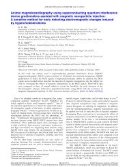
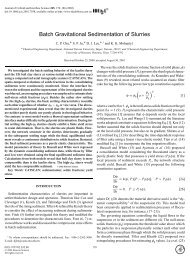
(Fig. 1). On further questioning, she reported having started<br />
Fig. 1. Reticular hyperpigmentation over the buttocks, upper thighs,<br />
lower legs and feet.<br />
Acta Derm Venereol 82<br />
to take a hot bath for 60 to 90 min in a bathtub nightly 6<br />
months previously. The bathtub was not deep, so her knees<br />
and chest remained <strong>ab</strong>ove the water when she was bathing,<br />
and distribution of the skin lesions corresponded to the areas<br />
that were immersed in the water. Skin biopsy specimens were<br />
taken from a lesion on her left buttock and left ankle. The<br />
microscopic ® ndings of the two specimens were similar, showing<br />
hyperkeratosis, epidermal thinning with loss of rete ridges,<br />
basal hyperpigmentation and dermal oedema. Slight pigment<br />
incontinence and a mild super- ® cial perivascular lymphocytic<br />
in® ltrate were also revealed. Stain for elastin showed an<br />
increased amount of elastic ® bres in the upper and middle<br />
dermis. Based on the clinical manifestations and microscopic<br />
® ndings, a diagnosis of erythema <strong>ab</strong> <strong>igne</strong> was made. The<br />
patient was instructed not to take hot baths. Her skin lesions<br />
gradually faded during the following months.<br />
DISCUSSION<br />
<strong>Erythema</strong> <strong>ab</strong> <strong>igne</strong> is a well-known adverse effect of repeated<br />
heat exposure. In the past, it has usually been seen on the<br />
inner aspects of the thighs and lower legs of women who sit in<br />
front of open ® res, ® replaces or furnaces. Since the introduction<br />
of central heating, it has become rare, but it can still be<br />
seen in people who use hot-water bottles or heating pads for<br />
pain relief. Skin lesions may not appear for 3 weeks after the<br />
start of exposure to heat (3). When the lesion ® rst appears, it<br />
may fade if not then submitted to heat exposure, but will tend<br />
to persist after prolonged exposure to heat (3). Furnitureinduced<br />
erythema <strong>ab</strong> <strong>igne</strong> has been reported in people who<br />
used a therapeutic chair with a built-in heating unit (4).<br />
Besides, erythema <strong>ab</strong> <strong>igne</strong> developing on unusual areas may<br />
be a clue to internal disease or internal malignancy (5, 6).<br />
Though taking a hot bath is not an uncommon h<strong>ab</strong>it, there<br />
has been no report of erythema <strong>ab</strong> <strong>igne</strong> developing on people<br />
h<strong>ab</strong>ituated to hot bathing. Our patient had taken a 60 to<br />
90-min hot bath nightly for 6 months. This length of time and<br />
frequency of hot bathing is unusual.<br />
<strong>Erythema</strong> <strong>ab</strong> <strong>igne</strong> is believed to carry a slightly increased<br />
risk of malignant transformation (1, 4, 7), especially when<br />
hydrocarbons are the source of heat (4). However, there has<br />
been no report of malignancy developing on erythema <strong>ab</strong> <strong>igne</strong><br />
caused <strong>by</strong> a warm bottle or hot pad. In addition to inducing<br />
erythema <strong>ab</strong> <strong>igne</strong>, infrared radiation may contribute to premature<br />
aging and enhance the carcinogenic effects of ultraviolet<br />
radiation (8). While a hot bath is both refreshing and<br />
relaxing, it may be harmful to our skin if taken too frequently.<br />
REFERENCES<br />
1. Page EH, Shear NH. Temperature-dependent skin disorders.<br />
J Am Acad Dermatol 1988; 18: 1003 ± 1019.<br />
2. Baruchin AM. <strong>Erythema</strong> <strong>ab</strong> <strong>igne</strong>: a neglected entity? Burns<br />
1994; 20: 460 ± 462.
3. Galvin SA, Buchness MR. Rectangular reticulate patches on<br />
the pretibial areas. <strong>Erythema</strong> <strong>ab</strong> <strong>igne</strong>. Arch Dermatol 1990;<br />
126: 386 ± 387.<br />
4. Meffert JJ, Davis BM. Furniture-induced erythema <strong>ab</strong><br />
<strong>igne</strong>. J Am Acad Dermatol 1996; 34: 516 ± 517.<br />
5. Butler ML. <strong>Erythema</strong> <strong>ab</strong> <strong>igne</strong>, a sign of pancreatic<br />
disease. Am J Gastroenterol 1977; 67: 77 ± 79.<br />
An Unusual Manifestation of Linear Atrophoderma of Moulin<br />
6. Ash<strong>by</strong> M. <strong>Erythema</strong> <strong>ab</strong> <strong>igne</strong> in cancer patients. J R Soc<br />
Med 1985; 78: 925 ± 927.<br />
7. Arrington JH 3rd, Lockman DS. Thermal keratoses and<br />
squamous cell carcinoma in situ associated with erythema<br />
<strong>ab</strong> <strong>igne</strong>. Arch Dermatol 1979; 115: 1226 ± 1228.<br />
8. Kligman LH, Kligman AM. Re¯ ections on heat. Br J<br />
Dermatol 1984; 110: 369 ± 375.<br />
Ljubka Miteva and E. Obreshkova<br />
Department of Dermatology and Venereology, Medical University, Zdrave 2 str., BG-1431 So® a, Bulgaria.<br />
E-mail: mitev@ns.medfac.acad.bg<br />
Accepted July 18, 2002.<br />
Sir,<br />
Moulin et al. (1) ® rst described linear atrophoderma in 1992<br />
and differentiated this uncommon condition from other linear<br />
dermatoses. Clinically, it is present as pigmented and atrophic<br />
bands or lines that follow Blaschko’s lines exactly, with no preceding<br />
in¯ ammation or subsequent induration or scleroderma.<br />
To date, 11 cases of linear atrophoderma of Moulin (LAM)<br />
have been reported in the literature (1 ± 7). Here, we present<br />
a woman with two types of skin lesions: linear atrophic<br />
lesions on the right arm and on the left mandibular region<br />
along with atrophic areas and telangiectatic macules in<br />
linear arrangement following the course of Blaschko’s lines<br />
on the right buttock and leg.<br />
The expression of the disease in our case does not conform<br />
exactly to any of the reported cases with classic cutaneous<br />
manifestations characteristic for LAM.<br />
CASE REPORT<br />
A 20-year-old woman presented with a 4-year history of<br />
asymptomatic, unilateral linear skin lesions following the<br />
appearance of Blaschko’s lines on the arms and legs and on<br />
the face. Her medical and family history was unremark<strong>ab</strong>le. The<br />
disease had a chronic course without progression or regression.<br />
Physical examination revealed two types of skin lesions:<br />
hypopigmented, atrophic, slightly depressed linear lesions on<br />
the medial aspect of the right arm, from the axilla to the wrist<br />
and on the left mandibular region, and 1-cm wide atrophic<br />
band-like lesions along with multiple telangiectatic macules<br />
following Blaschko’s lines on the right buttock and leg<br />
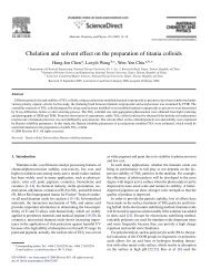
(Fig. 1).<br />
L<strong>ab</strong>oratory ® ndings, including a complete blood count,<br />
urinalysis and liver function test, were normal. Complete<br />
ophthalmologic, odontologic and radiologic surveys revealed<br />
no <strong>ab</strong>normalities.<br />
A skin biopsy taken from the atrophic lesion with<br />
telangiectasias on the right leg revealed moderate psoriasiform<br />
epidermal hyperplasia with hyaline eosinophilic bodies in the<br />
Letters to the Editor 479<br />
Fig. 1. Linear atrophic telangiectatic lesions on the right buttock<br />
and leg. The pattern is consistent with Blaschko’s lines.<br />
spinous layer; oedema and dilated small blood vessels in the<br />
papillary dermis, some with hyalinization; scant in¯ ammatory<br />
in® ltrate of lymphocytes and plasma cells and some<br />
hyalinization of the collagen in the upper dermis. The elastic<br />
tissue is diminished (Fig. 2). These histologic features were<br />
considered to be compatible with the diagnosis of LAM.<br />
Acta Derm Venereol 82