Cell Lab Report
Cell Lab Report
Cell Lab Report

Create successful ePaper yourself
Turn your PDF publications into a flip-book with our unique Google optimized e-Paper software.
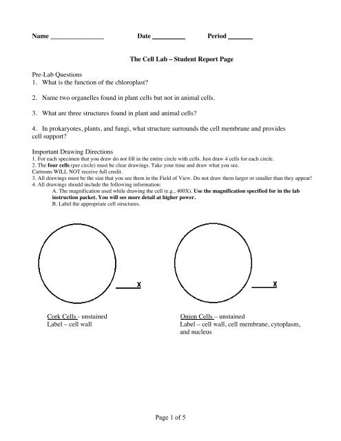
Name ________________ Date Period<br />
Pre-<strong>Lab</strong> Questions<br />
1. What is the function of the chloroplast?<br />
The <strong>Cell</strong> <strong>Lab</strong> – Student <strong>Report</strong> Page<br />
2. Name two organelles found in plant cells but not in animal cells.<br />
3. What are three structures found in plant and animal cells?<br />
4. In prokaryotes, plants, and fungi, what structure surrounds the cell membrane and provides<br />
cell support?<br />
Important Drawing Directions<br />
1. For each specimen that you draw do not fill in the entire circle with cells. Just draw 4 cells for each circle.<br />
2. The four cells (per circle) must be clear drawings. Take your time and draw what you see.<br />
Cartoons WILL NOT receive full credit.<br />
3. All drawings must be the size that you see them in the Field of View. Do not draw them larger or smaller than they appear!<br />
4. All drawings should include the following information:<br />
A. The magnification used while drawing the cell (e.g., 400X). Use the magnification specified for in the lab<br />
instruction packet. You will see more detail at higher power.<br />
B. <strong>Lab</strong>el the appropriate cell structures.<br />
Cork <strong>Cell</strong>s - unstained Onion <strong>Cell</strong>s – unstained<br />
<strong>Lab</strong>el – cell wall <strong>Lab</strong>el – cell wall, cell membrane, cytoplasm,<br />
and nucleus<br />
Page 1 of 5
Onion <strong>Cell</strong>s – stained Elodea <strong>Cell</strong>s - unstained<br />
<strong>Lab</strong>el – cell wall, cell membrane, <strong>Lab</strong>el – cell wall, cell membrane, cytoplasm,<br />
cytoplasm, and nucleus nucleus (if seen), chloroplast, and vacuole (if seen)<br />
Cheek <strong>Cell</strong>s - unstained Cheek <strong>Cell</strong>s - stained<br />
<strong>Lab</strong>el – cell membrane, cytoplasm, and <strong>Lab</strong>el – cell membrane, cytoplasm, and<br />
nucleus nucleus<br />
Potato <strong>Cell</strong>s - stained Bell Pepper <strong>Cell</strong>s – unstained<br />
<strong>Lab</strong>el – cell wall, cell membrane, <strong>Lab</strong>el – cell wall, cell membrane,<br />
cytoplasm, and amyloplast cytoplasm, and chromoplast<br />
Page 2 of 5
Frog Blood <strong>Cell</strong>s Bacteria <strong>Cell</strong>s<br />
(prepared slides) (prepared slides – label the type of bacteria)<br />
<strong>Lab</strong>el – cell membrane, cytoplasm, <strong>Lab</strong>el – capsule, cell wall, cell membrane,<br />
and nucleus and cytoplasm<br />
Questions (Use complete sentences for full credit!)<br />
A. Cork cells:<br />
1. What difference did you notice about the cells near the edge of your slice compared to the cells near<br />
the center of your slice? Explain!<br />
2. What cell structures do you see when looking at cork cells?<br />
3. Why do the cork cells appear to be empty?<br />
B. Onion cells:<br />
4. What microscopic evidence shows that the onion cell is a plant cell?<br />
5. What structures can be seen in an unstained onion cell.<br />
6. How do the stained onion cells appear differently than the unstained onion cells?<br />
7. Do the onion cells you looked at have chloroplasts? Why or why not?<br />
C. Elodea cells:<br />
8. How are Elodea cells the same and/or different than the onion cells?<br />
9. What are the functions of a cell wall?<br />
10. With regard to cytoplasmic streaming, what might be the function of this phenomenon?<br />
Page 3 of 5
D. Cheek cells:<br />
11. How are cheek cells different from the plant cells you have studied?<br />
12. What is the purpose for staining cells?<br />
E. Potato cells:<br />
13. What structure can be seen with the aid of the iodine stain?<br />
14. Why are potatoes a good source of carbohydrates? Be specific!<br />
F. Red Bell Pepper cells:<br />
15. What structure is visible in the bell pepper cell and not in any other cell in this investigation?<br />
16. What is a similarity in the functions of the chloroplast and chromoplast?<br />
G. Frog Blood cells:<br />
17. What structures were present in the frog blood cells that were present in your cheek cells?<br />
H. Bacteria cells:<br />
18. Were any internal cell structures present? Why or why not?<br />
19. Compare the size and shape (appearance) of a bacterium cell to any one of the other cells observed.<br />
20. Are bacteria single cell or multicellular organisms? What evidence did you observe to support your<br />
answer? Hint: compare them to other organisms observed in this investigation!<br />
Page 4 of 5
* Complete the following chart below based on “what you know” using “yes” or “no” and “E” or “P”<br />
Onion<br />
Cheek<br />
Elodea<br />
Cork<br />
Frog blood<br />
Bell pepper<br />
Potato<br />
Bacteria<br />
<strong>Cell</strong> wall<br />
<strong>Cell</strong> membrane<br />
Has Chloroplast, or Chromoplast,<br />
or Amyloplast<br />
(Please state which one if yes)<br />
Page 5 of 5<br />
Nucleus<br />
Eukaryotic<br />
or<br />
Prokaryotic