

orthopaedic i n s i g h t s - Cleveland Clinic
orthopaedic i n s i g h t s - Cleveland Clinic
orthopaedic i n s i g h t s - Cleveland Clinic

Create successful ePaper yourself
Turn your PDF publications into a flip-book with our unique Google optimized e-Paper software.
A physician’s newsletter from the Department of Orthopaedic Surgery<br />
THE CLEVELAND CLINIC<br />
FOUNDATION<br />
<strong>orthopaedic</strong> insights<br />
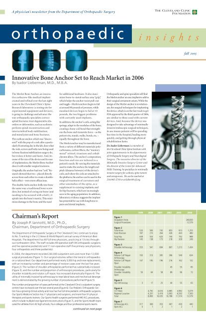
Innovative Bone Anchor Set to Reach Market in 2006<br />
By Isador Lieberman, M.D., M.B.A.<br />
The Merlot Bone Anchor, an innovative<br />
corkscrew-like medical implant<br />
created and refined over the last eight<br />
years in the <strong>Cleveland</strong> <strong>Clinic</strong>’s Spine<br />
Research laboratory (a testing site for<br />
experimental equipment and devices),<br />
is going to challenge and advance the<br />
way <strong>orthopaedic</strong> specialists correct<br />
spinal fractures; treat degenerative disorders<br />
or deformities, such as scoliosis;<br />
perform spinal reconstruction and<br />
intervertebral body stabilization;<br />
and mend joint and bone fractures.<br />
The curlicue anchor, which was “discovered”with<br />
the pop of a cork after a particularly<br />
frustrating day in the lab, does what<br />
the rods, screws and bolts now being used<br />
in bone fixation/stabilization surgery do,<br />
but it does it better and faster. And, because<br />
of the size of the device and its ease<br />
of implantation, the Merlot Bone Anchor<br />
does it with smaller surgical incisions.<br />
Originally, the anchor had one “coil.”Research<br />
showed that two – placed directly<br />
across from each other to create a doublehelix<br />
effect – were more efficacious.<br />
The double-helix anchor drills into bone<br />
the same way a traditional bone screw<br />
does, but instead of coring out bone (and<br />
needing to be secured with a bolt), it<br />
spirals into the bone’s matrix. This minimizes<br />
damage to the bone and the need<br />
Chairman’s Report<br />
for additional hardware. It also maximizes<br />
bone-to-metal surface area “grip”,<br />
which helps the anchor resist pull-out<br />
and toggle–Merlot anchors begin to fail<br />
at around 80 pounds of pressure, while<br />
standard devices begin to fail at 10<br />
pounds, the two biggest problems<br />
with currently used implants.<br />
In addition, the anchor’s coils, acting like<br />
springs, adapt to the modulus of the bone,<br />
creating a bone-coil bond that strengthens<br />
the bone and transmits force – as the<br />
patient sits, stands, walks, bends, etc. –<br />
equally throughout the bone.<br />
The Merlot anchor may be manufactured<br />
from a variety of different materials: polycarbonates,<br />
carbon fibers, the “memory<br />
metal” nitinol, titanium and cobalt<br />
chrome alloys. The anchor’s composition,<br />
function and size are tailored to a<br />
patient’s needs. By varying the materials<br />
used to make the anchor, the length and<br />
pitch (distance between) of the device’s<br />
coils, and where the coils are attached to<br />
the platform, the anchor can be used in the<br />
surgical treatment of curvature and<br />
other anomalies of the spine, as a<br />
supplement to existing implants, and<br />
for hip fractures, which are increasingly<br />
seen in the aging population. In addition,<br />
laboratory evidence suggests the implant<br />
has potential for use with long bone repairs<br />
and dental implants.<br />
By Joseph P. Iannotti, M.D., Ph.D.,<br />
Chairman, Department of Orthopaedic Surgery<br />
The Department of Orthopaedic Surgery at The <strong>Cleveland</strong> <strong>Clinic</strong> continues to enjoy<br />
its No. 5 ranking in the U.S.News & World Report’s annual survey of America’s Best<br />
Hospitals. The department has 60 full-time physicians, practicing at 13 sites throughout<br />
northeastern Ohio. The staff includes 49 operative staff (44 <strong>orthopaedic</strong> surgeons<br />
and five operative podiatrists) and 11 non-operative staff (five primary care physicians,<br />
four orthopedists and three podiatrists).<br />
In 2004, the department recorded 240,000 outpatient visits and more than 12,000<br />
surgical procedures (Figure 1). Our surgical volumes reflect the trend in <strong>orthopaedic</strong>s<br />
on a national level. Our department performed nearly 3,000 hip and knee replacements,<br />
with an increasing number and percentage of revision cases over the last five years<br />
(Figure 2). The number of shoulder arthroplasties performed has substantially increased<br />
(Figure 3), and the number and proportion of arthroscopic procedures, particularly for<br />
shoulder instability and rotator cuff repair, has increased dramatically (Figure 4). The<br />
utility of minimally invasive hip arthroscopy to treat labral tears and mechanical derangement<br />
is demonstrated by the growing number of procedures performed (Figure 5).<br />
The number and proportion of cases performed at the <strong>Cleveland</strong> <strong>Clinic</strong>’s outpatient surgery<br />
centers have increased over the last several years (Figure 6). Our Pediatric Orthopaedic Section<br />
has a growing clinical activity and now has five full-time pediatric <strong>orthopaedic</strong> surgeons.<br />
Our Sports Medicine Section has 11 physicians and surgeons, and more than 91 physical<br />
therapists and sports trainers. Our Sports Health surgeons performed 440 ACL procedures,<br />
which include multiple knee ligament reconstructions (Figure 7), and the Sports Health team<br />
cared for athletes from 42 high schools, four colleges and four professional sports teams.<br />
continued on next page<br />
Orthopaedic and spine specialists will find<br />
the Merlot anchor an easy implant to add to<br />
their surgical armament arium.While the<br />
design of the Merlot anchor is revolutionary,<br />
the surgical techniques for implanting<br />
the device, which is on the fast-track to get<br />
FDA approval by the third quarter of 2006,<br />
are similar to those used with current<br />
devices. And, because the device was<br />
designed to take advantage of minimally<br />
invasive/endoscopic surgical techniques,<br />
its use means patients will be spending<br />
less time in the hospital, healing more<br />
quickly, and getting through physical<br />
rehabilitation faster.<br />
Dr. Isador Lieberman is a member of<br />
the <strong>Cleveland</strong> <strong>Clinic</strong> Spine Institute with<br />
joint appointments in the departments<br />
of Orthopaedic Surgery and Neurological<br />
Surgery. The executive director of the<br />
Minimally Invasive Surgery Center and<br />
the director of the Center for Advanced<br />
Skills Training, he specializes in minimally<br />
invasive surgery for scoliosis, spine tumors<br />
and osteoporosis. He can be reached at<br />
216/445-2743 or at lieberi@ccf.org.<br />
fall 2005<br />
1999 2000 2001 2002 2003 2004<br />
Figure 1<br />
Outpatient Visits 240,000<br />
Surgical Procedures 12,579<br />
Figure 2<br />
Total Knee 533 593 745 853 913 1,255<br />
Knee Revision 187 215 264 426 443 480<br />
Total Hip 449 553 620 740 792 988<br />
Hip Revision 145 138 173 218 209 249<br />
Figure 3<br />
Shoulder Arthroscopy 353 541 694 807 1,213 1,434<br />
Figure 4<br />
Rotator Cuff Repair<br />
Arthroscopy<br />
193 313 395 396 549 634<br />
Rotator Cuff Repair<br />
Open<br />
147 195 196 216 162 132<br />
Shoulder Instability<br />
Arthroscopy<br />
45 55 85<br />
Shoulder Instability<br />
Open<br />
50 64 58 89 96 72<br />
Figure 5<br />
Hip Arthroscopy 3 17 25 40 55 65<br />
Figure 6<br />
CCF Main 4,792 4,616 4,885 4,982 5,079<br />
Regional Location 4,201 5,419 5,903 6,592 7,500<br />
Total 8,993 10,035 10,788 11,574 12,579<br />
Figure 7<br />
Arthroscopy ACL 317 369 353 407 430 440<br />
Open ACL 0 1 2 1 2 1
2<br />
continued from front page<br />
The Orthopaedic Research Center (ORC) now has 40 full-time Ph.D. scientists and 40<br />
<strong>orthopaedic</strong> surgeons involved in basic bench research. The ORC was formed four years<br />
ago and currently has more than $12 million in total direct funding, more than $6 million<br />
of which is from the NIH (Figure 8). In 2004, the ORC obtained its first NIH-funded training<br />
grant and Core Center Grant. The department also was successful in the recruitment<br />
of a director for its newly formed <strong>Clinic</strong>al Outcomes Research Center and will hire five research<br />
project managers over the next year. The <strong>Clinic</strong>al Outcomes Research Center will<br />
focus on arthroplasty surgery, adverse events and use of orthobiologic products.<br />
Dr. Joseph Iannotti is Chairman, Department of Orthopaedic Surgery, Chairman of<br />
the Orthopaedic Research Center and a member of the Section of Hand and Upper<br />
Extremity Surgery. He can be reached at 216/445-5151 or 800/553-5056, ext. 55151.<br />
Orthopaedic<br />
OR of the Future<br />
By Jonathan Schaffer,<br />
M.D., M.B.A.<br />
Are <strong>orthopaedic</strong> surgeons ready for the<br />
practical challenges our profession will<br />
face in the next 25 years? According to the<br />
National Institute on Aging, by 2030 the<br />
number of Americans aged 65 to 74 will<br />
increase from 19 million to an estimated<br />
36 million people, and the number of<br />
people aged 75 to 84 will grow from 16<br />
million to 25 million.<br />
Consequently, the population of patients<br />
with degenerative arthritis will continue<br />
double-digit growth well into the foreseeable<br />
future. This will mean equivalent<br />
growth in the demand for arthroplasty,<br />
which will exert considerable pressure on<br />
the <strong>orthopaedic</strong> operating room. In response,<br />
the <strong>orthopaedic</strong> OR of the future<br />
must be designed to handle a high volume<br />
of arthroplasty surgeries with more<br />
speed, precision and consistency in quality,<br />
outcomes, safety and value.<br />
To begin planning for these changes,<br />
The <strong>Cleveland</strong> <strong>Clinic</strong> has partnered with<br />
Stryker Corporation (Kalamazoo, Mich.)<br />
to model the <strong>orthopaedic</strong> OR of the future,<br />
on location at The <strong>Cleveland</strong> <strong>Clinic</strong>.<br />
This technology and commercialization<br />
agreement provides a unique opportunity<br />
for research and development, new technology<br />
testing and physician education.<br />
The <strong>orthopaedic</strong> OR of the future will<br />
optimize the surgical environment for<br />
greatest efficiency and highest quality<br />
in managing the increased surgical<br />
volume, while achieving significant<br />
cost reductions. To this end, every<br />
process, interrelationship and strategy<br />
related to arthroplasty is being scrutinized<br />
and re-engineered with extensive<br />
input from nurses and physicians.<br />
• Formed in 2001<br />
• 42 PhD and MD <strong>Clinic</strong>ian<br />
Investigators<br />
• 13 BME<br />
• 17 Orthopaedic Surgery<br />
• 13 Departments<br />
• Lerner Research Institute<br />
ND building<br />
Job analysis, a technique borrowed from industry,<br />
will determine the most efficient arrangement for<br />
supplies, equipment and personnel in the room.<br />
Through physical changes in room layout<br />
and process changes in tasks related<br />
to arthroplasty, we believe we can significantly<br />
reduce cycle time for the OR.<br />
Ultimately, every <strong>orthopaedic</strong> OR should<br />
be able to handle one more case per day,<br />
boosting efficiency and cutting costs.<br />
In the preliminary planning, The <strong>Cleveland</strong><br />
<strong>Clinic</strong>-Stryker team identified over<br />
400 distinct processes that take place<br />
between the time the patient is scheduled<br />
for surgery until discharge. The next<br />
objective is to create the most efficient<br />
and effective model for arthroplasty,<br />
then construct the OR to test it.<br />
What will that model look like? Standardization<br />
in processes and procedures<br />
will be the key to meeting the increased<br />
operative demand. Standardizing virtually<br />
every aspect of arthroplasty, from<br />
scheduling through discharge, allows for<br />
better planning and cost management;<br />
any variances that occur can be dealt<br />
with more easily.<br />
Standardization will take many forms.<br />
For example, every arthroplasty OR will<br />
be set up identically, down to the items in<br />
the drawers. The arthroplasty procedure<br />
itself is being dissected into discrete tasks,<br />
and each task, such as how a nurse hands<br />
an instrument to the surgeon, will be described<br />
in detail.<br />
The floor in the OR of the future will<br />
be clear of equipment and supplies, and<br />
unnecessary barriers, cabinets and storage<br />
areas will be removed or redesigned.<br />
Patient X-rays will be displayed on monitors<br />
located throughout the room. Surgical<br />
navigation will allow for more accurate<br />
implant placement. The stock room will<br />
13<br />
12<br />
11<br />
10<br />
9<br />
8<br />
7<br />
6<br />
5<br />
4<br />
Figure 8<br />
Orthopaedic Research Center<br />
2001-02<br />
Dollars in millions<br />
2002-03 2003-04<br />
Total<br />
run on a computerized inventory system<br />
to ensure supply availability and stock rotation.<br />
Job analysis, a technique borrowed<br />
from industry, will determine the most<br />
efficient arrangement for supplies, equipment<br />
and personnel in the room.<br />
Planning is under way to accommodate<br />
the advanced technology that will be<br />
an essential component of the new<br />
<strong>orthopaedic</strong> OR. Surgeons will have the<br />
capability of using computer simulation<br />
to visualize the path of an instrument<br />
before making an incision.<br />
The first of three prototype ORs based<br />
on this model is under construction.<br />
The <strong>Cleveland</strong> <strong>Clinic</strong>-Stryker team plan<br />
to have the prototype completed for the<br />
October Medical Innovation Summit<br />
here at The <strong>Cleveland</strong> <strong>Clinic</strong>, which this<br />
year showcases <strong>orthopaedic</strong> surgery. We<br />
consider this room the first step in an ongoing<br />
evolution in which we will continue<br />
to evaluate, learn and make further<br />
changes that will prepare us for the future.<br />
Dr. Jonathan Schaffer is an <strong>orthopaedic</strong><br />
surgeon with special interest in arthroplasty<br />
and <strong>orthopaedic</strong> research. He also is the<br />
Managing Director of e-<strong>Cleveland</strong> <strong>Clinic</strong>.<br />
He can be reached at 216/444-8960 or by<br />
e-mail at schaffj@ccf.org.
Commercializing Orthopaedic Innovations<br />
By Christopher Coburn<br />
<strong>Cleveland</strong> <strong>Clinic</strong> <strong>orthopaedic</strong> surgeons<br />
currently have more than two dozen<br />
<strong>orthopaedic</strong> technologies or devices in<br />
the commercialization pipeline through<br />
CCF Innovations, the <strong>Clinic</strong>’s technology<br />
commercialization arm. Established in<br />
2000, CCF Innovations assists physicians<br />
in translating therapies, devices and diagnostics<br />
into medical products through<br />
spin-off companies, licensees and equity<br />
partnerships. Orthopaedic surgery is one<br />
of 12 specialty areas that work hand<br />
in hand with CCF Innovations and has<br />
been one of the most fruitful in terms of<br />
number of inventions. <strong>Cleveland</strong> <strong>Clinic</strong><br />
<strong>orthopaedic</strong> surgeons who have launched<br />
products through CCF innovations<br />
attribute their success to working in an<br />
environment that promotes the interchange<br />
of ideas and experience between<br />
the clinical and basic science arenas.<br />
By bringing new ideas to market and<br />
developing an efficient distribution<br />
system for them, CCF Innovations’ goal<br />
is to benefit the largest number of patients<br />
possible. CCF Innovations develops a<br />
team that includes physicians and their<br />
inventions, deal makers, patent attorneys<br />
and corporate partners to turn an invention<br />
into a product. CCF Innovations has<br />
a successful record in achieving this, and<br />
the <strong>Cleveland</strong> <strong>Clinic</strong>’s ratio of inventions<br />
to dollar of research money is among the<br />
highest in the United States.<br />
The <strong>orthopaedic</strong> devices and<br />
technologies registered with<br />
CCF Innovations are at varying<br />
stages of development. If<br />
a new technology is fully developed,<br />
CCF Innovations often can<br />
create a spin-off company or license<br />
the technology within six months.<br />
Before committing to a physician and<br />
an invention, CCF Innovations experts<br />
conduct a careful analysis that includes<br />
evaluation of the product’s clinical efficacy<br />
and its market potential. Every<br />
new product must have an identifiable<br />
patient group. The responsibility of<br />
CCF Innovations is to step back and<br />
see the full potential that a new product<br />
has to offer.<br />
Ohio BioGel, one of 13 <strong>Cleveland</strong> <strong>Clinic</strong><br />
spin-off companies established in the<br />
past three years, illustrates the benefits<br />
of this approach for patients and the<br />
inventor. A biomaterial in development<br />
with <strong>Clinic</strong> researcher Anthony Calabro,<br />
Ph.D., for two and a half years, BioGel<br />
is a platform technology with applications<br />
across multiple specialties. In<br />
<strong>orthopaedic</strong>s, BioGel can be used to<br />
reconstruct cartilage and intervebral<br />
discs, as a tissue filler and as an arthrosurfacer<br />
to create a more natural interaction<br />
between a joint prosthesis and<br />
native bone.<br />
Novel Hydrogel Technology Moves<br />
Closer Toward Commercialization<br />
By Anthony Calabro, Ph.D.<br />
Localized damage to the articular cartilage surface as a result of a sports or work-related<br />
injury can be severely disabling and lead to osteoarthritis. We have developed novel glycosaminoglycan-based<br />
(GAG-based) hydrogels for use as biomaterials in a wide variety of<br />
tissue engineering and repair applications including repair of localized cartilage defects in<br />
the field of <strong>orthopaedic</strong> surgery.<br />
In developing these novel hydrogels, we have shifted the paradigm used in most tissue engineering<br />
research from a cell-based approach to a biomaterial-based approach that relies<br />
heavily on the insights provided by nature’s own macromolecular design for a healthy,<br />
functional articular cartilage. Articular cartilage is the resilient load-bearing tissue that<br />
forms the articulating surfaces of diarthrodial joints. The cartilage functions to absorb<br />
mechanical shock and spread applied load onto subchondral bone.<br />
Normal articular cartilage exhibits its viscoelastic properties, as well as its ability to<br />
resist deformation and absorb compressive loads, principally as a result of creating<br />
a macromolecular network with low porosity and a high density of fixed negative<br />
charges. The mechanical properties of such a macromolecular network manifest<br />
themselves in a concentration-dependent fashion through the well-known mechanisms<br />
associated with Donnan, electrorepulsion and stress-shielding effects. In cartilage,<br />
this is accomplished by the fixation of COO- and SO42- groups on resident<br />
hyaluronan (HA) and chondroitin sulfate (CS) chains at the high concentration of<br />
these GAGs normally found in cartilage (50-100 mg/ml).<br />
In designing biomaterials for cartilage repair, our laboratory has taken the approach of<br />
using the same molecular scaffold materials (e.g., HA, CS) already proved to produce a<br />
mechanically functional cartilage tissue. The use of these GAGs as scaffold materials,<br />
when formed into the proper 3-D architecture, assures a biologically compatible product<br />
that reproduces the desired mechanical function of cartilage.<br />
A novel enzyme-driven method for cross-linking these GAGs into solid matrices has<br />
been developed by our laboratory, which: 1) preserves sufficient negative-charge density<br />
on the GAGs to recreate both Donnan and electrorepulsion effects and, thus, the mechanical<br />
properties of cartilage; 2) cross-links at GAG concentrations equivalent to those<br />
in cartilage – required to recreate the stress-shielding effect (50-100 mg/ml) and, thus,<br />
protect the biomaterial during normal physiological loading; and 3) is biocompatible,<br />
allowing in vivo cross-linking within the void volume of a cartilage defect.<br />
Our laboratory also has taken a novel approach to cartilage repair. Conventional<br />
cartilage tissue engineering involves seeding chondrocytes on a mechanically inferior<br />
biomaterial and then relying on the de novo synthesis of a mechanically functional cartilage<br />
extracellular matrix (ECM) by the seeded chondrocytes as the inferior biomaterial<br />
The <strong>orthopaedic</strong> devices and technologies registered with<br />
CCF Innovations are at varying stages of development. If a<br />
new technology is fully developed, CCF Innovations often<br />
can create a spin-off company or license it within six months.<br />
Beyond <strong>orthopaedic</strong>s, potential applications<br />
for BioGel also have been identified<br />
in plastic surgery, ophthalmology,<br />
otolaryngology, cardiology and gastroenterology.<br />
One of the most versatile<br />
inventions to come out of Orthopaedic<br />
Surgery, the BioGel technology eventually<br />
may be licensed to other companies<br />
for specific applications.<br />
Orthopaedic surgeons Isador Lieberman,<br />
M.D., M.B.A., and George Muschler,<br />
M.D., currently have 17 and 8 inventions,<br />
respectively, in various stages of development<br />
with CCF Innovations. The majority<br />
of Dr. Lieberman’s projects are related to<br />
spinal surgery, including innovative fixation<br />
devices and methods and apparatus<br />
for correcting scoliosis and stabilizing<br />
adjacent bones. Dr. Lieberman’s Merlot<br />
Bone Anchor device for endoscopic scoliosis<br />
surgery is nearing release after eight<br />
years in development.<br />
Dr. Muschler, who holds a joint appointment<br />
with the Department of Biomedical<br />
Engineering, has specialty interests in<br />
fracture nonunion and reconstructive<br />
surgery, leading to a concomitant interest<br />
in tissue engineering. His Cellect Selective<br />
Figure 1–Hydrogel repaired cartilage lesion<br />
Retention Technology uses minimally<br />
invasive bone marrow harvesting to<br />
populate graft material, with a three- to<br />
fourfold increase in osteoprogenitor cell<br />
concentrations. It has been licensed to<br />
Johnson & Johnson since 2004 and is<br />
commercially available. Continuing<br />
his interest in this area, Dr.Muschler’s<br />
current inventions also are related to<br />
bone marrow harvesting, bone grafting<br />
and limb lengthening.<br />
Other <strong>Cleveland</strong> <strong>Clinic</strong> <strong>orthopaedic</strong> surgeons<br />
and their inventions in progress<br />
with CCF Innovations include Viktor<br />
Krebs, M.D., and Wael Barsoum, M.D.,<br />
expanding material fastening system;<br />
Kenneth Marks, M.D., acetabular cup,<br />
in partnership with Dr. Lieberman;<br />
Peter Evans, M.D., Ph.D., carpal tunnel<br />
guide; Robert Biscup, D.O., bone processing<br />
device; and Robert McLain,<br />
M.D., cannulated ultrasound device<br />
for pedicle screw placement.<br />
Chris Coburn is Executive Director of CCF<br />
Innovations. He can be reached at 216/445-<br />
4008 or at coburnc@ccf.org.<br />
Figure 2–Uncross-linked hydrogel<br />
scaffold degrades. This approach has failed, predominantly due to an inability to<br />
successfully coordinate new cartilage synthesis with scaffold degradation as well as an<br />
inability to maintain an anabolic phenotype for seeded chondrocytes in an essentially<br />
catabolic or diseased environment.<br />
Our approach has been to employ a biomaterial with the functional mechanical properties<br />
of cartilage, which has been minimally modified to make it resistant to normal<br />
proteolytic pathways of cartilage degradation. In essence, the paradigm shift is designed<br />
to: 1) fill the wound site with a mechanically sound material from the outset; 2) rely on<br />
chondrocytes, if needed, for maintenance of the ECM – not its de novo synthesis; and 3)<br />
utilize modification of the carbohydrate-based ECM itself to ensure its in vivo survival.<br />
Currently, these novel GAG-based biomaterials are being tested for repair of localized cartilage<br />
defects in a porcine animal model (see Figures 1 and 2). CCF Innovations, the <strong>Cleveland</strong><br />
<strong>Clinic</strong>’s technology commercialization arm, is currently working to commercialize<br />
this novel GAG-based hydrogel technology. Due to the wide potential clinical application<br />
of this platform technology, the current strategy involves development of a <strong>Cleveland</strong><br />
<strong>Clinic</strong>-controlled spin-off company, Ohio BioGel, to commercialize certain clinical applications,<br />
while licensing to a wide spectrum of corporate partners for other clinical application.<br />
Chris Coburn, Executive Director of CCF Innovations, is directing the project.<br />
CCF Innovations has received funds from the state of Ohio through its Ohio Validation<br />
and Seed Fund Initiative to establish a <strong>Cleveland</strong> <strong>Clinic</strong>-controlled BioValidation Fund.<br />
This new fund will make investments in early-stage <strong>Cleveland</strong> <strong>Clinic</strong> biotechnology spinoff<br />
companies, including Ohio BioGel, based on the novel hydrogel technology. Investments<br />
in Ohio BioGel by the BioValidation Fund will be used to advance the technology<br />
toward human trials and FDA approval.<br />
Dr. Anthony Calabro has joint appointments with the departments of Biomedical Engineering,<br />
Orthopaedic Surgery, and the Head and Neck Institute. He can be reached at 216/445-3273 or<br />
800/553-5056, extension 53273.<br />
3
4<br />
Volar Plating of Distal Radius Fractures<br />
By Jeffrey N. Lawton, M.D., and Peter J. Evans, M.D., Ph.D., FRCSC<br />
Numerous surgical options for addressing<br />
displaced distal radius fractures exist,<br />
including internal and external fixation.<br />
While volar plating for certain types of<br />
distal radius fractures has been used in the<br />
past, dorsally placed plates and external<br />
fixators have dominated. Recently, newer<br />
constructs that provide a locked volar<br />
plate have expanded the indications for<br />
plating distal radius fractures.<br />
Today, volar locked plates are being used<br />
in a majority of distal radius fracture<br />
types. Although many of these fractures<br />
exhibit dorsal displacement and dorsal<br />
comminution, which are better biomechanically<br />
addressed with a dorsal plate<br />
in buttress mode, enthusiasm for routine<br />
dorsal plating of distal radius fractures<br />
has been limited by complications associated<br />
with extensor tendons. Extensor<br />
tendon adhesions, tendonitis and<br />
tendon ruptures are thought to occur<br />
because of the limited soft tissue interposed<br />
between the dorsally applied<br />
plates and the extensor tendons.<br />
With the ability to lock the screws at various<br />
angles, a blade plate-type rigid construct<br />
is created. Therefore, despite being<br />
placed on the volar side of a dorsally displaced<br />
fracture, the articular surface and<br />
subchondral bone can be maintained in<br />
a reduced position during healing. Depending<br />
on the system, the angle that the<br />
distal fixed screws/pegs make with the<br />
plate allows various fracture fragments<br />
to be addressed individually.<br />
Perceived concerns with the volar approach<br />
include injury to the radial artery<br />
and/or median nerve. A modified Henry<br />
approach with incision of the flexor<br />
carpi radialis (FCR) sheath allows a re-<br />
Simple wounds in the diabetic foot can<br />
rapidly progress from minor problems<br />
to limb-threatening ulcers due to the<br />
presence of diabetic vascular disorders<br />
and neuropathy. The <strong>Cleveland</strong> <strong>Clinic</strong><br />
Diabetic Foot Care Program, established<br />
in 2003, applies the latest treatments,<br />
technology and computer approaches to<br />
prevent and manage wounds and ulcers<br />
in the diabetic foot.<br />
People with diabetes have a lifetime risk of<br />
15 percent of developing a foot ulcer, and<br />
a non-healing ulcer is the most common<br />
antecedent to lower-limb amputation.<br />
Our aggressive team approach includes<br />
ulcer prevention and treatment, regular<br />
foot examinations, patient education,<br />
and therapeutic intervention to reduce<br />
the risk of ulcers and lower-extremity<br />
amputations.<br />
Patients with emergent problems are<br />
scheduled for immediate consultation<br />
in our Diabetic Foot Care <strong>Clinic</strong> within<br />
the Department of Orthopaedic Surgery<br />
so that appropriate treatment and intervention<br />
can be initiated rapidly. Other<br />
patients receive a comprehensive evalua-<br />
producible exposure with protection<br />
of the neurovascular structures. The surgeon<br />
makes a longitudinal incision overlying<br />
the FCR tendon. The FCR tendon<br />
is then retracted radially, and the surgeon<br />
incises the floor of the FCR sheath. Blunt<br />
dissection allows identification of the<br />
pronator quadratus, which is then incised<br />
off the radial aspect of the radius,<br />
leaving a cuff of tissue for later repair.<br />
This approach exposes both the fracture<br />
site and the area for the volarly placed<br />
plate. To allow for further mobilization,<br />
or in the subacute setting, the brachioradialis<br />
can be subperiosteally elevated and<br />
released, thus eliminating its deforming<br />
force upon the distal fragment.<br />
The fracture is then reduced and<br />
provisionally stabilized with a styloid<br />
K-wire or, alternatively, with the aid<br />
of the plate and the provisional K-wire<br />
holes provided in some systems.<br />
In Step with Diabetic Foot Care<br />
By Peter Cavanagh, Ph.D., D.Sc., and Georgeanne Botek, D.P.M<br />
tion that includes foot pressure mapping<br />
together with vascular and neurologic<br />
workups to identify risk factors and<br />
to define the cause of ulcers and other<br />
problems.<br />
Physicians apply the PEDIS (perfusion,<br />
extent/size, depth/tissue loss, infection<br />
and sensation) classification system<br />
to assess ulcers and evaluate progress<br />
toward healing so that staging and<br />
description of ulcers is consistent. To<br />
complement this standardization, our<br />
electronic medical records system has<br />
been enhanced to capture photographs<br />
of wounds as part of the patient’s<br />
medical record. This innovation allows<br />
clinicians to track wound healing more<br />
effectively and consistently.<br />
Relief of weight bearing is critical in the<br />
healing of diabetic foot ulcers and is well<br />
documented in the literature. Yet it is frequently<br />
overlooked or undertreated by<br />
many clinicians and, therefore, remains<br />
the single most significant reason that<br />
ulcers in the diabetic foot fail to heal.<br />
Our clinicians often use a total contact<br />
cast, which encloses the foot, as an<br />
Following final plate placement, bone<br />
grafting through a small incision dorsally<br />
can be made to fill the void left by<br />
the implanted cancellous bone.<br />
Based upon stability, a typical postoperative<br />
course involves seven to 10 days in a<br />
postoperative splint, followed by a thermal<br />
plastic splint. An early supervised<br />
protocol of wrist and forearm range of<br />
motion as well as maintained digital and<br />
shoulder motion and edema control also<br />
are recommended. At approximately<br />
eight to 10 weeks, based upon clinical and<br />
radiographic progress, one may begin<br />
progressive strengthening.<br />
Dr. Jeffrey Lawton, a member of the Section<br />
of Hand and Upper Extremity Surgery,<br />
can be reached at 216/445-6915.<br />
Dr. Peter Evans, is head of Hand and<br />
Upper Extremity Surgery and a member<br />
of the Lerner Research Institute. He can<br />
be reached at 216/444-7973.<br />
integral part of the treatment plan.<br />
This device effectively relieves pressure<br />
to allow the ulcer to heal.<br />
With a total contact cast, changed<br />
regularly and closely followed by the<br />
physician, ulcers heal in an average of<br />
42 days, compared with healing times<br />
as long as a year with treatments that<br />
do not address the weight relief issue.<br />
We have achieved similar rapid healing<br />
times in ulcers that have remained open<br />
for as long as two years.<br />
After an ulcer is healed, managing<br />
the foot’s mechanical interaction with<br />
the world is critical to preventing ulcer<br />
recurrence, which, in various studies,<br />
has been shown to occur in 50 percent<br />
to 100 percent of patients within three<br />
years. Proper footwear can reduce this<br />
risk significantly. Our research team uses<br />
computer modeling based on MRI images<br />
to digitally reconstruct feet in three<br />
dimensions (see accompanying article<br />
on p. 12). This will eventually enable<br />
individualized footwear recommendations<br />
to be made for each patient.<br />
Reduced distal radius fracture<br />
with volar locked plate<br />
A map of pressure underneath the foot during<br />
walking is collected as part of the patient evaluation<br />
in the Diabetic Foot Care Program.<br />
An IRB registry is maintained, and,<br />
through this database, we have been able<br />
to pursue a number of clinical research<br />
projects during the past two years.<br />
Current projects include a study of<br />
methicillin-resistant bacteria in foot<br />
ulcers; footwear management in patients<br />
with Charcot fractures, and studies<br />
of new approaches to ulcer healing.<br />
Patients can enroll in clinical trials of<br />
these new approaches to wound care.<br />
Dr. Peter Cavanagh is Chairman of the<br />
Department of Biomedical Engineering<br />
and Academic Director of Diabetic Foot<br />
Care. His specialty interests include foot<br />
disorders in diabetic foot disease and bone<br />
loss in long-duration spaceflight. He can<br />
be reached at 216/445-6980 or via e-mail<br />
at cavanap@ccf.org.<br />
Dr. Georgeanne Botek is Medical Director<br />
of the Diabetic Foot Care Program and<br />
has a strong interest in diabetic foot care.<br />
She can be reached at 216/445-8152 or via<br />
e-mail at botekg@ccf.org.
Artificial Discs:<br />
Long-awaited Alternative to Lumbar Fusion<br />
Lumbar fusion continues to be one of the most prevalent spinal surgeries in the<br />
United States, yet data reveal that not all of those with degenerative disc disease who<br />
undergo the surgery will experience significant pain relief and functional restoration.<br />
Total disc replacement with an artificial disc is considered the most promising alternative<br />
to spinal fusion, and <strong>orthopaedic</strong> surgeons have worked<br />
for 20 years to develop a successful prosthesis.<br />
Until recently, the ideal artificial disc has eluded developers.<br />
Now, the latest generation of these prostheses draws on<br />
advances in technology, materials and biomechanics,<br />
allowing the prostheses to closely imitate the function<br />
of the native disc. <strong>Cleveland</strong> <strong>Clinic</strong> spine surgeons are<br />
involved in the development, testing and early adoption<br />
of two of the most promising new disc replacements.<br />
Freedom Lumbar Disc<br />
By Isador Lieberman, M.D.<br />
The Freedom Lumbar Disc (AxioMed Spine Corp., Beachwood,<br />
Ohio) is an innovative new artificial disc comprising a<br />
polymeric gel core bonded to titanium retaining plates. The<br />
polymer’s material characteristics, in combination with the<br />
implant design, provide 3-D motion, load sharing, shock<br />
absorption and dynamic stiffness as needed in response to<br />
varying loads in a fashion that replicates the function of a<br />
normal, healthy disc.<br />
European trials of the Freedom disc are scheduled<br />
to begin in the fall of 2005, and U.S. trials are<br />
scheduled to begin six months later. The prosthesis<br />
is designed for patients 30 to 50 years old with<br />
early- to mid-stage degenerative disc changes and<br />
mechanical back pain who have not responded to<br />
nonoperative treatment.<br />
We are not yet sure of precisely what the disc’s role<br />
will be in the treatment of symptomatic degenerative<br />
discs. The decision of when to use it will depend on a balance<br />
of patient expectations, the appropriate timing for surgery<br />
and the results that realistically can be achieved. We do know that<br />
the Freedom disc is not suitable for patients with end-stage degenerative<br />
discs; these patients still will require fusion.<br />
Under laboratory conditions’ worst-case scenario – simulating a person<br />
jumping 1,000 times a day and landing on his heels – the Freedom disc had<br />
a service life of 40 years.<br />
Exercise across the lifespan is a key<br />
component of bone health. Adult<br />
premenopausal exercisers demonstrate increased<br />
lean mass compared with sedentary<br />
controls, and those exercising with higher<br />
impact have greater leg strength and higher<br />
whole-body and regional bone mineral<br />
density (BMD). Eumenorrheic athletes<br />
in the highest impact and jumping sports<br />
have the highest BMD in weight-bearing<br />
areas and demonstrate the highest levels<br />
of bone-formation markers.<br />
A positive association exists between<br />
BMD and resistance (e.g., strength,<br />
weight) training. Novel strength training<br />
introduced to postmenopausal women<br />
positively affects BMD, and the effect at<br />
specific sites correlates with the cumulative<br />
amount of weight lifted.<br />
Although highly trained and elite female<br />
athletes demonstrate increased BMD in<br />
loaded regions, the effect of high levels<br />
of activity on bone can be detrimental.<br />
The literature consistently supports the<br />
contention that the osteogenic effects of<br />
exercise are modulated in a complex<br />
manner by the nutritional and hormonal<br />
status of the athlete. For example,<br />
female elite-level winter sports athletes<br />
with normal menstrual function have<br />
higher BMD compared with age- and body<br />
mass-matched controls, a difference that<br />
remains significant after controlling for<br />
lean mass. However, those athletes with<br />
a history of oligo- or amenorrhea seem<br />
to lose the osteogenic benefit of their<br />
sport participation and demonstrate<br />
similar BMD as eumenorrheic, nonathletic<br />
controls.<br />
Dancers and gymnasts exhibit this same<br />
effect. Amenorrheic dancers more often<br />
report delayed menarche and a higher incidence<br />
of dieting behaviors than eumenorrheic<br />
dancers. They also demonstrate<br />
significantly lower BMD over time and<br />
may have an increased risk of stress fracture.<br />
Collegiate female gymnasts have<br />
higher BMD at all sites prior to and during<br />
the competitive season than runners,<br />
despite a greater reported prevalence of<br />
The Charite Artificial Disc<br />
By Robert McLain, M.D.<br />
The <strong>Cleveland</strong> <strong>Clinic</strong> is among the first institutions in the United States to use the Charite<br />
artificial disc (DePuy Spine, Raynham, Mass.) in clinical practice. Approved by the U.S.<br />
FDA in October 2004, the Charite disc is a metal/polyethylene insert that restores alignment<br />
and allows controlled motion in the spine.<br />
The ideal patient is 30 to 50 years old, with good bone quality and no significant<br />
nerve compression. Although the FDA has set age 65<br />
as the upper limit, bone quality and degeneration are the<br />
determining factors in patient selection, and the disc will<br />
find its best applications in younger patients.<br />
The Charite disc offers significant advantages over lumbar<br />
fusion in that it maintains spinal flexibility and reduces the risk<br />
of repeat fusions, but that does not change the fact that patient<br />
selection is paramount. If a patient is not a fusion candidate,<br />
the Charite artificial disc is not an appropriate treatment.<br />
DuPuy Spine has designated The <strong>Cleveland</strong> <strong>Clinic</strong> as a training<br />
center for teaching <strong>orthopaedic</strong> spine surgeons the implantation<br />
technique for the Charite disc. The procedure is best<br />
reserved for spine surgeons experienced in an anterior<br />
approach to vertebral surgery because of the complexities<br />
involved. In some cases, a vascular surgeon may assist in the<br />
exposure of the anterior vertebral body and disc space.<br />
When the vessels and organs have been displaced and the<br />
vertebra is in view, the spine surgeon excises the degenerated<br />
disc. The Charite artificial disc is press-fit into<br />
position, where it is held in place by the natural vertebral<br />
tension. In the hands of an experienced surgeon,<br />
operative time is shorter than for a lumbar fusion and<br />
blood loss is minimal.<br />
Most patients require a two- to four-day hospital stay and<br />
are ambulatory postoperatively without a brace. Following<br />
surgical healing and rehabilitation to regain strength and balance,<br />
activity is virtually unrestricted.<br />
Dr. Robert McLain, of the departments of Orthopaedic Surgery and<br />
Neurological Surgery, has been designated by DuPuy Spine as a clinical<br />
instructor for the Charite artificial disc. He can be reached at 216/444-2744<br />
or at mclainr@ccf.org.<br />
Dr. Isador Lieberman, of the Departments of Orthopaedic Surgery and Neurological Surgery,<br />
is one of the developers of the Freedom Lumbar Disc and will participate in the U.S. trials. He<br />
can be reached at 216/445-2743 or at lieberi@ccf.org.<br />
Bone Mineral Density (BMD) in Women: Effects of Exercise<br />
By Susan Joy, M.D<br />
Photo courtesy of DePuy Spine, Inc.<br />
menstrual dysfunction. Because of the<br />
tremendous repetitive loads applied to<br />
various skeletal sites in the sport of gymnastics,<br />
dramatic responses in bone formation<br />
can be observed at multiple sites<br />
associated with greater size-adjusted<br />
strength measures.<br />
The Female Athlete Triad<br />
When energy intake does not keep up<br />
with the demands imposed upon the<br />
body by strenuous exercise, an energy<br />
deficit can result. Research has suggested<br />
that the negative observed effects are not<br />
from the exercise itself but rather from the<br />
energy imbalance created in this scenario.<br />
Whether or not caloric restriction is intentional,<br />
a relative energy deficit in relation<br />
to demand is understood to be the<br />
trigger of a biochemical and hormonal<br />
cascade that ultimately deleteriously<br />
affects bone in the setting of the Female<br />
Athlete Triad.<br />
The Female Athlete Triad syndrome, as<br />
originally defined, results from disordered<br />
eating, which in turn causes hypothalamic<br />
Photo courtesy of AxioMed Spine Corp.<br />
amenorrhea and subsequent osteoporosis<br />
in athletic women. It is now understood<br />
to represent more of a continuum involving<br />
energy balance, menstrual dysfunction<br />
and bone mineralization. All athletic<br />
women should be routinely screened for<br />
such behaviors, risk factors or possible<br />
sequelae of the Triad.<br />
The effects of exercise on female bone<br />
health are likely modulated by a number<br />
of complex factors including genetics, age,<br />
lifetime history of activity, current history<br />
of activity, diet and nutritional status, and<br />
contraceptive use, to name a few. It is important<br />
to lend some consideration to all<br />
these issues when providing comprehensive<br />
care to female athletes of all ages.<br />
Dr. Susan Joy, Director of Women’s Sports<br />
Health and a member of the Orthopaedic<br />
Surgery Department’s Section of Sports<br />
Health, can be reached at 216/986-4000<br />
or toll-free at 877/440-TEAM (8326).<br />
5
6<br />
Alternative Bearings in Hip Replacement<br />
By Wael K. Barsoum, M.D., Robert Molloy, M.D., and Viktor Krebs, M.D.<br />
Seniors are the fastest growing subset of the American population. It is estimated that by<br />
2030, 70 million people will be over age 65, up from 35 million in 2000. For the arthritic<br />
population, many of whom are seniors, alternatives to improve quality of life are essential.<br />
In the absence of a cure, total joint replacement is one of the most successful interventions<br />
available to those with arthritis. The prognosis of this disease has significantly improved<br />
with hip replacements and advances in prosthetic technology.<br />
A great deal of research, including studies performed at The <strong>Cleveland</strong> <strong>Clinic</strong>, has been<br />
conducted with an eye toward developing materials with extended wear resistance. In<br />
theory, if we can decrease the wear particles generated in a hip replacement, the prosthesis<br />
should last longer. In our clinical experience, most patients we see initially believe a hip<br />
prosthesis will last 10 to 15 years. However, in the last five years, new technologies have<br />
allowed us to utilize materials that may change this perception dramatically. In addition,<br />
new materials have been introduced that may live up to the promise of extended wear<br />
resistance, while more are being developed.<br />
Hip replacements depend on a bearing surface to replace the worn-out portion of<br />
the joint. As with any type of bearing, the surfaces in the hip are subject to friction,<br />
which, after time, leads to wearing of the bearing surface. The wear particles generated<br />
cause an inflammatory reaction by the body’s immune system. In an effort to “wall off”<br />
these wear particles, the immune cells actually trigger bone loss; thus, the joint wears out.<br />
As new materials become available, hip replacements are offered to patients earlier,<br />
allowing them to get back to their lifestyles as soon as possible. Newly developed materials<br />
and bearings that show promise of extended wear resistance include highly cross-linked<br />
polyethylene, metal-on-metal bearings, and ceramic-on-ceramic bearings. A method of<br />
increasing the bonds within a polyethylene bearing, highly cross-linking polyethylene has<br />
been shown to decrease wear by 90 percent in some laboratory studies. An improved<br />
metal-on-metal bearing, already showing impressive short-term results, has enjoyed<br />
a recent rebirth in the United States.<br />
<strong>Cleveland</strong> <strong>Clinic</strong> Scientist Wins Academy Award<br />
Antonie (“Ton”) van den Bogert, Ph.D., of the <strong>Cleveland</strong> <strong>Clinic</strong> Department of Biomedical<br />
Engineering, was presented with an Academy Award from the Academy of Motion<br />
Picture Arts and Sciences in Los Angeles earlier this year. The award specifically was for<br />
Technical Achievement, honoring Dr. van den Bogert and three colleagues from outside<br />
the <strong>Clinic</strong> for their work with motion capture software.<br />
Motion capture technology uses special cameras that track the motion of reflective<br />
markers attached to the human body and convert this motion into animation using data<br />
captured from the cameras. Once the captured data are stored in the computer, are can be<br />
processed, analyzed or used to create animation.<br />
Motion capture technology has been used since the mid-1980s as a research tool to<br />
study the biomechanics of human movement, but, until recently, the technology often<br />
produced results that did not have the quality required for movies and computer games.<br />
“Often one could see feet sliding on the floor or bones changing length,”says Dr. van den<br />
Bogert. This prompted him to invent a new algorithm, based on mathematical models of<br />
the human skeleton, to refine and smooth the motions, making the animation look more<br />
like real human movement.<br />
Using ECMs as Regenerative Scaffolds for Tendon Repair<br />
By Kathleen Derwin, Ph.D., and Joseph P. Iannotti, M.D., Ph.D.<br />
In recent years, natural extracellular matrices (ECMs) have been marketed for clinical<br />
use as patches to reinforce soft tissue repair, several with a specific indication for rotator<br />
cuff surgery. The ECMs are derived from dermis (GraftJacket,Permacol,OrthoMend)<br />
and small intestine submucosa, or SIS (Restore, CuffPatch).<br />
Limited peer-reviewed literature exists on the efficacy of these products for tendon<br />
repair or augmentation, and no retrospective or prospective clinical trials have been<br />
published using these materials for tendon applications. Consequently, <strong>Cleveland</strong><br />
<strong>Clinic</strong> <strong>orthopaedic</strong> physicians and researchers have been evaluating these commercial<br />
products for tendon repair. Our work has demonstrated that these ECMs are significantly<br />
more compliant than tendon, especially at physiologic strains for tendon (~5%),<br />
suggesting that these ECMs should be stretched at implantation in order to realize their<br />
mechanical potential. The primary advantage of these commercial ECMs, however,<br />
may be more biologic than mechanical.<br />
Auto- and allograft fascia lata have been used clinically for tendon repair for several<br />
decades; however, the concept of optimizing human fascia lata as a more standardized,<br />
processed, off-the-shelf product for soft tissue repair has not been developed. Fascia<br />
lata is an attractive natural ECM for tendon repair because its biomechanical properties<br />
are on the same order as tendon. Structurally a bi-layer of tendon fascicles, fascia lata<br />
ECM also has similar composition to tendon.<br />
The most recent FDA-approved material in the United States is ceramic bearing. These<br />
bearings have shown up to a 99 percent decrease in the generation of wear particles in the<br />
laboratory. It is hoped that ceramic bearings will offer a longer life span for the prosthesis<br />
and decrease revision rates.<br />
In terms of wear resistance, ultimately time is the test. While we follow our patients<br />
and track the results of new bearings, we are constantly looking to advance to newer<br />
technologies so that we may offer patients hip replacements that last a lifetime.<br />
As of this writing, a new bearing is being developed that will actually use a synthetic<br />
diamond. Imagine that!<br />
Dr. Victor Krebs, Head of the Section of Adult Reconstruction at the <strong>Cleveland</strong> <strong>Clinic</strong>’s<br />
main campus, specializes in total joint replacement, revision total joint replacement and<br />
hip arthroscopy. He can be reached at 216/445-3834 or 800/553-5056, ext. 53834.<br />
Dr. Wael K. Barsoum, a member of the Section of Adult Reconstruction, specializes in<br />
arthroscopy, reconstruction and replacement of hip, knee and shoulder joints at <strong>Cleveland</strong><br />
<strong>Clinic</strong> Westlake. He can be reached at 440/808-4682.<br />
In the biomechanics laboratory in the Department of Biomedical Engineering at<br />
the <strong>Cleveland</strong> <strong>Clinic</strong> Lerner Research Institute, Dr. van den Bogert applied his new<br />
algorithms to allow a much more accurate analysis of skeletal motion during sport<br />
movements than was previously possible. For example, a recent study on knee ligament<br />
injuries involved recording the movements of a group of elite-level basketball players<br />
and recreational athletes using high-speed motion analysis equipment – six cameras<br />
track motion at a speed of 240 frames per second. Dr. van den Bogert then generated<br />
random “mistakes” in their movements to simulate injuries and identify the attributes<br />
that contributed to the ACL injuries.<br />
In addition to biomechanical applications, Dr. van den Bogert’s motion capture software<br />
has been used to produce computer games such as “NBA Live 2004”and “Tiger Woods<br />
PGA Tour 2004,”as well as in major motion pictures such as Lord of the Rings, I, Robot,<br />
the remake of King Kong, The Matrix Reloaded, and The Matrix Revolutions.<br />
Dr. Ton van den Bogert, of the Department of Biomedical Engineering and the Orthopaedic<br />
Research Center, is interested in the areas of biomechanics, joint injury and motor control.<br />
He can be reached at 216/444-5566.<br />
At The <strong>Cleveland</strong> <strong>Clinic</strong>, we are developing<br />
human fascia lata as a regenerative scaffold<br />
for musculoskeletal soft tissue repair. We envision<br />
a fascia product that will offer both<br />
sufficient mechanical strength as well as an<br />
appropriate milieu to “kick-start” the woundhealing<br />
process. Fascia ECM may provide an<br />
attractive alternative to using a dermis- or<br />
SIS-derived ECM for tendon repair.<br />
Dr. Joseph Iannotti, Chairman of the Department<br />
of Orthopaedic Surgery, is Co-chairman of<br />
the Orthopaedic Research Center and a member<br />
of the Section of Hand and Upper Extremity<br />
Surgery. He can be reached at 216/445-5151.<br />
Dr. Kathleen Derwin, a member of the<br />
departments of Biomedical Engineering and<br />
Orthopaedic Surgery, is interested in tendon<br />
mechanobiology and tissue engineering. She<br />
can be reached at 216/445-5982.<br />
Accolade® TMZF®<br />
Femoral Hip Stem<br />
with Trident® Ceramic<br />
Acetabular System<br />
Photo courtesy of<br />
Stryker Orthopaedics<br />
At The <strong>Cleveland</strong> <strong>Clinic</strong>, we are developing human<br />
fascia lata as a regenerative scaffold for musculoskeletal<br />
soft tissue repair.
Total Shoulder Replacement:<br />
Improving Outcomes with Proper Prosthetic Selection<br />
By Joseph P. Iannotti, M.D., Ph.D.<br />
In 2004, upper extremity surgeons within the <strong>Cleveland</strong> <strong>Clinic</strong>’s Department of<br />
Orthopaedic Surgery performed nearly 300 shoulder replacements for a wide variety<br />
of shoulder arthritic and traumatic disorders. The ability to obtain optimal results is a<br />
combination of surgical technique, prosthetic design and postoperative rehabilitation.<br />
Charles S. Neer II, M.D., developed the first modern shoulder prosthesis 40 years ago. In<br />
the 1980s, shoulder replacement was demonstrated to be durable and effective for many<br />
types of arthritis but was most effective for the treatment of the older, more sedentary<br />
patient with advanced osteoarthritis with an intact rotator cuff.<br />
Technical and disease-related challenges remained when treating young, active patients<br />
that had higher functional demands and longer life expectations. In these cases, there was<br />
justifiable concern for premature implant failure. In the late 1990s, shoulder prosthetics<br />
that were more bone sparing were developed and helped to improve clinical failures<br />
related to the high loads associated with the young, active patient.<br />
In patients with severe rotator cuff damage, the standard prosthetics that proved to<br />
be so effective for treatment of arthritis with an intact rotator cuff demonstrated more<br />
variable and less predictable functional results. Other prosthetics were designed for<br />
the patient with severe loss of function associated with severe arthritis and massive<br />
irreparable rotator cuff tears.<br />
Currently, the type of shoulder prosthesis used is tailored to the type of shoulder arthritis<br />
as well as to the patient’s age and anticipated activity level. Thus, patients have greater<br />
options for long-term function and for optimizing the results of surgery.<br />
A standard shoulder prosthesis (Figure 1) has a stemmed humeral component that is fitted<br />
or cemented into the humeral shaft. Third-generation stemmed humeral implants have a<br />
modular humeral head component that is sized to the patient’s normal anatomy and is<br />
adjustable to compensate for variations in humeral head position with respect to the<br />
anatomic axis of the stem. The glenoid and humeral components are unconstrained,<br />
and the function of the shoulder is dependent upon the function of the rotator cuff.<br />
The polyethylene component is commonly fixed with a set of pegs (Figure 2). The Anchor<br />
Peg glenoid component currently used at The <strong>Cleveland</strong> <strong>Clinic</strong> has very low incidence of<br />
glenoid loosening, but concern remains for wear debris resulting in particulate-generated<br />
component loosening. Glenoid wear is a greater concern with the younger and active<br />
patient due to the patient’s activity level and life expectancy.<br />
The young and active patient has high functional demands and a long life expectancy,<br />
making any prosthesis likely to fail over the life of the patient. In these circumstances, a<br />
more conservative humeral replacement uses a reaming tool to reshape the arthritic head<br />
and place the resurfacing non-stemmed humeral component (Figure 3). Without humeral<br />
head resection, access to the glenoid is limited, and a polyethylene component is not easily<br />
placed, nor is it desirable in the younger active patient.<br />
In cases when glenoid resurfacing is required, an autogenous soft tissue glenoplasty is<br />
obtained from the resected joint capsule or from allograft tissue, most commonly taken<br />
from the Achilles tendon or knee lateral meniscus. Suture anchors are placed circumferentially<br />
around the glenoid at its rim, and the soft tissue is secured to the glenoid (Figure 4).<br />
It is anticipated that the soft tissue will improve the functional result over hemiarthroplasty<br />
alone and will prevent the issues associated with wear and loosening of the polyethylene<br />
component. This bone-conservative approach used in the first replacement<br />
will allow for a standard replacement to be used in a way that is similar to that which<br />
would be seen with a first-time surgery.<br />
In an older patient with a massive irreparable rotator cuff tear, standard non-constrained<br />
humeral hemiarthroplasty is likely to yield a poor functional result because the containment<br />
and centering function of the rotator cuff is lost, and the deltoid is incapable of elevating the<br />
arm without this centering function. In this clinical scenario, a rench-designed constrained<br />
reverse total shoulder arthroplasty can substitute for the function of the absent rotator cuff<br />
and allow the deltoid to elevate the arm above shoulder level. In the design used at The<br />
<strong>Cleveland</strong> <strong>Clinic</strong>, the center or rotation is moved medially to the interface between the<br />
glenoid and prosthetic thereby markedly decreasing the stress and likelihood of glenoid<br />
component loosening. This also improves the strength of the deltoid by increasing its<br />
moment arm (Figure 5).<br />
The European experience with this prosthesis over the last 10 years has demonstrated<br />
excellent clinical results. This prosthesis was first FDA approved in the United States<br />
in March 2004. Since that time, <strong>Cleveland</strong> <strong>Clinic</strong> shoulder surgeons have implanted<br />
over 60 reverse implants, 40 of which have been in cases with other failed prosthetic<br />
implants. Our early results correlate well with the excellent clinical results seen by<br />
the European surgeons (Figure 6).<br />
Current clinical practice now allows for selection of a prosthetic design that is best suited for<br />
the patient’s pathology, age and anticipated activity level. Although not all patients and pathology<br />
are a best fit for these three prosthetic designs, they nonetheless allow best options for the<br />
improvement of functional results and longevity of the prosthesis for many patients.<br />
Dr. Joseph Iannotti, Chairman of the Department of Orthopaedic Surgery, and Staff,<br />
Section of Upper Extremity Surgery, can be reached at 216/445-5151.<br />
2a<br />
Figure 1 Standard total shoulder arthroplasty<br />
with a stemmed humeral component and an<br />
all-polyethylene glenoid.<br />
Figure 2 (a)<br />
Intraoperative<br />
preparation of<br />
the glenoid for<br />
the anchor<br />
peg glenoid<br />
component;<br />
(b) anchor<br />
peg glenoid<br />
component.<br />
Figure 3 (a) Preoperative X-ray of a young patient (less than 20 years of age) with post-surgical arthritis<br />
secondary to acute chondrolysis; (b) intraoperative view of the humeral head prior to reaming;<br />
(c) intraoperative view of the reamed humeral head showing a bone cyst; (d) and after bone grafting<br />
(e): postoperative X-ray after placement of a C.A.P. resurfacing hemiarthroplasty.<br />
3a 3b 3c<br />
3d 3e<br />
Figure 4 (a) Allograft lateral meniscus with the anterior and posterior horns sewn together to form<br />
a ring; (b) the posterior horn of the meniscus is sewn to the posterior rim of the prepared glenoid using<br />
suture anchors; (c) the allograft meniscus sewn into place to resurface the glenoid and thereby avoiding<br />
a polyethylene component.<br />
Figure 5 A Reverse Delta Prosthesis for management of<br />
massive rotator cuff tears with arthritis. The center<br />
of rotation is within the center of the spherical glenoid<br />
component. Medialization of the center of rotation in<br />
this prosthetic design results in low forces at the screwbone<br />
interface, thereby decreasing the risk of loosening.<br />
Medialization of the center of rotation also increases<br />
the moment arm for the deltoid, thereby increasing its<br />
strength for elevation of the shoulder.<br />
6a 6b 6c 6d 6e<br />
4a 4b 4c<br />
2b<br />
Figure 6 (a) Preoperative X-ray and (b) MRI<br />
showing a massive rotator cuff tear and severe<br />
arthritis; (c) preoperative forward elevation in<br />
this patient; (d) postoperative X-ray after Delta<br />
Reverse shoulder arthroplasty and (e) postoperative<br />
function nine months after surgery.<br />
7
8<br />
Joint Preserving Surgery for Pediatric<br />
and Adolescent Hip Disease<br />
By Ryan C. Goodwin, M.D.<br />
Residual hip dysplasia remains a significant<br />
cause of degenerative joint disease in the<br />
adult hip. Relatively minor alterations in<br />
hip mechanics due to a shallow, dysplastic<br />
acetabulum produce increased shear<br />
forces, point loading and, ultimately, rapid<br />
degeneration of the native hip joint.<br />
While recent advances in materials<br />
technology and surgical techniques can<br />
theoretically prolong the life of a total hip<br />
prosthesis, no substitute for the native hip<br />
joint exists; thus, prolonging its survival<br />
remains paramount. Correction of<br />
mechanical abnormalities due to residual<br />
dysplasia during the childhood and<br />
adolescent years has been shown to reduce<br />
symptoms and prolong the life of the<br />
native hip joint.<br />
Residual dysplasia in the adolescent years<br />
typically presents with symptoms such as<br />
a limp or Trendelenburg gait, groin pain or<br />
other subtle functional limitations. Some<br />
patients may only report difficulty with<br />
sports or other aggressive activities. Others<br />
may be asymptomatic. Positive findings<br />
on physical exam include mild asymmetry<br />
in hip motion, positive Trendelenburg gait<br />
or sign, or pain with hip range of motion.<br />
Sometimes no abnormalities are found.<br />
Plain radiographs are typically sufficient<br />
to make the diagnosis of acetabular dysplasia.<br />
3-D CT scans are valuable in defining<br />
the extent and location of dysplasia<br />
and are critical in preoperative planning.<br />
Evidence of degenerative joint disease or<br />
incongruence of the hip joint should be<br />
noted if present. Closure of the triradiate<br />
cartilage varies with patient age and<br />
gender and should also be noted.<br />
Surgical treatment is indicated in select<br />
patients with residual acetabular dysplasia<br />
that produces symptoms or in those with<br />
concomitant subluxation. The goals<br />
of surgery include relief of the patient’s<br />
symptoms, normalization of acetabular<br />
anatomy and prolonging the life of the<br />
native hip joint. Redirectional pelvic<br />
osteotomies are typically the main surgical<br />
option for achieving these goals.<br />
The presence of a congruent hip joint<br />
is essential for the success of these procedures.<br />
Patients with more than early<br />
arthritis or incongruent hip joint are not<br />
candidates for redirectional osteotomy<br />
of the acetabulum. The triple innominate<br />
osteotomy (TIO) and the Bernese periacetabular<br />
osteotomy (PAO) are two welldescribed<br />
procedures that address this<br />
clinical problem.<br />
The TIO involves three cuts in the<br />
pelvis: one in the ilium, one in the<br />
pubis and one in the ischium. Internal<br />
fixation is typically applied to the iliac<br />
and pubic osteotomies, after which patients<br />
can be mobilized without a spica<br />
cast, provided the fixation is satisfactory.<br />
The procedure typically requires<br />
two incisions, and direct visualization<br />
of the osteotomies is possible.<br />
The PAO is accomplished through a<br />
single larger incision and involves more<br />
precise juxta-acetabular cuts. Specialized<br />
osteotomies are used and image intensification<br />
is critical intraoperatively because<br />
some of the cuts are not directly<br />
visualized in the surgical field. The PAO<br />
should not be considered if the triradiate<br />
cartilage remains open.<br />
Aftercare involves protected weight<br />
bearing and gentle mobilization until<br />
radiographic union is present. Most adolescents<br />
do not require implant removal<br />
unless they become symptomatic. In most<br />
cases, patients can resume full activities,<br />
Orthopaedic Surgery Residency Update<br />
By Thomas Kuivila, M.D.<br />
The <strong>Cleveland</strong> <strong>Clinic</strong> Orthopaedic Surgery Residency Program continues to thrive and<br />
produce <strong>orthopaedic</strong> surgeons ready to lead the specialty into the 21st century. Our departmental<br />
educational mission is to educate residents who will provide outstanding patient<br />
care, lead the <strong>orthopaedic</strong> community of the future and contribute to the<br />
<strong>orthopaedic</strong> fund of knowledge through clinical and basic science research.<br />
• We are currently in the process of applying to the ACGME for an increase in our resident<br />
class size. Our department has had four residents per year since the 1970s, and<br />
yet our clinical productivity has grown several-fold and the <strong>orthopaedic</strong> surgery staff<br />
has nearly quintupled. The goal is to increase our resident class to six residents per year.<br />
This will not only afford the residents greater educational opportunities at the main<br />
campus, but it also will allow greater participation by residents in the growing educational<br />
and clinical activities at Lutheran Hospital, a community hospital within the<br />
<strong>Cleveland</strong> <strong>Clinic</strong> Health System.<br />
• Alumni Director, Blaine McCoy, M.D., hosted an outstanding alumni program at the inaugural<br />
Orthopaedic Alumni Day held at the <strong>Cleveland</strong> <strong>Clinic</strong> main campus in September<br />
2004. There was excellent participation by <strong>orthopaedic</strong> surgery alumni from around the<br />
country, and we look forward to our next non-AAOS alumni gathering with anticipation.<br />
• A warm welcome from the Department of Orthopaedic Surgery is extended to the incoming<br />
class of Orthopaedic Surgery interns. Those selected for the incoming class beginning<br />
July 1, 2005, are: John Ryan, 2005 graduate of Case Western Reserve<br />
University School of Medicine; John Bottros, 2005 graduate of the State University of<br />
New York at Syracuse College of Medicine; Ryan Patterson, 2005 graduate of the University<br />
of California-Irvine College of Medicine; and Carlos Higuera, 1999 graduate of<br />
including sports.<br />
The major difference between<br />
the PAO and the<br />
TIO is that the posterior<br />
column is left intact with<br />
a PAO and earlier weight<br />
bearing can be permitted.<br />
Again, the PAO is<br />
contraindicated in patients<br />
with an open triradiate<br />
cartilage, making<br />
the TIO an excellent<br />
choice in these patients. The learning<br />
curve for both procedures is significant;<br />
thus, surgeons should<br />
obtain special training, including<br />
observation and cadaver work, before<br />
using these procedures extensively in<br />
any given practice.<br />
Residual acetabular dysplasia<br />
continues to pose<br />
a unique clinical problem<br />
in adolescent patients.<br />
While it remains controversial<br />
as to whether or<br />
not patients with residual<br />
dysplasia should be offered<br />
surgery in the absence<br />
of symptoms, it<br />
seems clear that in symptomatic<br />
patients, joint<br />
preserving procedures<br />
such as the TIO and PAO<br />
are effective in correcting abnormalities<br />
of hip biomechanics and, ultimately,<br />
prolonging the life of the native hip.<br />
Dr. Ryan Goodwin is a member of the<br />
Section of Pediatric Orthopaedic Surgery,<br />
specializing in hip disorders, scoliosis and<br />
trauma. He can be reached at 216/445-4024.<br />
AP pelvis radiograph of a 14 year-old female<br />
with residual left acetabular dysplasia and subluxation.<br />
Center-edge angle measures 12 degrees<br />
on the affected hip. (The contralateral<br />
normal hip measures 35 degrees.)<br />
AP pelvis radiograph of the same patient immediately<br />
following TIO. Note the rigid internal<br />
fixation of the iliac and pubic osteotomies and<br />
the corrected acetabulum. The teardrop is horizontalized<br />
and the post-op center-edge angle<br />
measures 37 degrees.<br />
Pontificia Universidad Javeriana in Bogota, Columbia. Dr. Higuera is currently a General<br />
Surgery intern at The <strong>Cleveland</strong> <strong>Clinic</strong>. We are exceptionally pleased with our entering<br />
class of 2005. These gentlemen represent the cream of our applicant crop. We<br />
received in excess of 400 applications and interviewed 48 outstanding individuals.<br />
The four selectees were in our top-10 ranked applicants.<br />
• Five years ago the residency expanded from a five-year program to a six-year program.<br />
The residents who graduated this year mark the last group of five-year <strong>orthopaedic</strong><br />
surgery residents. We bid a fond adieu to this year’s graduates:<br />
David Gurd, M.D., a Miami University graduate and a 2000 graduate of the Ohio State<br />
University College of Medicine who will be a fellow in pediatric <strong>orthopaedic</strong> surgery at<br />
Texas Scottish Rite Hospital in Dallas next year.<br />
Richard Dal Canto, M.D., Ph.D., a Northwestern University alumnus and a 2000 graduate<br />
of Stanford University School of Medicine, has accepted a position as a fellow in<br />
<strong>orthopaedic</strong> spine surgery at the University of Utah School of Medicine in Salt Lake City.<br />
John Polousky, M.D., who graduated from Colorado State University and is a 2000<br />
graduate of the University of Southern California School of Medicine, will be a pediatric<br />
<strong>orthopaedic</strong> surgery fellow at the University of Colorado School of Medicine in Denver.<br />
Bob Molloy, M.D., who did his undergraduate work at Michigan State University and is<br />
a 2000 graduate of Wayne State University School of Medicine, will be the Otto AuFranc<br />
fellow in adult reconstructive surgery at New England Baptist Hospital in Boston.<br />
Dr. Thomas Kuivila, of the Section of Pediatric Orthopaedics, is the Residency Program<br />
Director. He can be reached at 216/444-2741 or 800/553-5056, ext. 42741.
Shoulder Arthrosis in the Young Patient:<br />
Treatment Options<br />
By Anthony Miniaci, M.D.<br />
Osteoarthrosis of the shoulder is being recognized more frequently as a condition<br />
for which treatment can offer significant improvement in pain relief and function.<br />
A better understanding of shoulder anatomy and function as well as improvements in<br />
implant design have resulted in more treatment options for patients with arthritic shoulders.<br />
Across the world, shoulder surgery in general and shoulder arthroplasty surgery<br />
specifically, are two of the most rapidly expanding areas in <strong>orthopaedic</strong> surgery.<br />
Patients younger than 50 can have significant disability with arthritic shoulders, and<br />
few options have existed for them. Many can get arthritis as a result of trauma or repetitive<br />
stress from heavy lifting, or following stiffness as a result of surgery or injury. Many<br />
treatments, including various arthroscopic techniques and cartilage resurfacing procedures,<br />
have been described for treating arthritis in younger patients. Various biological<br />
resurfacings of the joint have been attempted in the shoulder of younger patients with<br />
some success. Recently, some newer implant designs and concepts have allowed us to<br />
combine biological glenoid resurfacing with metal partial or full-surface replacements<br />
that are less disruptive to the patient’s own anatomy and leave options for the future.<br />
Surgical Options<br />
Arthroscopic Lavage, Debridement, and Microfracture, Capsular Releases<br />
Joint debridement, lavage and drilling were described as early as 1941 for the knee joint.<br />
The rationale of treatment was to remove loose debris and mechanical obstruction from<br />
the joint. Although patients experience early relief of symptoms, long-term benefits were<br />
not sustained and no repair of the defects occurred.<br />
Procedures such as abrasion and microfracture were developed to try to stimulate a healing<br />
response via a vascular repair by perforating the subchondral bone. These procedures<br />
result in a fibrocartilage repair that gives good early clinical results but is less promising in<br />
the long term, likely because the biomechanical properties of the fibrocartilage repair are<br />
inferior to those of normal articular cartilage.<br />
Patients that develop arthritic problems in the shoulder usually get tightening of their<br />
ligamentous structures (the capsule). By releasing these tight ligaments, patients can get<br />
some short-term relief of their symptoms. This release results in less compression in the<br />
joint and may slow down the progression of arthritis and give some symptom relief.<br />
Therefore, any arthroscopic “cleanout” should be combined with release of the capsule<br />
in an arthritic knee.<br />
Osteochondral Autograft and Allograft Transplantation<br />
Restoration of the joint surface with osteochondral grafts is the only procedure that actually<br />
replaces the defect with intact hyaline cartilage, although the areas between the grafts<br />
are filled with fibrous tissue. The entire repaired defect is not hyaline cartilage. In reality,<br />
it is islands of hyaline cartilage surrounded by fibrous tissue. Joint surfaces and contours<br />
can be restored; however, the size of defects is a limiting factor because only a limited<br />
number of autografts are available. For larger defects, allografts are an option, although<br />
tissue viability and cell survival are not as consistent as with autogenous material.<br />
The procedure is technically demanding and requires significant practice at delivering<br />
plugs in the appropriate position. In addition, this procedure is not advocated when the<br />
joint is arthritic; it is more often used when the defects are isolated. This procedure has<br />
been more commonly used in the knee. Results in the shoulder, although sparse, have<br />
not been as good.<br />
Interposition Arthroplasty<br />
The concept of interpositional athroplasty is one where a “Biological Buffer” is placed<br />
between the joint surfaces so the arthritic surfaces do not abut, thereby, theoretically, reducing<br />
pain. Different graft materials, such as fascia lata or meniscus, have been used to<br />
create this interposition with some variable results. When performing these procedures,<br />
it is necessary to debride the joint, release the capsule and restore soft tissue balance.<br />
Focal or Complete Joint Prosthetic Resurfacing<br />
Recently, joint resurfacing implants have been developed for younger patients. By resurfacing<br />
the joint, bone is preserved. The goal of this procedure is to provide pain relief by<br />
restoring a smooth and continuous load-bearing surface. Various implants have been designed<br />
to achieve this goal. Some early designs were made spherical. Although the humeral<br />
head is by and large spherical in its central portion, it becomes more ovoid at its edges.<br />
The HemiCAP is a new prosthetic implant designed to resurface the joint. The implant<br />
has the ability to better match the patient’s anatomy with its asymmetric options for<br />
implantation. The procedure is minimally invasive, offers reduced morbidity, and involves<br />
little bone resection, so future total joint procedures aren’t compromised.<br />
The <strong>Cleveland</strong> <strong>Clinic</strong> Orthopaedic CME Calendar<br />
The technology allows for the mapping of the articular surface based on a titanium<br />
screw inserted into the base of the defect. With accurate measurements, one can then<br />
select the size, shape and contour of the implant necessary to reconstruct the defect.<br />
The procedure is amenable to an arthroscopic-assisted approach to joint resurfacing<br />
with the advantages of lower morbidity and fewer complications.<br />
Animal studies have demonstrated no adverse effects on the opposite articulating<br />
surface and no loosening or disruption of the implant. <strong>Clinic</strong>al trials involving the shoulder<br />
are progressing. <strong>Cleveland</strong> <strong>Clinic</strong> <strong>orthopaedic</strong> physicians are working on a minimally<br />
invasive arthroscopic technique.<br />
Total Joint Resurfacing with Bone Preserving Techniques<br />
With excellent results thus far, we have combined interpositional arthroplasty with joint<br />
resurfacing. The procedure involves using a meniscus allograft on the glenoid side and a<br />
HemiCAP on the humerus surface. Our preliminary results have been quite encouraging.<br />
Patients have had significant pain relief and more improvement in function than with<br />
conventional shoulder arthroplasties. Many patients have demonstrated restoration of<br />
full range of motion, even when the arthritic changes were quite severe.<br />
The reasons for these excellent results are not fully delineated. However, they likely<br />
involve the ability to maintain the patient’s own anatomy and to restore the humeral<br />
surface with a design that more closely resembles the surface being replaced, due to<br />
the availability of aspherical and spherical shapes. Another reason may be the placement<br />
of a smooth articulating surface against the biological graft.<br />
We are performing these procedures on an open basis but have minimized muscle<br />
damage and bone resection during exposure. Most patients require less than a 24-hour<br />
hospital stay, and recovery is faster than with conventional shoulder replacements.<br />
Dr. Anthony Miniaci, Department of Orthopaedic Surgery and Executive Director,<br />
<strong>Cleveland</strong> <strong>Clinic</strong> Sports Health, specializes in management of sports injuries, shoulder<br />
and knee reconstruction and cartilage resurfacing. He can be reached at 216/444-2625<br />
or 800/553-5056, extension 42625.<br />
Physicians are welcome to attend the following upcoming clinical and basic science symposia:<br />
A Knee Summit from Cradle to Rocker:<br />
An International Exchange of Solutions<br />
for Contemporary Knee Problems<br />
Sept. 14-17, 2005<br />
InterContinental Hotel & MBNA<br />
Conference Center<br />
<strong>Cleveland</strong>, OH<br />
Diabetic Lower Extremity Summit:<br />
An International Summit on Research<br />
and Treatment<br />
Oct. 20-22, 2005<br />
InterContinental Hotel & MBNA<br />
Conference Center<br />
<strong>Cleveland</strong>, OH<br />
A postraumatic focal<br />
osteochondral defect in<br />
a young active patient.<br />
Follow-up X-rays show excellent<br />
positioning of the implant.<br />
3rd Medical Innovation Summit:<br />
Orthopaedics Medical Innovations<br />
Oct. 24-26, 2005<br />
InterContinental Hotel & MBNA<br />
Conference Center<br />
<strong>Cleveland</strong>, OH<br />
Visit: clevelandclinic.org/innovations<br />
Defect resurfaced with a<br />
“Hemicap” Implant.<br />
<strong>Cleveland</strong> <strong>Clinic</strong> Bone Summit 2006<br />
May 4-6, 2006<br />
InterContinental Hotel & MBNA<br />
Conference Center<br />
<strong>Cleveland</strong>, OH<br />
For more information, call the Department of Continuing Education at 216/444-5696 or 800/762-8173, or visit www.clevelandclinicmeded.com<br />
9
10<br />
Bioabsorbable Interference Screw Fixation of Tendons in Foot & Ankle Surgery<br />
By James J. Sferra, M.D., and Brian G. Donley, M.D.<br />
When performing reconstructive surgery in the foot and ankle, one often has to perform<br />
tendon transfers for various reasons. Traditional tendon transfer techniques have been<br />
well established, but there can be associated graft length limitations as well as extensive<br />
surgical approaches required to harvest such grafts. In response to these pitfalls, various<br />
modes of graft fixation have been utilized. The bioabsorbable interference screw is<br />
becoming the fixation technique of choice in foot and ankle surgery.<br />
Traditional methods of tendon transfer fixation to bone are multitudinous and have<br />
included direct suture of the tendon to the periosteum, suture of the tendon into bone<br />
troughs, suture anchors (both metal and bioabsorbable), and passage through bone<br />
tunnels. These techniques often require sufficient tendon length, which is not often possible,<br />
or a length that demands exposure into “hazardous” areas, which is best to avoid.<br />
Attempts to harvest the flexor hallucis longus or flexor digitorum longus tendons in<br />
the plantar midfoot serve as an example. Bioabsorbable interference screws allow<br />
a shorter length of tendon to be transferred and, thus, avoid the extra length and<br />
exposure requirements.<br />
A vast experience with bioabsorbable screws has been gained in anterior cruciate<br />
ligament surgery of the knee. The principles that have been established in the knee have<br />
recently been applied to tendon transfers in the foot and ankle. Type III collagen fibers<br />
(Sharpey's fibers) interconnect the interface between the tendon and bone at six weeks<br />
postoperatively. Thus, bone blocks at the end of the transferred tendons are no longer<br />
necessary. Also, a recent study has shown that fixation using these interference screws<br />
results in initial strengths similar to osseus tunnel techniques. Once one gains surgical<br />
acumen with these devices, they can save a considerable amount of intraoperative time.<br />
Common foot and ankle procedures that require the use of tendon grafts include<br />
transfer of the (FDL into the navicular; transfer of the flexor hallucis longus FHL) into<br />
the calcaneus; anterior and posterior tibialis tendon transfers for muscle imbalance<br />
in neuromuscular disorders; and lateral ankle ligament reconstructions utilizing free<br />
tendon segments.<br />
Flexor Digitorum Longus Transfer for Posterior Tibial Tendon Insufficiency<br />
The most common cause of adult-onset flatfoot is posterior tibial tendon insufficiency.<br />
Transfer of the FDL tendon into the navicular or medial cuneiform, in combination with<br />
some type of bony procedure, is the mainstay of treatment in the early stages.<br />
Deep dissection into the plantar aspect of the foot to harvest the FDL distally is required<br />
if one uses the traditional “tunnel” method. Here, the FDL is passed from plantar to<br />
dorsal through a bone tunnel within the respective bone, and then sewn to itself and the<br />
surrounding periosteum and nearby posterior tibial tendon. By using the bioabsorbable<br />
interference screw, one avoids the extensive periosteal dissection of the bone and the<br />
deep dissection through the venous plexus because much less tendon is needed.<br />
The screws come in various sizes and are cannulated. Because the FDL diameter<br />
averages 5 mm, a slightly larger screw is employed. The tendon is placed into the depth<br />
of the hole at the desired tension and held firmly while the interference screw is inserted.<br />
The foot is plantar flexed and inverted while the screw is placed. The screw affords<br />
a shorter tendon length requirement, less distal dissection into the venous region,<br />
a smaller incision, less likelihood of bone fracture and a shorter operative time.<br />
Mechanical testing has shown that fixation strength with the screws for this application<br />
is one and a half to three times the requisite strength. Ideal drill hole size for each screw<br />
has not been firmly established. In recent testing conducted at The <strong>Cleveland</strong> <strong>Clinic</strong>,<br />
the effect of pilot hole size on the pull-out strength of an FDL tendon secured with a<br />
bone analog using a 5.5 mm (Arthrex, Inc., Naples, Florida) bioabsorbable screw was<br />
studied. Thirty FDL specimens were tested, with pilot hole sizes of 5.0, 5.5 and 6.0 mm.<br />
Pull-out tests were performed using an MTS Servohydraulic testing frame. The pull-out<br />
strength for each construct was determined by recording the peak of the load-versustime<br />
curve. The results revealed that the pull-out loads and stresses were the same for<br />
all three pilot hole sizes. All specimens failed at the interface between the FDL and the<br />
bioabsorbable screw.<br />
Specialized Training Program<br />
Open to Practicing Physicians<br />
In 2004, The <strong>Cleveland</strong> <strong>Clinic</strong> established<br />
the Center for Advanced Skills<br />
Training (CAST) under the leadership<br />
of <strong>orthopaedic</strong> and spine surgeon<br />
Isador Lieberman, M.D.<br />
The CAST program offers highly focused<br />
short-term programs designed to train<br />
practicing physicians in new, innovative<br />
and effective surgical or therapeutic<br />
techniques.<br />
The courses offered through CAST<br />
stretch across disciplines. Within<br />
Orthopaedic Surgery and Spine Surgery,<br />
for example, short-term preceptorships<br />
in kyphoplasty and meniscal surgery<br />
currently are available.<br />
The CAST program involves direct<br />
teacher-to-participant interaction and<br />
hands-on training through one to four<br />
weeklong preceptorships. Allowing<br />
physicians to exhaust their learning<br />
curve under the direct supervision of<br />
leaders in the field makes the CAST<br />
program unique.<br />
Other unique aspects of the CAST<br />
program include schedule flexibility and<br />
curriculum tailoring. For example, to<br />
meet the specific needs of participants<br />
who are willing to take time off from<br />
their practice to enhance their knowledge<br />
and skills, the CAST Advisory<br />
Board accepts requests from physicians<br />
Step 1<br />
Step 3<br />
Flexor Hallucis Longus Transfer for Achilles Problems<br />
Several Achilles tendon problems can be addressed by augmenting the Achilles repair<br />
with an FHL transfer. Chronic tendinosis, missed ruptures, and extensive insertional calcific<br />
tendinitis are a few examples. The FHL transfer is thought to mechanically strengthen<br />
the diseased Achilles tendon and possibly provide blood supply to the degenerative<br />
segment of that tendon. Harvest of the FHL through a separate medial midfoot incision<br />
to allow adequate length for passage through a calcaneal trough and then back<br />
onto the FHL<br />
or Achilles tendon is the most traditional method. However, use of suture anchors<br />
and, more recently, inference bioabsorbable screws has obviated the need for this separate<br />
incision. Rather, with maximal plantar flexion of the ankle and hallux, enough<br />
tendon can be obtained through the posterior incision for it to be transferred into the<br />
calcaneus with either a suture anchor or an interference screw.<br />
Dr. Brian Donley, Vice Chairman of the Department of Orthopaedic Surgery and a<br />
member of the Section of Foot and Ankle Surgery, has a strong interest in research<br />
and in the treatment of fractures and bunions, reconstruction after foot and ankle<br />
trauma, acquired deformities, including flat feet, and arthritis and sports injuries in the<br />
foot and ankle. He can be reached at 216/445-2570 or 800/553-5056, ext. 52570.<br />
Dr. James Sferra, Head of the Section of Foot and Ankle Surgery, specializes in all aspects<br />
of foot and ankle reconstruction. He can be reached at 216/445-8507 or 800/553-5056,<br />
ext. 58507.<br />
who are interested in training in a specialized<br />
technique. Thus, if a particular<br />
technique is not offered in the catalog,<br />
interested physicians can make a special<br />
request. The Advisory Board will then<br />
identify <strong>Cleveland</strong> <strong>Clinic</strong> preceptors<br />
who are willing to structure programs<br />
around their interests.<br />
For more information about the Center<br />
for Advanced Skills Training or to view the<br />
catalog, visit www.clevelandclinic.org/<br />
education/cast. Contact the CAST office<br />
at 216/445-5601, or e-mail Nancy Farrow<br />
at farrown@ccf.org.<br />
Step 2<br />
Images courtesy of Arthrex, Inc.<br />
Step 1: A blue marker highlights where the tendon<br />
should enter the bone tunnel. A #2 FiberWire Loop<br />
is made through the tenodesis driver system. The<br />
loop should snare the tendon 2mm behind the tip<br />
as shown.<br />
Step 2: The driver is inserted into the bone tunnel<br />
(drilled 2 mm longer than the length of the screw<br />
used), holding firm tension on the back of the tenodesis<br />
driver handle. The blue marker should now<br />
line up with the cortical bone.<br />
Step 3: The screw is advanced until it is flush with<br />
or slightly below the cortical rim. The remaining<br />
sutures can be tied together as added fixation.<br />
Center for Advanced<br />
Skills Training<br />
Because every physician deserves<br />
world-class training<br />
The mission of the Center for<br />
Advanced Skills Training is to provide<br />
practicing physicians nationally<br />
and internationally with the opportunity,<br />
environment and facilities to<br />
learn new or enhance existing procedural<br />
and clinical skills. The <strong>Cleveland</strong><br />
<strong>Clinic</strong> has a long history of<br />
training postgraduate physicians in<br />
innovative technology and procedures,<br />
and the CAST program opens<br />
new avenues for physicians to acquire<br />
world-class training.
Unicompartmental Arthroplasty Gaining Favor;<br />
Long-Term Success Yet to be Proved<br />
By Mark Froimson, M.D.<br />
Knee arthritis is one of the most common<br />
causes of disability for which patients seek<br />
advice from primary care physicians and<br />
<strong>orthopaedic</strong> surgeons. This disease can<br />
present with a wide range of severity and<br />
can involve different compartments of<br />
the joint with various degrees of severity.<br />
Global involvement of the knee with<br />
arthritic degeneration or destruction is<br />
well treated with total knee replacement.<br />
Controversy exists regarding the necessity<br />
of total knee replacement when damage<br />
is restricted to one area or compartment<br />
of the knee and the remaining cartilage<br />
is uninvolved.<br />
Unicompartmental arthroplasty is a<br />
viable and attractive alternative to total<br />
knee arthroplasty when the arthritis<br />
mainly involves one compartment of<br />
the knee, with relative<br />
sparing of the remaining<br />
joint. In<br />
such instances, it is<br />
reasonable to propose<br />
resurfacing of<br />
only the diseased<br />
compartment, thus<br />
restoring knee alignment<br />
and allowing for load sharing between<br />
the replaced and original compartments.<br />
While intuitively attractive, this procedure<br />
has a history of controversy, due to<br />
both early reports of inferior survivorship<br />
from loosening or disease progression<br />
and the high rates of success for<br />
total knee arthroplasty. As new evidence<br />
emerges, newer techniques and patient<br />
demand have driven a resurgence of<br />
interest, with convincing evidence of<br />
superior short-term benefits of partial<br />
knee replacement without significant<br />
compromise to survivorship rates.<br />
Proper indications and meticulous<br />
surgical technique are predictors of success<br />
for any surgical procedure and are<br />
particularly essential for partial knee replacement.<br />
Pain should be well localized<br />
to the compartment exhibiting disease,<br />
and often the patient can point to the<br />
offending area with one finger. Diffuse<br />
or global pain is less likely to respond to<br />
replacement of one compartment. The<br />
status of the patellofemoral joint has not<br />
been consistently correlated with success,<br />
but the presence of pain in the lateral or<br />
patellofemoral region of the joint preoperatively<br />
is a predictor of persistent pain<br />
postoperatively. Contraindications to<br />
this procedure include inflammatory<br />
One of America’s Best<br />
arthritis, severe fixed deformity, previous<br />
opposite compartment meniscectomy<br />
and tricompartmental arthritis.<br />
Surgical considerations include appropriate<br />
implant-to-implant alignment<br />
and adequate correction of deformity<br />
to allow proper load sharing to prevent<br />
premature failure. Minimally invasive<br />
approaches should not compromise<br />
visualization or the ability to accomplish<br />
either of these essential goals.<br />
Failure of a unicompartmental arthroplasty<br />
may occur due to implant wear,<br />
loosening or subsidence, or progression<br />
of symptomatic arthritis in the lateral or<br />
patellar compartments. Malaligned implants<br />
lead to increased peak stress in the<br />
polyethylene and, consequently, early<br />
wear and loosening. Similarly, under-<br />
LOS Rehab MS04 Days Day with- ROM at<br />
facility PCA to 90º out device 1year<br />
UKA 1.2 3% 90 mg 2.4 12 125<br />
TKA 3.3 50% 150 mg 6.2 25 114<br />
correction of joint malalignment can<br />
lead to increased stress on the implant<br />
and early loosening, while over correction<br />
risks adverse load conditions in the<br />
retained compartment that can lead to<br />
symptoms of progressive arthritis and<br />
early failure.<br />
Recommended correction of the varus<br />
knee depends on the optimum predisease<br />
alignment for the given patient and<br />
may range from one to five degrees of<br />
postoperative valgus. Patellar impingement<br />
against the femoral implant can be<br />
symptomatic and lead to revision. Patellar<br />
arthritis generally is well tolerated in<br />
unicompartmental arthroplasty.<br />
In a recent review of our early experience<br />
with this procedure, the short-term<br />
results of a group of fifty patients (age<br />
40 to 75) undergoing unicompartmental<br />
arthroplasty compared favorably with a<br />
matched group of patients who had<br />
undergone total knee arthroplasty for<br />
similar unicompartmental disease.<br />
Although follow up was only two to four<br />
years, the focus of the comparison was<br />
on the early postoperative experience<br />
and initial return to function. The patients<br />
with unicompartmental arthroplasty<br />
fared better in terms of length of<br />
stay, need for rehab facility, postopera-<br />
tive morphine use, time to achieve 90<br />
degrees of flexion, ability to ambulate<br />
without assistive device and final<br />
achieved range of motion (see table).<br />
No patients in either group experienced<br />
early or late infections, and one patient<br />
in each group developed deep venous<br />
thrombosis. There have been no failures<br />
or revisions in either group.<br />
This early experience and early data have<br />
suggested that the proposed advantages<br />
of unicondylar arthroplasty, including<br />
quicker recovery, fewer short-term complications<br />
and better functional outcome,<br />
may have some validity. This group of<br />
patients reports a high rate of short- to<br />
mid-term satisfaction. Clearly,these data reflect<br />
a relatively small group,and long-term<br />
functional improvement and survivorship<br />
have yet to be proven.<br />
In addition, although<br />
revision of a unicompartmental<br />
arthroplasty has<br />
been shown to be<br />
less complex than<br />
total knee revision, this<br />
has yet to be corroborated for this group<br />
of patients. Further follow up of any<br />
failures will be necessary to determine<br />
whether defects in the replaced compartment<br />
necessitate the use of augments,<br />
bone grafting and/or stems.<br />
Newer instrumentation has increased<br />
the appeal of the unicompartmental<br />
arthroplasty by allowing for small incisions<br />
with attendant lower patient morbidity<br />
and shorter hospital stays. Care<br />
must be taken when employing a minimally<br />
invasive approach to ensure that<br />
final implant fixation and alignment,<br />
keys to longevity of the construct, are<br />
not compromised. The long-term<br />
results of these techniques are not yet<br />
available, and common sense suggests<br />
that incision size should not be the<br />
dominant outcome measure of this<br />
technique. Similarly, as this procedure<br />
gains popularity, and despite the early<br />
experience at our institution, its routine<br />
application to younger patients awaits<br />
long-term documentation of success<br />
in this population.<br />
Dr. Mark Froimson of the Department<br />
of Orthopaedic Surgery specializes in hip<br />
and knee replacement and minimally<br />
invasive techniques. He can be reached<br />
at 216/444-8784.<br />
The <strong>Cleveland</strong> <strong>Clinic</strong> Department of Orthopaedic Surgery has a long history of excellence and<br />
innovation in medical and surgical care for those with musculoskeletal injuries and diseases. For the<br />
past several years, U.S.News & World Report has consistently ranked the Department of Orthopaedic<br />
Surgery among the nation’s top five <strong>orthopaedic</strong> programs in a survey combining physician polling with<br />
mortality rates and other data. The <strong>Cleveland</strong> <strong>Clinic</strong> has been designated as one of the top five hospitals<br />
in America since 1990 by U.S.News.<br />
Pre-op<br />
Post-op<br />
Post-op<br />
11
12<br />
The Man in the Plastic Mask<br />
By Richard D. Parker, M.D.<br />
When <strong>Cleveland</strong> Cavaliers star forward LeBron James was elbowed in the face during the<br />
heat of a professional basketball game last December, the force was sufficient to fracture<br />
a facial bone. Moments after the injury, James underwent a physical examination by<br />
<strong>Cleveland</strong> <strong>Clinic</strong> team physicians, and within 30 minutes of the accident, James was undergoing<br />
a radiograph and CT scans of his face and skull at The <strong>Cleveland</strong> <strong>Clinic</strong>. Plastic<br />
surgeon Frank Papay, M.D., a specialist in facial fractures, was consulted, and he determined<br />
that surgical intervention would not be needed.<br />
James was determined to return to play as soon as possible, so key to his treatment<br />
would be protecting the fractured facial bone and preventing pain and further injury<br />
or displacement. Through experience with these injuries in other athletes, <strong>Clinic</strong> physicians<br />
determined that the optimal treatment would be to design a protective orthosis<br />
to meet these goals and to allow James to continue playing without compromising his<br />
peripheral vision.<br />
As soon as the swelling was reduced and absent was evidence of infection in the sinus or<br />
orbital areas, James was referred to Chris Piel, C.O., Chief of Orthotics and Prosthetics at<br />
the <strong>Clinic</strong>. In consultation with <strong>Cleveland</strong> <strong>Clinic</strong> <strong>orthopaedic</strong> specialists, Piel’s department<br />
regularly creates custom orthoses for a diverse range of adult and pediatric problems, including<br />
arthritis, spina bifida, cerebral palsy and sports injuries to professional and amateur<br />
athletes.<br />
For James, Piel designed a mask that fit snugly to protect and immobilize the facial bone<br />
without creating pressure points in other areas of the face. Constructed of a lightweight<br />
polycarbonate, the mask was sufficiently durable to withstand the rigors of a professional<br />
basketball game yet lightweight enough to be comfortable during wear.<br />
The Department of Orthotics and Prosthetics was able to fabricate James’ mask within<br />
one day, facilitating his rapid return to action. James wore the mask for six weeks, playing<br />
in more than 20 games under careful medical supervision. He was permitted to discontinue<br />
use of the orthosis when the facial area could be palpated without causing him pain.<br />
Dr. Richard Parker is Education and Fellowship Director of <strong>Cleveland</strong> <strong>Clinic</strong> Sports<br />
Health at The <strong>Cleveland</strong> <strong>Clinic</strong>. He also is the Head Team Physician to the <strong>Cleveland</strong><br />
Cavaliers. He can be reached at 216/444-2992 or at parkerr@ccf.org.<br />
Therapeutic Footwear Design: Effective Solutions using Computational Models<br />
By Ahmet Erdemir, Ph.D. and Peter R. Cavanagh, Ph.D., D.Sc.<br />
<strong>Cleveland</strong> <strong>Clinic</strong> <strong>orthopaedic</strong> specialists are using computational modeling to test therapeutic<br />
footwear designs for use in patients at high risk of ulceration, most commonly<br />
diabetic patients with peripheral neuropathy. Elevated and repetitive loading underneath<br />
the foot in these patients can cause skin breakdown, possibly leading to ulceration.<br />
Therapeutic footwear can offer relief of plantar pressures. However, scientific guidelines<br />
for footwear prescription are not well defined. Barefoot and in-shoe plantar pressure<br />
measurements can be used to evaluate the efficacy of a footwear intervention once the<br />
shoe has been manufactured, but systematic testing of multiple design variables is difficult.<br />
For each design change, a new intervention needs to be manufactured and tested<br />
in a number of subjects. As a result, footwear selection in practice still relies on the<br />
experience of the health care provider and on trial and error.<br />
Computational modeling offers an effective platform through which a large variety of<br />
footwear designs can be examined without the burden of high-volume human subject<br />
experimentation. The models can simulate variations in anatomy and footwear. For example,<br />
the insole geometry and material can be systematically varied, and the plantar tissue<br />
thickness can be changed to guide strategies for the prescription of therapeutic footwear.<br />
One viable approach is finite element modeling, where complex structures are represented<br />
by many small geometric elements. When boundary conditions, loading and friction at<br />
the foot-shoe interface are defined, the models can predict stresses under the foot.<br />
An integrated approach lays down the foundations of finite element simulations by<br />
computational and experimental research that provides the necessary input to develop<br />
models (see figure). The models include representations of foot anatomy, footwear<br />
geometry, soft tissue properties, footwear materials, foot kinematics and loading, and<br />
foot-footwear-ground interactions. Magnetic resonance imaging quantifies the anatomy<br />
of the foot to generate solid models of soft tissue and bones. Ultrasound provides the<br />
material properties of plantar soft tissue. Mechanical tests are used to obtain nonlinear<br />
stress-strain responses of footwear materials. Barefoot pressure measurements of<br />
a subject provide information on the baseline state of the individual foot and give input<br />
loading data for the model. Subject specificity is established by optimization protocols to<br />
map model-predicted pressure measurements to those measured experimentally. In-shoe<br />
pressure measurements using footwear manufactured according to the best model-predicted<br />
interventions are used directly in human experiments to validate the most effective<br />
designs for a given subject.<br />
Design principles for primary prevention in low-risk diabetic feet (without significant<br />
complications) can be established by manipulating design variables in standard interventions<br />
such as thickness, layering and material for flat insoles, arch supports and off-the-shelf<br />
conforming insoles. For high-risk diabetic feet (complex feet with deformity and high focal<br />
pressure), guidelines for plantar pressure relief can be provided by simulating individualized<br />
designs, including metatarsal pads, bars, insole plugs, depressions, elevated arch supports<br />
and rigid rocker-bottom shoes.<br />
Photo courtesy of The <strong>Cleveland</strong> Cavaliers<br />
An integrated approach for effective therapeutic footwear<br />
design based on computational modeling.<br />
Two-dimensional models have already provided guidelines for selection of insole<br />
thickness, material and conformity level to relieve heel pressures and for plug design<br />
for local pressure relief underneath the metatarsal heads. Using these models, it is<br />
possible to test new interventions within minutes. Designs for plantar pressure relief<br />
underneath the forefoot are evaluated by 3-D models, which can address load redistribution<br />
between the metatarsal heads and the longitudinal arch of the foot. These<br />
models provide a cost-effective framework for generating principles for therapeutic<br />
footwear design, allowing design to proceed without the lengthy trial-and-error<br />
process that often is conducted with the patient.<br />
Dr. Ahmet Erdemir is a member of the Department of Biomedical Engineering. He can<br />
be reached at 216/445-9523.<br />
Dr. Peter Cavanagh is Chairman of the Department of Biomedical Engineering and<br />
is Academic Director of the <strong>Cleveland</strong> <strong>Clinic</strong> Diabetic Foot Care Program. He has been<br />
involved in the study of diabetic foot complications for a number of years, including a<br />
term as chair of the American Diabetes Association Council on Foot Care. To speak with<br />
Dr. Peter Cavanagh, please call 216/445-6980 or 800/553-5056, ext. 56980. To refer<br />
patients to the Diabetic Foot Care Program, call 888/931-FOOT (3668).
Selective Cell Retention Technologies Offer Lower<br />
Morbidity than Autograft for Spinal Applications<br />
By George Muschler, M.D.<br />
Novel technologies, including improved<br />
bone graft substitutes, are continually<br />
being developed to overcome the limitation<br />
of autologous bone while offering<br />
fusion rates that are equivalent to autograft.<br />
To date, preclinical and clinical data<br />
demonstrate that selective cell retention<br />
technology, used with suitable graft matrices,<br />
represents an effective and lowmorbidity<br />
alternative to autograft for<br />
spinal applications and fracture care.<br />
The CELLECT Selective Cell Retention<br />
system was introduced in 2002. Since then,<br />
its adoption rate by both <strong>orthopaedic</strong> and<br />
spine surgeons has grown substantially.<br />
Selective cell retention technology relies<br />
on the principles of an affinity column to<br />
populate graft material with high proportions<br />
of osteoprogenitor cells.<br />
The CELLECT system was developed<br />
to be used with demineralized bone<br />
(Optium DBM), hydroxyapatite-coated<br />
collagen (HEALOS) Bone Graft Replacement)<br />
or tricalcium phosphate granules<br />
(CONDUIT). Autologous bone marrow<br />
processed through these graft materials<br />
at a controlled specific rate allows for the<br />
attachment of osteoprogenitors to the<br />
graft matrix. A three- to four-fold increase<br />
in osteoprogenitor cell concentrations is<br />
typically captured within the graft materials<br />
compared with native bone marrow<br />
aspirate.<br />
The CELLECTTM system has been evaluated<br />
with demineralized bone and HEALOS in<br />
preclinical and clinical studies. Preclinical<br />
evaluations were performed in a canine<br />
model for spine fusion and in critical-size<br />
femoral defects. In this animal model,<br />
autograft generally heals successfully, but<br />
a non-grafted defect will result in a fibrous<br />
non-union. Similar healing patterns and<br />
rates were observed in defects filled with<br />
the CELLECT-enriched grafts.<br />
More recently, human clinical studies<br />
were initiated to evaluate the use of graft<br />
enriched through CELLECT in spinal<br />
fusion applications. Fifty-one patients<br />
were enrolled in an IRB-approved pilot<br />
study at five centers. 1 All patients underwent<br />
a one- or two-level posterolateral<br />
fusion, with all but one having a concomitant<br />
interbody fusion. Bone marrow<br />
was aspirated from the iliac crest perioperatively<br />
and passed through a mix of<br />
demineralized and mineralized allograft<br />
using the CELLECT system.<br />
Thus far, patient-reported outcomes<br />
demonstrate significant improvement in<br />
preoperative pain and disability. Fusion<br />
rates at 12 and 24 months are comparable<br />
with rates obtained with autologous bone<br />
graft. To date, 38 patients have completed<br />
a 12-month clinical follow-up including<br />
independent radiographic evaluation<br />
using plain lumbar radiographs. Eight patients<br />
have completed 24-month followup<br />
with both radiographic evaluation and<br />
CT scans with sagittal reconstruction. No<br />
serious adverse events resulted from the<br />
aspiration procedure.<br />
At 12 months, visual analog scale (VAS)<br />
scores decreased by an average of 56.2<br />
percent, 54.7 percent and 65.2 percent for<br />
Orthopaedic Training Grant<br />
Prepares Physician-Scientists<br />
By Peter Cavanagh, Ph.D., D.Sc.,<br />
and Joseph Iannotti, M.D., Ph.D.<br />
The <strong>Cleveland</strong> <strong>Clinic</strong> is the recipient of a Training Grant in Orthopaedic Science from<br />
the National Institutes of Health (NIH), establishing the Department of Orthopaedic<br />
Surgery as a training ground for forward-thinking, innovative leaders who will define<br />
the treatments of the future. The <strong>Clinic</strong> is one of the few NIH-funded centers to focus<br />
on the training of clinician-scientists in <strong>orthopaedic</strong>s.<br />
The grant provides for a number of research/clinical study opportunities each year. At<br />
the entry level, four medical students with an interest in <strong>orthopaedic</strong>s spend eight to 10<br />
weeks in the department and at the Lerner Research Institute over the summer. This has<br />
proven to be an ideal interface between the Department of Orthopaedics and <strong>Cleveland</strong><br />
<strong>Clinic</strong> Lerner College of Medicine, which has defined the training of physician-scientists<br />
as its goal.<br />
At a higher level, the grant provides for two one-year opportunities for doctoral students<br />
in <strong>orthopaedic</strong>s; two one- to three-year experiences for post-doctoral students<br />
with MDs or PhDs who are invited to work in a specific area of <strong>orthopaedic</strong>s with a<br />
<strong>Cleveland</strong> <strong>Clinic</strong> mentor; and one two-year experience for a fellow who has completed<br />
his or her <strong>orthopaedic</strong> research and wants to pursue further study in a specialty area.<br />
The grant recognizes The <strong>Cleveland</strong> <strong>Clinic</strong>’s strong interdisciplinary program in <strong>orthopaedic</strong>s.<br />
The department has a long tradition of collaboration among clinicians,<br />
clinician scientists and basic scientists on jointly funded and staffed projects in areas<br />
of clinically relevant, translational research. We have found this to be an environment<br />
in which <strong>orthopaedic</strong> residents and fellows who are committed to developing the skills<br />
to pursue a career in academic medicine can thrive.<br />
Dr. Peter Cavanagh, Chairman of the Department of Biomechanical Engineering, and<br />
Dr. Joseph Iannotti, Chairman of the Department of Orthopaedic Surgery, are co-directors<br />
of the <strong>orthopaedic</strong> training program. They share a commitment to preparing young<br />
physicians for careers as clinician-researchers in <strong>orthopaedic</strong>s. Dr. Cavanagh may be<br />
reached at 216/445-6980 or at cavanap@ccf.org, and Dr. Iannotti may be reached at<br />
216/445-5151 or at iannotj@ccf.org.<br />
back, right leg and left leg pain, respectively.<br />
Mean Oswestry Disability Index<br />
(ODI) scores decreased by an average of<br />
53.7 percent compared with baseline. The<br />
posterolateral fusion rate at 12 months<br />
was 84.2 percent (32/38). At 24 months,<br />
VAS scores decreased by an average of<br />
36.3 percent, 52.0 percent and 68.0 percent<br />
for back, right leg and left leg pain,<br />
respectively. Mean ODI scores decreased<br />
by an average of 56.7 percent compared<br />
with baseline. The posterolateral fusion<br />
rate at 24 months was 87.5 percent (7/8)<br />
based on CT scan. Two patients have<br />
required reoperation at a mean of 15<br />
months post-op.<br />
Dr. George Muschler holds appointments<br />
in the departments of Orthopaedic Surgery<br />
and Biomedical Engineering as well as in<br />
the Taussig Cancer Center and the Transplant<br />
Center. His specialty interests include<br />
adult reconstructive and joint replacement<br />
<strong>orthopaedic</strong> surgery, treatment of fracture<br />
nonunion and research in musculoskeletal<br />
tissue engineering. He can be reached at<br />
216/444-5338 or 800/553-5056, ext. 45338.<br />
1) Lauryssen C, Youssef JA, Wang JC, Lieberman<br />
IH, Brodke DS, Haynesworth SE, Patel T, Muschler<br />
GF. Selective Osteoprogenitor Cell Retention for<br />
Enhancement of Lumbar Spinal Fusion: A Preliminary<br />
Report. Presented at the 20th Annual Meeting<br />
of the AANS/CNS Joint Section on Disorders of the<br />
Spine & Peripheral Nerves. March 11, 2005,<br />
Phoenix, Arizona.<br />
Photograph of CELLECT TM Device<br />
Mechanism of Action of CELLECT TM<br />
Device. Marrow is passed twice<br />
through a graft material; most osteoprogenitor<br />
cells attach to the matrix.<br />
<strong>Cleveland</strong> <strong>Clinic</strong> to Host 3rd<br />
Annual Medical Innovation Summit:<br />
Bench to Bedside and Back,<br />
October 24-26, 2005<br />
The <strong>Cleveland</strong> <strong>Clinic</strong> will once again host the Medical Innovation Summit, which is<br />
expected to draw over 800 top-level industry executives, entrepreneurs, investors<br />
and clinicians. This year’s Summit, scheduled for October 24 through 26, will focus on<br />
<strong>orthopaedic</strong> medical innovations.<br />
World-class presenters will highlight new technologies and trends in <strong>orthopaedic</strong>s and<br />
other high-growth clinical areas, venture investing and the challenges related to getting<br />
products to market. Additionally, for the third year in a row, live interactive surgery will<br />
be presented.<br />
The 2005 Medical Innovation Summit, titled Bench to Bedside and Back, will feature<br />
internationally recognized speakers and will explore in detail the blurring of traditional<br />
boundaries between medical industry segments and clinical specialties that are driving<br />
increased market consolidation, the emergence of new business models and the<br />
evolution of imaginative forms of collaboration.<br />
The <strong>Cleveland</strong> <strong>Clinic</strong> Medical Innovation Summit will include presentations and<br />
interactive panel sessions on specific medical innovations, technology drawn from<br />
multiple disciplines and convergence across traditional boundaries. Speakers include<br />
Newt Gingrich, Former Speaker, U.S. House of Representatives; Michael Porter, Ph.D.,<br />
Harvard Business School; Arthur Collins, CEO, Medtronic; Steve MacMillan, CEO,<br />
Stryker; Michael Dormer, Worldwide Chairman of Medical Devices for Johnson&Johnson;<br />
and Sir Christopher O’Donnell, CEO, Smith & Nephew, plus many others.<br />
The event will be held at the InterContinental Hotel & MBNA Conference Center<br />
on the main campus of The <strong>Cleveland</strong> <strong>Clinic</strong> (ranked among the top 4 hospitals in<br />
U.S.News & World Report’s annual “America’s Best Hospitals” survey), in <strong>Cleveland</strong>,<br />
Ohio.<br />
For more information on the Summit, including a complete list of speakers for 2005,<br />
visit www.clevelandclinic.org/innovations.<br />
13
14<br />
Joseph P. Iannotti, M.D., Ph.D.<br />
Chairman, Department of<br />
Orthopaedic Surgery;<br />
Professor, <strong>Cleveland</strong> <strong>Clinic</strong><br />
Lerner College of Medicine<br />
Office: 216/445-5151<br />
Specialty Interests: Shoulder<br />
disorders, including sports<br />
injuries; arthroscopic surgery;<br />
complex and revision surgery<br />
of the shoulder; shoulder biomechanics;<br />
and tendon tissue<br />
engineering<br />
Thomas E. Anderson, M.D.<br />
Office: 216/444-2581<br />
Specialty Interests: Sports<br />
medicine and upper extremity<br />
surgery, excluding the hand<br />
Jack T. Andrish, M.D.<br />
Basic Science Research Director,<br />
<strong>Cleveland</strong> <strong>Clinic</strong> Sports Health<br />
Office: 216/444-2629<br />
Specialty Interests: Pediatric<br />
<strong>orthopaedic</strong>s, scoliosis and<br />
sports medicine<br />
Suneel Apte, M.B.B.S., D.Phil.<br />
Office: 216/445-3278<br />
Specialty Interests:<br />
Connective tissue biology and<br />
<strong>orthopaedic</strong> research<br />
George P. Balis, M.D.<br />
Office: 216/445-0934<br />
Specialty Interests: Osteoarthritis,<br />
prevention and treatment<br />
of injuries in the aging<br />
athlete, pre- and post-op use<br />
of intra-articular viscosupplementation,<br />
healthy lifestyle as<br />
part of <strong>orthopaedic</strong> treatment<br />
plan, and treatment of hip,<br />
knee and shoulder problems<br />
R. Tracy Ballock, M.D.<br />
Head, Section of Pediatric<br />
Orthopaedic Surgery<br />
Office: 216/444-5775<br />
Specialty Interests: Pediatric<br />
<strong>orthopaedic</strong>s, <strong>orthopaedic</strong> research<br />
and skeletal development<br />
Wael K. Barsoum, M.D.<br />
Associate Director,<br />
Residency Program<br />
Office: 440/808-4682<br />
Specialty Interests: Hip, knee,<br />
elbow and shoulder reconstruction<br />
and replacement; hip,<br />
knee and shoulder arthroscopy<br />
Thomas W. Bauer, M.D., Ph.D.<br />
Office: 216/444-6830<br />
Specialty Interests: Orthopaedic<br />
pathology, metabolic bone<br />
disease, surgical pathology and<br />
biomaterials<br />
George H. Belhobek, M.D.<br />
Office: 216/444-6584<br />
Specialty Interests: Sports<br />
medicine radiology, arthrography,<br />
<strong>orthopaedic</strong> oncology,<br />
musculoskeletal MRI and radiology<br />
of joint replacements<br />
Gordon R. Bell, M.D.<br />
Head, Section of Spine Surgery;<br />
Vice Chairman, Department of<br />
Orthopaedic Surgery<br />
Office: 216/444-8126<br />
Specialty Interests: Spinal<br />
surgery, adult reconstructive<br />
surgery and fractures<br />
staff directory<br />
John A. Bergfeld, M.D.<br />
Director, Medical Affairs,<br />
<strong>Cleveland</strong> <strong>Clinic</strong> Sports Health<br />
Office: 216/444-2618<br />
Specialty Interests: Sports medicine,<br />
knee joint disorders and<br />
replacement, and traumatic<br />
injuries related to sports activity<br />
Boris Bershadsky, Ph.D.<br />
Office: 216/444-5955<br />
Specialty Interests: Low-cost<br />
integrated outcome systems,<br />
<strong>orthopaedic</strong> outcomes, data<br />
quality, statistical and relational<br />
data systems<br />
Allan M. Boike, D.P.M.<br />
Office: 216/444-2614<br />
Specialty Interests: Bunion<br />
surgery, forefoot surgery and<br />
general podiatric conditions<br />
Lester S. Borden, M.D.<br />
Office: 216/444-2627<br />
Specialty Interests: Primary and<br />
revision total joint replacement<br />
of the hip and knee<br />
Georgeanne Botek, D.P.M.<br />
Medical Director,<br />
Diabetic Foot Care Program<br />
Office: 216/445-8152<br />
Specialty Interests: Diabetic foot<br />
care, forefoot surgery (elective,<br />
reconstructive and trauma),<br />
rheumatoid foot, geriatric foot,<br />
and musculoskeletal foot and<br />
ankle pathology<br />
John J. Brems, M.D.<br />
Head, Section of Shoulder<br />
and Elbow Surgery<br />
Office: 216/692-7774<br />
Specialty Interests: Surgery of the<br />
shoulder and elbow, joint replacement<br />
and arthritis surgery<br />
Peter J. Brooks, M.D.<br />
Office: 216/444-4284<br />
Specialty Interest:<br />
Total joint replacement and<br />
revision surgery<br />
Anthony Calabro Jr., Ph.D.<br />
Office: 216/445-3273<br />
Specialty Interests: Carbohydrate<br />
chemistry, especially<br />
glycosaminoglycan biology,<br />
and tissue-engineering studies<br />
of environmental factors affecting<br />
cartilage metabolism and<br />
differentiation<br />
John E. Cann, D.P.M.<br />
Office: 440/204-7800<br />
Specialty Interests: Podiatry,<br />
all aspects of foot and ankle<br />
reconstruction, care of the<br />
diabetic foot<br />
Peter Cavanagh, Ph.D., D.Sc.<br />
Professor and Chairman,<br />
Department of Biomedical<br />
Engineering<br />
Office: 216/445-6980<br />
Specialty Interests: Human biomechanics,<br />
kinesiology and<br />
locomotion, focusing on lowerextremity<br />
complications of<br />
diabetes and bone mineral loss<br />
during long-term spaceflight<br />
Alfred J. Cianflocco, M.D.<br />
Office: 216/692-7771<br />
Specialty Interests: Neck<br />
and back problems, sports<br />
medicine and general<br />
medical <strong>orthopaedic</strong>s<br />
Wayne Daum, M.D.<br />
Office: 440/899-5613<br />
Specialty Interests: Nonoperative<br />
adult <strong>orthopaedic</strong>s, hip<br />
disease, low back pain, sacroiliac<br />
pain, and evidence-based<br />
medicine<br />
Alan W. Davis, M.D.<br />
Office: 440/899-5613<br />
Specialty Interests: Foot and<br />
ankle surgery, sports medicine,<br />
care of the diabetic foot, arthroscopy,<br />
general <strong>orthopaedic</strong>s<br />
Brian Davis, Ph.D.<br />
Office: 216/444-1055<br />
Specialty Interests: Gait analysis,<br />
<strong>orthopaedic</strong> biomechanics,<br />
biomedical instrumentation<br />
and diabetic foot ulceration<br />
Kathleen Derwin, Ph.D.<br />
Office: 216/445-5982<br />
Specialty Interests: Tendon and<br />
ligament biomechanics and<br />
biology/tissue engineering<br />
Robert J. Dimeff, M.D.<br />
Primary Care Director,<br />
<strong>Cleveland</strong> <strong>Clinic</strong> Sports Health<br />
Office: 216/444-2185<br />
Specialty Interests: Sports<br />
medicine, anabolic steroid<br />
and nutritional supplement use<br />
among athletes, and exercise-<br />
induced medical disorders<br />
Brian G. Donley, M.D.<br />
Vice Chairman,<br />
Department of<br />
Orhopaedic Surgery<br />
Office: 216/445-2570<br />
Specialty Interest: General<br />
and sports-related foot and<br />
ankle surgery.<br />
Robert N. Dushin, D.P.M.<br />
Office: 440/808-4682<br />
Specialty Interests: Diabetic<br />
foot care, general podiatry<br />
Peter Evans, M.D., Ph.D.<br />
Head, Section of Hand & Upper<br />
Extemity Surgery; Director,<br />
<strong>Cleveland</strong> Combined Hand<br />
Fellowship; Director CCF<br />
Peripheral Nerve Center<br />
Office: 216/444-7973<br />
Specialty Interests: Elbow, hand<br />
and shoulder surgery, arthroscopic<br />
hand and upper extremity<br />
surgery, and peripheral nerve<br />
regeneration and transplantation<br />
Avrum I. Froimson, M.D.<br />
Office: 216/839-3700<br />
Specialty Interests: Surgery<br />
of the hand, wrist and elbow<br />
Mark I. Froimson, M.D.<br />
Quality Review Officer<br />
Office: 216/444-8784<br />
Specialty Interests: Hip<br />
and knee replacement and<br />
arthroscopy<br />
Ryan Goodwin, M.D.<br />
Office: 216/444-4024<br />
Specialty Interests: Pediatric<br />
<strong>orthopaedic</strong>s, pediatric and<br />
adolescent hip disorders,<br />
scoliosis and spinal deformity,<br />
pediatric <strong>orthopaedic</strong> trauma,<br />
foot and lower extremity<br />
disorders<br />
A. Seth Greenwald, D.Phil.<br />
Office: 216/523-7004<br />
Specialty Interests:<br />
Biomechanics and biomaterials<br />
of artificial implants<br />
Alan R. Gurd, M.D.<br />
Office: 216/444-2616<br />
Specialty Interests: Pediatric<br />
<strong>orthopaedic</strong>s, scoliosis, cerebral<br />
palsy and spina bifida<br />
James O. Hall, D.P.M.<br />
Office: 216/444-9230<br />
Specialty Interests: General<br />
podiatric conditions, diabetic<br />
foot care, infections of the foot,<br />
and forefoot surgery<br />
Vincent Hascall, Ph.D.<br />
Co-Director,<br />
Orthopaedic Research Center<br />
Office: 216/445-5676<br />
Specialty Interests:<br />
Connective tissue biology<br />
and role of hyaluronan in<br />
inflammation<br />
Christopher Herbert, D.P.M.<br />
Office: 440/985-3113<br />
Specialty Interests: Podiatry,<br />
diabetic foot management,<br />
foot and ankle trauma, reconstructive<br />
foot and ankle<br />
surgery, pediatric podiatry<br />
and congenital deformities,<br />
and sports medicine<br />
David Hicks, M.D.<br />
Office: 216/444-5466<br />
Specialty Interests: Anatomical<br />
pathology, <strong>orthopaedic</strong> pathology<br />
Susan Joy, M.D.<br />
Women’s Sports Health Director,<br />
<strong>Cleveland</strong> <strong>Clinic</strong> Sports Health<br />
Office: 216/986-4000<br />
Specialty Interests: Sports medicine,<br />
women’s sports health,<br />
comprehensive stress fracture<br />
management, medical problems<br />
in athletes, pediatric/adolescent<br />
athletic injuries<br />
Michael J. Joyce, M.D.<br />
Office: 216/444-4282<br />
Specialty Interests: Trauma,<br />
oncology, total joint replacement<br />
and musculoskeletal<br />
tissue banking
Ulf Knothe, M.D.<br />
Office: 216/445-6513<br />
Specialty Interests: Adult reconstruction,<br />
hip and knee<br />
replacement, and <strong>orthopaedic</strong><br />
research<br />
Michael C. Kolczun II, M.D.<br />
Office: 440/204-7800<br />
Specialty Interests:<br />
Total joint replacement and<br />
sports medicine<br />
Viktor E. Krebs, M.D.<br />
Head, Section of Adult<br />
Reconstruction<br />
Office: 216/445-3834<br />
Specialty Interests: Total joint<br />
replacement, revision total<br />
joint replacement and hip<br />
arthroscopy<br />
Thomas E. Kuivila, M.D.<br />
Director, Residency Program<br />
Office: 216/444-2741<br />
Specialty Interests: Pediatric<br />
and adolescent <strong>orthopaedic</strong>s,<br />
scoliosis surgery, Ilizarov techniques<br />
and pediatric trauma<br />
Jeffrey Lawton, M.D.<br />
Office: 216/445-6915<br />
Specialty Interests: Hand, wrist,<br />
forearm and elbow surgery,<br />
including congenital upper<br />
extremity difference and traumatic<br />
reconstructions, athletic<br />
injuries, carpal tunnel and<br />
repetitive injuries, and arthritis<br />
Veronique Lefebvre, Ph.D.<br />
Office: 216/445-0762<br />
Specialty Interests:<br />
Skeletogenesis, transcriptional<br />
regulation, cell differentiation,<br />
developmental biology and<br />
molecular genetics<br />
Isador H. Lieberman, M.D.<br />
Office: 216/445-2743<br />
Specialty Interests: Orthopaedic<br />
spine surgery, minimally invasive<br />
surgery, scoliosis, spine<br />
tumors, and osteoporosis and<br />
spine fractures<br />
Steven A. Lietman, M.D.<br />
Office: 216/445-2742<br />
Specialty Interests: Adult reconstruction,<br />
trauma, tumors<br />
Kenneth E. Marks, M.D.<br />
Office: 216/444-2637<br />
Specialty Interests:<br />
Musculoskeletal tumors<br />
and joint replacement<br />
Mary Blair Matejczyk, M.D.<br />
Office: 216/444-2608<br />
Specialty Interests: Joint<br />
replacement surgery of the hip<br />
and knee, and <strong>orthopaedic</strong><br />
problems in the elderly<br />
Cahir McDevitt, Ph.D.<br />
Office: 216/444-7692<br />
Specialty Interests: Biochemistry<br />
of connective tissues in<br />
aging and osteoarthritis, intervertebral<br />
disc diseases, biochemistry<br />
of adhesion proteins, and<br />
proteoglycans and collagens<br />
Patrick J. McKee, D.P.M.<br />
Director, Podiatric Medical<br />
Residency Program<br />
Office: 216/445-5161<br />
Specialty Interests: Foot<br />
and ankle trauma, painful<br />
heel conditions, forefoot<br />
deformities and general<br />
preventive care<br />
Robert F. McLain, M.D.<br />
Office: 216/444-2744<br />
Specialty Interests: Back and<br />
neck surgery, reconstruction<br />
and lower disc surgery<br />
Ronald Midura, Ph.D.<br />
Office: 216/445-3212<br />
Specialty Interests: Bone<br />
mineral production, bone<br />
and cartilage biochemistry<br />
in aging and disease, and<br />
hormonal control of the<br />
connective tissue matrix<br />
Anthony Miniaci, M.D.<br />
Executive Director and<br />
Section Head, <strong>Cleveland</strong><br />
<strong>Clinic</strong> Sports Health<br />
Office: 216/444-2625<br />
Specialty Interests: Sports<br />
medicine, shoulder reconstruction,<br />
knee reconstruction and<br />
cartilage resurfacing<br />
Michael E. Moore, M.D.<br />
Office: 440/519-3003<br />
Specialty Interest:<br />
General <strong>orthopaedic</strong>s<br />
Thomas Mroz, M.D.<br />
Office: 216/445-9232<br />
Specialty Interests: Spinal surgery,<br />
minimally invasive spinal<br />
surgery, <strong>orthopaedic</strong> surgery<br />
George F. Muschler, M.D.<br />
Director, Orthopaedic Surgery;<br />
Vice Chair, Biomedical<br />
Engineering<br />
Office: 216/444-5338<br />
Specialty Interests:<br />
Musculoskeletal oncology, adult<br />
reconstructive <strong>orthopaedic</strong> surgery,<br />
fracture nonunions, and<br />
research in bone healing and<br />
bone grafting materials<br />
R. Douglas Orr, M.D.<br />
Office: 216/363-2410<br />
Specialty Interests: Spine<br />
surgery, spinal deformity and<br />
spinal tumors<br />
Richard D. Parker, M.D.<br />
Education Director,<br />
<strong>Cleveland</strong> <strong>Clinic</strong> Sports Health<br />
Office: 216/444-2992<br />
Specialty Interests: Sports medicine,<br />
arthroscopy, and knee<br />
and shoulder surgery<br />
David W. Piraino, M.D.<br />
Office: 216/444-4845<br />
Specialty Interests:<br />
Arthrography, bone biopsies,<br />
MRI, musculoskeletal radiology<br />
and three-dimensional image<br />
processing<br />
Michael P. Recht, M.D.<br />
Chairman,<br />
Department of eRadiology<br />
Office: 216/444-2285<br />
Specialty Interests:<br />
Musculoskeletal radiology<br />
and MRI<br />
Bradford J. Richmond, M.D.<br />
Office: 216/444-3931<br />
Specialty Interests: Bone<br />
densitometry, arthrography,<br />
bone biopsies, CT, MRI and<br />
musculoskeletal radiology<br />
Kelly Richter, M.D.<br />
Office: 440/878-2500<br />
Specialty Interests:<br />
Sports medicine and pediatric<br />
<strong>orthopaedic</strong>s<br />
Takao Sakai, M.D., Ph.D.<br />
Office: 216/445-3223<br />
Specialty Interests: Adult<br />
reconstructive surgery and<br />
<strong>orthopaedic</strong> research<br />
Jonathan L. Schaffer, M.D.<br />
Director, e<strong>Cleveland</strong> <strong>Clinic</strong><br />
Office: 216/444-8960<br />
Specialty Interests: Adult<br />
reconstructive surgery and<br />
<strong>orthopaedic</strong> research<br />
Jean P. Schils, M.D.<br />
Chairman, Department of<br />
Musculoskeletal Radiology<br />
Office: 216/444-0775<br />
Specialty Interest:<br />
Musculoskeletal radiology<br />
William H. Seitz Jr., M.D.<br />
Office: 216/839-3700<br />
Specialty Interests:<br />
Management of all upper<br />
extremity problems, including<br />
shoulder,elbow,wrist and hand,<br />
from congenital,arthritic,traumatic,<br />
neurologic and acquired<br />
conditions<br />
Alfred Serna, M.D.<br />
Office: 440/204-7800<br />
Specialty Interests:<br />
Sports medicine and total joint<br />
replacement<br />
James J. Sferra, M.D.<br />
Head, Section of Foot and<br />
Ankle Surgery<br />
Office: 216/445-8507<br />
Specialty Interest:<br />
All aspects of foot and<br />
ankle reconstruction<br />
David B. Shapiro, M.D.<br />
Office: 440/204-7800<br />
Specialty Interest: Surgery<br />
of the hand and wrist<br />
Maria Siemionow, M.D., Ph.D.<br />
Office: 216/444-2405<br />
Specialty Interests:<br />
Microsurgery, hand surgery,<br />
peripheral surgery, transplantation<br />
and microcirculation<br />
research<br />
Daniel J. Single, M.D.<br />
Office: 440/204-7800<br />
Specialty Interests:<br />
General <strong>orthopaedic</strong>s, adult<br />
reconstruction<br />
Dina Stock, D.P.M.<br />
Office: 216/444-9230<br />
Specialty Interests: General<br />
adult and pediatric podiatry;<br />
forefoot, rearfoot and ankle<br />
conditions; dermatologic conditions<br />
related to the foot; and<br />
diabetic foot care<br />
Paul S. Treuhaft, M.D.<br />
Office: 440/204-7800<br />
Specialty Interests:<br />
General non-surgical<br />
<strong>orthopaedic</strong>s<br />
Antonie van den Bogert, Ph.D.<br />
Office: 216/444-5566<br />
Specialty Interests:<br />
Biomechanics, joint injuries<br />
and motor control<br />
James H. Walker, M.D.<br />
Office: 216/986-4000<br />
Specialty Interests: Total joint<br />
replacement and trauma<br />
Michael Walker, M.D.<br />
Office: 216/444-3603<br />
Specialty Interests:<br />
Conservative care of back pain,<br />
and nonoperative care of<br />
degenerative joint disease<br />
and sports injuries<br />
Garron G. Weiker, M.D.<br />
Office: 216/986-4000<br />
Specialty Interests:<br />
Sports medicine, and gymnastics<br />
and dance injuries<br />
James S. Williams Jr., M.D.<br />
Surgical Skills Training Director,<br />
<strong>Cleveland</strong> <strong>Clinic</strong> Sports Health<br />
Office: 216/692-7770<br />
Specialty Interests: Sports<br />
medicine, knee and shoulder<br />
surgery, arthroscopy and cartilage<br />
repair transplantation<br />
Guang Yue, Ph.D.<br />
Office: 216/445-9336<br />
Specialty Interests:<br />
Neural control of the hand<br />
and fingers, skeletal muscle<br />
physiology and biomechanics,<br />
neuroimaging, human motor<br />
control, and neural adaptations<br />
in injuries and diseases<br />
15
in this issue:<br />
Innovative Bone<br />
Anchor Set to Reach<br />
Market in 2006. . . . . . . . . . . . . 1<br />
Volar Plating of<br />
Distal Radius Fractures. . . . . . . 4<br />
Artificial Discs:<br />
Long-awaited Alternative<br />
to Lumbar Fusion . . . . . . . . . . . 5<br />
Alternative Bearings<br />
in Hip Replacement . . . . . . . . . 6<br />
Total Shoulder Replacement:<br />
Improving Outcomes with<br />
Proper Prosthetic Selection . . . 7<br />
Shoulder Arthrosis in<br />
the Young Patient:<br />
Treatment Options. . . . . . . . . . 9<br />
Bioabsorbable Interference<br />
Screw Fixation of Tendons<br />
in Foot & Ankle Surgery . . . . 10<br />
Orthopaedic Insights is a publication<br />
of the <strong>Cleveland</strong> <strong>Clinic</strong> Department<br />
of Orthopaedic Surgery.<br />
Joseph P.Iannotti,M.D.,Ph.D.<br />
Chairman,Department of<br />
Orthopaedic Surgery<br />
Brian G.Donley,M.D.,Editor-in-Chief<br />
Editorial Board:<br />
Gordon Bell,M.D.<br />
John Brems,M.D.<br />
Mark Froimson, M.D.<br />
Ken Marks,M.D.<br />
Kathleen Derwin,Ph.D.<br />
Richard Parker,M.D.<br />
Yen Izanec, Marketing Manager<br />
Laura R.Greenwald,Managing Editor<br />
Michael Viars,Graphic Designer<br />
The <strong>Cleveland</strong> <strong>Clinic</strong> Foundation is an independent,<br />
not-for-profit multispecialty academic<br />
medical center,recognized as a National Referral<br />
Center and an international health resource.<br />
It is dedicated to providing quality specialized<br />
care and includes an outpatient <strong>Clinic</strong>,a Hospital<br />
with more than 1,000 staffed beds,an Education<br />
Division and a Research Institute.<br />
www.clevelandclinic.org/ortho<br />
To refer a patient, call<br />
216/444-BONE or 800/553-5056.<br />
Bulk Rate<br />
U.S. Postage<br />
PAID<br />
<strong>Cleveland</strong>, Ohio<br />
Permit No. 4184<br />
eRadiology: World Class<br />
Imaging at Your Doorstep<br />
By Michael Recht, M.D.<br />
Subspecialized medical care is the standard of care in medicine. No one other than an<br />
<strong>orthopaedic</strong> surgeon would perform an anterior cruciate ligament reconstruction or<br />
arthroscopic repair of a rotator cuff tear. However, it is still common for general radiologists<br />
with no subspecialized <strong>orthopaedic</strong> training to interpret the images that lead to<br />
these procedures.<br />
This lack of subspecialized radiology interpretations has been a cause of concern for<br />
many <strong>orthopaedic</strong> surgeons and can result in suboptimal care. Now, through the use of<br />
new developments in information technology systems, the <strong>Cleveland</strong> <strong>Clinic</strong> Department<br />
of eRadiology offers subspecialized radiology interpretations to <strong>orthopaedic</strong> surgeons<br />
anywhere in the country. In addition, we work with <strong>orthopaedic</strong> surgeons to provide<br />
these services as part of their practice, maximizing convenience for the patient while<br />
maintaining the highest standards of patient care.<br />
The Department of eRadiology began as a natural progression of the <strong>Cleveland</strong> <strong>Clinic</strong>’s<br />
need to integrate seven hospitals, 14 family health centers and several outpatient imaging<br />
centers into one cohesive radiology service model. The result was the installation of<br />
an integrated Radiology Information System (RIS) and Picture Archival Communications<br />
System (PACS), thus enabling the easy movement of patient data and images anywhere<br />
within the network.<br />
Over the past five years this model has grown to include more than 30 separate imaging<br />
locations in 12 states, and radiologists perform in excess of 50,000 exams annually.<br />
Modalities include MRI, CT and nuclear medicine, including PET scans.<br />
Each of our client locations is integrated with our radiologists via a high-speed virtual<br />
private network that allows <strong>Cleveland</strong> <strong>Clinic</strong> radiologists to receive each exam in real<br />
time, while conforming to HIPAA regulations. We train technologists at our client locations<br />
in the use of the appropriate modalities, as well as in our RIS and PACS systems. We<br />
provide exam protocols for every procedure based upon patients’ medical indications<br />
and histories. If the technologists uncover an unexpected or major finding, they send us<br />
images while the patient is having the exam. This way, the exam can be modified<br />
without having to call the patient back for additional imaging. In addition, the referring<br />
physician can be notified before the patient leaves the imaging center.<br />
Radiology reports are typically available within 24 hours, with STAT reports available<br />
upon request. If unexpected findings surface, our radiologists call the referring physician<br />
immediately. Although our client locations can print films, our PACS system enables the<br />
site to burn a CD of the images. The CD can be used for on-site archival or given to the<br />
patient. In addition, the images and reports can be made available to the referring physician<br />
via Internet access using a secure password and ID number, enabling the referring<br />
physician to review their patients’ results from anywhere.<br />
Many <strong>orthopaedic</strong> practices are bringing imaging services in-house to improve access,<br />
scheduling and patient convenience. eRadiology can ease this process by providing site<br />
design and construction, equipment selection and procurement, technologist training,<br />
informatics and examination interpretations.<br />
The radiologists associated with the Department of eRadiology provide services full<br />
time to our client locations; they are available throughout the day for consultations and<br />
follow-up with the referring physician, making our services truly integrated with the<br />
physician practice.<br />
Dr. Michael Recht, Chairman of the Department of eRadiology at The <strong>Cleveland</strong> <strong>Clinic</strong>,<br />
is available to discuss these services with you. Physicians may call 216/444-2285 or<br />
e-mail at rechtm@ccf.org.<br />
Director of <strong>Clinic</strong>al Outcomes<br />
Joins the Department of Orthopaedic Surgery<br />
<strong>Cleveland</strong> <strong>Clinic</strong><br />
subspecialty radiologists<br />
work with <strong>orthopaedic</strong><br />
surgeons to provide<br />
radiology interpretations,<br />
maximizing patient convenience<br />
and maintaining<br />
the highest standards of<br />
patient care.<br />
Boris Bershadsky, Ph.D., joined the Department of Orthopaedic Surgery in June as the Director of <strong>Clinic</strong>al Outcomes. Dr. Bershadsky<br />
brings 20-plus years of academic and business experience in biomedical, bioengineering and health care research to the department to<br />
greatly expand and further develop an organized program and infrastructure for the collection, analysis and publication of clinical outcome<br />
data. This outcome program will assess all patients having surgery in the Department of Orthopaedic Surgery, focusing on joint<br />
replacement, sports injuries, systemwide monitoring of complications and adverse events and development of clinical trials to evaluate<br />
new surgical treatments and devices. The goal over the next couple of years will be to establish a clinical outcomes program across the<br />
entire <strong>Cleveland</strong> <strong>Clinic</strong> Health System. Dr. Boris Bershadsky can be reached at 216/444-5955.<br />
<strong>Cleveland</strong>, Ohio 44195<br />
9500 Euclid Avenue W/14<br />
THE CLEVELAND CLINIC<br />
FOUNDATION


