for personal use only - Chair of Materials Science and ...
for personal use only - Chair of Materials Science and ...
for personal use only - Chair of Materials Science and ...

You also want an ePaper? Increase the reach of your titles
YUMPU automatically turns print PDFs into web optimized ePapers that Google loves.
Home Search Collections Journals About Contact us My IOPscience<br />
Underst<strong>and</strong>ing the growth <strong>of</strong> amorphous SiO 2 nan<strong>of</strong>ibers <strong>and</strong> crystalline binary nanoparticles<br />
produced by laser ablation<br />
This article has been downloaded from IOPscience. Please scroll down to see the full text article.<br />
2012 Nanotechnology 23 035601<br />
(http://iopscience.iop.org/0957-4484/23/3/035601)<br />
View the table <strong>of</strong> contents <strong>for</strong> this issue, or go to the journal homepage <strong>for</strong> more<br />
Download details:<br />
IP Address: 141.30.233.229<br />
The article was downloaded on 21/12/2011 at 11:36<br />
Please note that terms <strong>and</strong> conditions apply.
IOP PUBLISHING NANOTECHNOLOGY<br />
Nanotechnology 23 (2012) 035601 (8pp) doi:10.1088/0957-4484/23/3/035601<br />
Underst<strong>and</strong>ing the growth <strong>of</strong> amorphous<br />
SiO2 nan<strong>of</strong>ibers <strong>and</strong> crystalline binary<br />
nanoparticles produced by laser ablation<br />
Maria Dimitrakopoulou 1 , S<strong>and</strong>eep Gorantla 1 ,Jürgen Thomas 1 ,<br />
Thomas Gemming 1 , Gianaurelio Cuniberti 1,3 , Bernd Büchner 1,2<br />
<strong>and</strong> Mark H Rümmeli 1,4<br />
1 Leibniz Institute <strong>for</strong> Solid State <strong>and</strong> <strong>Materials</strong> Research (IFW), Helmholtzstraße 20,<br />
D-01069 Dresden, Germany<br />
2 Technische Universität Dresden, D-01062, Dresden, Germany<br />
E-mail: m.dimitrakopoulou@ifw-dresden.de <strong>and</strong> m.ruemmeli@ifw-dresden.de<br />
Received 10 September 2011, in final <strong>for</strong>m 4 November 2011<br />
Published 16 December 2011<br />
Online at stacks.iop.org/Nano/23/035601<br />
Abstract<br />
The pulsed-laser evaporation synthesis <strong>of</strong> silica nan<strong>of</strong>ibers <strong>and</strong> crystalline binary nanoparticles<br />
is investigated in detail. By careful adjustment <strong>of</strong> the synthesis parameters one can tailor the<br />
product to <strong>for</strong>m high yield nan<strong>of</strong>ibers or binary nanoparticles. Some control on their diameters<br />
is also possible through the synthesis parameters. Oxidation <strong>of</strong> the nan<strong>of</strong>ibers occurs upon<br />
exposure to air after the reaction.<br />
S Online supplementary data available from stacks.iop.org/Nano/23/035601/mmedia<br />
(Some figures may appear in colour <strong>only</strong> in the online journal)<br />
1. Introduction<br />
One-dimensional (1D) nanostructures have gained tremendous<br />
attention over the past ten years due to their exciting <strong>and</strong> <strong>use</strong>ful<br />
properties, <strong>for</strong> example, electrical, optical, mechanical <strong>and</strong><br />
chemical properties. Many <strong>of</strong> the changes in their physical<br />
properties arise from the confinement <strong>of</strong> electrons in two<br />
dimensions. The ability to change the physical properties <strong>of</strong><br />
1D structures makes them promising c<strong>and</strong>idates <strong>for</strong> application<br />
in various fields, including electronics, photonics, sensors <strong>and</strong><br />
energy applications [1–4]. A variety <strong>of</strong> synthesis approaches<br />
including bottom-up <strong>and</strong> top-down techniques have been<br />
developed <strong>for</strong> their fabrication [5–10]. Most <strong>of</strong> these methods<br />
have foc<strong>use</strong>d on the synthesis <strong>of</strong> semiconductor nanowires<br />
such as Si, Ge [11, 12] <strong>and</strong> III–V semiconductors, e.g. GaN,<br />
GaAs, GaP <strong>and</strong> InAs [13–15]. Silicon oxide nanowires <strong>and</strong><br />
nan<strong>of</strong>ibers are also <strong>of</strong> interest. They emit blue light over<br />
100 times brighter than their porous Si counterparts. This<br />
3 Institute <strong>for</strong> <strong>Materials</strong> <strong>Science</strong> <strong>and</strong> Max Bergmann Center <strong>of</strong> Biomaterials<br />
& <strong>Materials</strong> <strong>Science</strong> <strong>and</strong> Nanotechnology, Technische Universität Dresden,<br />
D-01062, Germany.<br />
4 Fakultät Mathematik & Naturwissenschaften, Technische Universität<br />
Dresden, D-01062, Germany.<br />
makes them attractive <strong>for</strong> high resolution optical heads in nearfield<br />
optical microscopes or optical interconnects in integrated<br />
optical devices [16]. In addition, they were reported to<br />
have very high mechanical strength <strong>and</strong> can be manufactured<br />
with very long lengths due to their glassy nature [17].<br />
Recently, films embedded with silica nanowires were shown<br />
to exhibit super-hydrophilic properties <strong>use</strong>ful <strong>for</strong> anti-fogging<br />
functionality [18]. They also <strong>of</strong>fer excellent bio-compatibility,<br />
making them enticing <strong>for</strong> <strong>use</strong> in biomedical applications [19].<br />
SiOx nanowires can be synthesized in different ways,<br />
including chemical vapor deposition (CVD), laser ablation,<br />
thermal evaporation, sol–gel, thermal-carbon reduction, flame<br />
spray pyrolysis, ion implantation <strong>and</strong> water-assisted synthesis<br />
under supercritically hydrothermal conditions [20–27].<br />
Here we systematically investigate the <strong>for</strong>mation <strong>of</strong><br />
amorphous silica nanowires or silica nan<strong>of</strong>ibers (NFs) using<br />
laser evaporation. The developed route <strong>use</strong>s Si as the feedstock<br />
<strong>and</strong> transition metals as the catalyst. No oxygen is provided<br />
during the reaction. The oxidation process <strong>of</strong> the NFs is<br />
shown to occur upon exposure to air. In addition, binary<br />
FeSi/Si nanoparticles <strong>for</strong>m in the reaction. Through careful<br />
adjustment <strong>of</strong> the synthesis parameters one can optimize the<br />
system <strong>for</strong> high yield silica NF production or high yield binary<br />
0957-4484/12/035601+08$33.00 1<br />
© 2012 IOP Publishing Ltd Printed in the UK & the USA
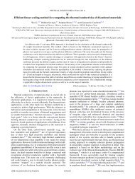
Nanotechnology 23 (2012) 035601 M Dimitrakopoulou et al<br />
Figure 1. (a) TEM overview <strong>of</strong> a high yield NF sample (produced at 1000 mbar, Fe content: 2 wt%, flow 0.5 slmp). (b) High yield binary NP<br />
sample (synthesis pressure 250 mbar, Fe content: 20 wt%, 0.5 slmp). (c) TEM micrograph typifying an NF. (d) Typical micrograph <strong>of</strong> (Fe)<br />
catalyst particle at the tip <strong>of</strong> an NF. The core–shell structure is clearly visible. (e) Representative TEM <strong>of</strong> a binary NP. The different contrast<br />
highlighting the two phases is clearly visible. (f) Magnification <strong>of</strong> a box in (e). Insets show the FFT <strong>for</strong> each side <strong>of</strong> the binary NP. All<br />
samples were synthesized at 1150 ◦ C.<br />
nanoparticle <strong>for</strong>mation. The FeSi/Si nanoparticles may also<br />
have <strong>use</strong>ful properties. β-FeSi2 is promising as a near-infrared<br />
detector or emitter with a b<strong>and</strong>gap <strong>of</strong> 0.83–0.87 eV [28]. In<br />
addition, it has a strong absorption coefficient [29] <strong>and</strong>ahigh<br />
Seebeck coefficient [30].<br />
2. Experimental methods<br />
The amorphous silicon dioxide nan<strong>of</strong>ibers <strong>and</strong> binary<br />
nanoparticles were synthesized by a laser ablation system. An<br />
Nd:YAG laser (Model SL 803, Spectron Laser Systems, with<br />
an output <strong>of</strong> 1400 mJ <strong>and</strong> repletion rate <strong>of</strong> 10 Hz) was <strong>use</strong>d<br />
to drive the ablation process. The laser spot size on the target<br />
was 6 mm. The reactor consisted <strong>of</strong> a 45 mm diameter quartz<br />
tube placed in a horizontal tube furnace (Linn High Therm<br />
2<br />
FRH-40/500/1100). At one end <strong>of</strong> the tube a gas feed system<br />
<strong>and</strong> laser transmission window was mounted while at the other<br />
(rear) end a gas exit system <strong>and</strong> Cu water-cooled cold finger<br />
(to collect the product) was mounted. A Mo target holder is<br />
then placed on the end <strong>of</strong> the cold finger so that the target sits<br />
at the center <strong>of</strong> the oven. The targets were prepared by mixing<br />
powdered catalyst metals (Fe, Ni, Co <strong>and</strong> Mo) with powdered<br />
Si <strong>and</strong> then pressing them in a 20 mm dye. The thickness <strong>of</strong><br />
the target after pressing was approx. 5 mm. After production<br />
<strong>of</strong> the target pellets they were annealed <strong>for</strong> 60 min at 1250 ◦ C<br />
in N2 at atmospheric pressure to help sinter the pellet <strong>and</strong> thus<br />
<strong>for</strong>m a stable target. The purities <strong>of</strong> the precursor materials was<br />
>99.9%. Once the targets were ready they were loaded into the<br />
reactor. The system was first thoroughly flushed with argon <strong>for</strong><br />
at least 20 min with a flow <strong>of</strong> 1 slpm. The laser evaporation
Nanotechnology 23 (2012) 035601 M Dimitrakopoulou et al<br />
Figure 2. (a) TEM overview <strong>of</strong> a high yield NF sample <strong>and</strong> (b) complementary EDX analysis. (c) High angle annular dark-field (HAADF)<br />
image <strong>of</strong> an NF <strong>and</strong> catalyst particle residing at its tip. The line indicates the local EDX scan presented in panel (d).<br />
reaction was run <strong>for</strong> 10 min. The temperature, gas pressure<br />
<strong>and</strong> flow were varied according to the experiment in progress.<br />
The structure <strong>and</strong> morphology <strong>of</strong> the as-grown nanostructures<br />
were examined using an FEI Tecnai F30 transmission<br />
electron microscopy (TEM) operating at 300 kV equipped<br />
with an electron energy dispersive x-ray analyzer (EDX). TEM<br />
samples were prepared by either directly pressing a small<br />
amount <strong>of</strong> sample onto a st<strong>and</strong>ard Cu grid with Al foil or<br />
by dispersing material in ethanol <strong>and</strong> dropping a little <strong>of</strong> the<br />
solution on a Cu TEM grid.<br />
The IR spectroscopic studies were per<strong>for</strong>med using a<br />
Bruker IFS 113V/88 spectrometer. In this case the samples<br />
were dispersed on KBr crystals. Complimentary Raman<br />
spectroscopic investigation made using a Thermo-Fisher DXR<br />
Raman spectrometer (excitation lasers: 532/633/780 nm). For<br />
the Raman investigations a small quantity <strong>of</strong> material was<br />
smeared on Al foil.<br />
3. Results <strong>and</strong> discussion<br />
The employed laser evaporation route generally leads to a<br />
product consisting <strong>of</strong> amorphous silica nan<strong>of</strong>ibers <strong>and</strong> binary<br />
(metal–silicide/Si) nanocrystals. We primarily focus on<br />
the data obtained with Fe as a catalyst. However, brief<br />
comparative studies with Ni, Co <strong>and</strong> Mo are also presented.<br />
By varying the synthesis parameters (gas pressure, gas flow<br />
rate <strong>and</strong> catalyst content) one can tailor the system to yield<br />
high yield amorphous NF samples (e.g. figure 1(a)) or high<br />
yield binary nanocrystal samples (e.g. figure 1(b)). The<br />
temperature throughout these studies remained at 1150 ◦ C.<br />
Closer examinations <strong>of</strong> the nanostructures clearly show the<br />
nan<strong>of</strong>ibers are amorphous as can be seen in figure 1(c).<br />
3<br />
Moreover at one end <strong>of</strong> the amorphous NF a core–shell catalyst<br />
particle resides <strong>and</strong> its diameter closely matches that <strong>of</strong> the<br />
NF. A typical example is shown in figure 1(d). Lattice<br />
in<strong>for</strong>mation derived from the fast Fourier trans<strong>for</strong>m (FFT)<br />
from micrographs <strong>and</strong> direct lattice fringe measurement <strong>of</strong> the<br />
catalyst suggest they are mostly <strong>of</strong> a pure Fe phase in the core<br />
with an iron oxide outer layer. In the case <strong>of</strong> the crystalline<br />
nanoparticles two phases are observed as shown by the clear<br />
difference in contrast in the two halves <strong>of</strong> the nanoparticle<br />
shown in figure 1(e). Closer examinations <strong>of</strong> these binary NPs<br />
show one side <strong>of</strong> the nanoparticle comprises a pure Si phase<br />
<strong>and</strong> the other an iron silicide phase. A typical example is<br />
presented in figure 1(f) in which the different crystalline phases<br />
are visibly obvious. FFT studies <strong>for</strong> each half identified lattice<br />
planes with spacings <strong>of</strong> 0.276 <strong>and</strong> 0.31 nm. These match<br />
well with (022) <strong>and</strong> (111) planes <strong>of</strong> β-FeSi2 <strong>and</strong> crystalline<br />
Si, respectively. The mean diameter <strong>of</strong> the produced NFs,<br />
depending on the growth conditions, ranges from 10 to 19 nm,<br />
while their length is <strong>of</strong> the order <strong>of</strong> μm. The mean diameter <strong>of</strong><br />
thebinaryNPsvaryfrom25to60nm.<br />
The purity <strong>of</strong> the samples was evaluated by EDX. The<br />
high yield binary NP samples showed primarily Fe <strong>and</strong> Si<br />
with traces <strong>of</strong> O present; the Cu signal arises from the grid<br />
<strong>and</strong> the Al from the Al foil <strong>use</strong>d in the transfer process<br />
(see supplementary in<strong>for</strong>mation figure S1 available at stacks.<br />
iop.org/Nano/23/035601/mmedia). However, in the case <strong>of</strong><br />
samples where NFs are present significant oxygen levels are<br />
detected (see figures 2(a) <strong>and</strong> (b)). In order to investigate<br />
this aspect in greater detail local EDX measurements in<br />
scanning transmission electron microscopy (STEM) mode<br />
were conducted. The studies showed the amorphous nan<strong>of</strong>ibers<br />
consist <strong>of</strong> Si <strong>and</strong> O with Si:O atomic ratios close to 1:2,
Nanotechnology 23 (2012) 035601 M Dimitrakopoulou et al<br />
Figure 3. (a) <strong>and</strong> (b) TEM overview <strong>of</strong> Co-catalyzed NFs <strong>and</strong> catalyst particle at NF tip, respectively. (c) <strong>and</strong> (d) TEM overview <strong>of</strong><br />
Ni-catalyzed NFs <strong>and</strong> catalyst particle at NF tip, respectively. (e) <strong>and</strong> (f) Overview <strong>and</strong> close-up image <strong>of</strong> binary NPs <strong>for</strong>med from Mo<br />
catalyst. All the samples were produced at 1150 ◦ C, with a pressure <strong>of</strong> 1000 mbar <strong>and</strong> an Ar flow <strong>of</strong> 0.5 slpm Ar flow rate.<br />
suggesting the fibers are amorphous SiO2. With regards to the<br />
catalyst at the ends <strong>of</strong> the NFs, the local EDX measurements<br />
revealed that they consist primarily <strong>of</strong> the catalyst (Fe) <strong>and</strong> a<br />
little O. The oxygen probably arises from the surface oxidation<br />
<strong>of</strong> the catalyst particle upon exposure to air. This can explain<br />
the core–shell structure typically observed on catalyst particles<br />
at the ends <strong>of</strong> the NFs (e.g. figure 1(d)). The silicon levels are<br />
at the noise level indicating the Si content is minuscule or nonexistent.<br />
Figures 2(c) <strong>and</strong> (d) show an archetypal example <strong>of</strong><br />
the local studies showing the above-discussed features.<br />
In addition to exploring Fe as a catalyst, we also<br />
investigated Ni, Co <strong>and</strong> Mo catalysts. These catalysts are able<br />
to <strong>for</strong>m one or more silicide phases with eutectic temperatures<br />
above 800 ◦ C. These comparative catalyst studies were<br />
conducted at 1000 mbar with an Ar flow <strong>of</strong> 0.5 slpm. The<br />
catalyst content was 2 wt%. These conditions were chosen<br />
as they correspond to the optimum conditions <strong>for</strong> high yield<br />
NF production with Fe catalysts (discussed later). TEM<br />
4<br />
analysis showed that the produced NFs catalyzed either by Co<br />
or Ni were also amorphous with a polycrystalline core–shell<br />
catalyst particle at their ends (figures 3(a)–(d)). However, the<br />
Mo-catalyzed samples resulted in the <strong>for</strong>mation <strong>of</strong> crystalline<br />
binary NPs (figures 3(e)–(f)).<br />
Complimentary local STEM-EDX studies (e.g. figure S2<br />
supplementary in<strong>for</strong>mation available at stacks.iop.org/Nano/<br />
23/035601/mmedia) were conducted. In the case <strong>of</strong> Ni <strong>and</strong><br />
Co the NFs are found to consist <strong>of</strong> Si <strong>and</strong> O as found with Fe<br />
catalysts. The NFs are produced in high yield, slightly higher<br />
than <strong>for</strong> Fe (>80%). Here we refer to yield as the ratio <strong>of</strong> NFs<br />
to free NPs.<br />
We now turn to detailed vibrational spectroscopic <strong>and</strong><br />
systematic statistical studies <strong>of</strong> the amorphous nan<strong>of</strong>ibers <strong>and</strong><br />
binary nanoparticles <strong>for</strong>med from the Fe catalyst system. We<br />
begin with the IR <strong>and</strong> Raman spectroscopy investigations.<br />
The infrared spectrum can be <strong>use</strong>d as a fingerprint<br />
<strong>for</strong> identification by the comparison <strong>of</strong> the spectrum <strong>of</strong> an
Nanotechnology 23 (2012) 035601 M Dimitrakopoulou et al<br />
Figure 4. Normalized <strong>and</strong> strapped IR absorption spectra <strong>for</strong><br />
reference SiO2 powder, high yield NFs, reference Si powder <strong>and</strong> high<br />
yield binary NPs (from bottom to top).<br />
unknown sample with that <strong>of</strong> a reference spectrum. Here we<br />
compare our high yield binary NP <strong>and</strong> NF samples with pure<br />
(powdered) Si <strong>and</strong> SiO2 samples. The data are presented in<br />
figure 4. The spectra have been normalized <strong>and</strong> the background<br />
strapped. The top two spectra in figure 4 correspond to the high<br />
yield binary NP sample at the top <strong>and</strong> the Si reference below.<br />
The similarities between both are obvious <strong>and</strong> confirm the<br />
TEM investigations showing part <strong>of</strong> the binary NP to comprise<br />
crystalline Si. The spectra <strong>for</strong> these two samples show three<br />
major absorption b<strong>and</strong>s in the range <strong>of</strong> 400–500, 770–810 <strong>and</strong><br />
1000–1306 cm −1 . These three absorption peaks are ascribed<br />
to the Si–O–Si rocking vibrational, the Si–O–Si symmetrical<br />
stretching vibrational mode (SS) <strong>and</strong> the Si–O asymmetric<br />
stretching vibrational mode (AS), respectively. In this latter AS<br />
mode two longitudinal optical (LO)—transverse optical (TO)<br />
pairs are infrared-active. This splitting is dependent on heat<br />
treatment [31, 32]. Moreover, they are dependent on the oxide<br />
layer thickness [33, 34]. The sensitivity <strong>of</strong> the LO–TO pairs<br />
to the factors can explain the observed differences between the<br />
high yield (optimum) binary NP sample <strong>and</strong> the reference Si<br />
sample. The oxide modes <strong>for</strong> both these samples arise from<br />
surface oxide states. In this spectral region no Si crystal peaks<br />
are observed beca<strong>use</strong> the large absorption coefficient <strong>of</strong> bulk<br />
SiO2, even <strong>for</strong> very thin layers, masks the Si contribution [35].<br />
The bottom two spectra in figure 4 correspond to the high yield<br />
(optimum) amorphous silica NFs <strong>and</strong> a reference silica sample<br />
(bottom). Both spectra are remarkably similar <strong>and</strong> confirm the<br />
amorphous nan<strong>of</strong>ibers are indeed <strong>for</strong>med from silica. Since<br />
the silica samples are now much thicker, as expected the Si–O<br />
(AS) mode is much stronger relative to the other modes.<br />
Raman spectroscopy is a powerful <strong>and</strong> reliable tool <strong>for</strong> the<br />
characterization <strong>of</strong> Si species <strong>and</strong> can nicely compliment IR<br />
spectroscopy. In contrast to the IR spectra, here the dominant<br />
response arises from Si modes. Around 520 cm −1 a strong<br />
silicon peak is observed <strong>for</strong> all samples. For discussion we<br />
present the Raman spectra <strong>for</strong> the high yield NF <strong>and</strong> binary<br />
NP samples <strong>and</strong> compare them to bulk Si as shown in figure 5.<br />
Both the Si peaks (518 cm −1 ) <strong>for</strong> the NP <strong>and</strong> NF samples are<br />
5<br />
Figure 5. Raman spectra <strong>for</strong> reference bulk Si, high yield binary NPs<br />
<strong>and</strong> high yield NFs (from bottom to top). Inset: magnification <strong>of</strong> the<br />
Si peaks to illustrate the redshift <strong>for</strong> the binary NPs <strong>and</strong> NFs relative<br />
to bulk Si.<br />
asymmetrical, broadened <strong>and</strong> redshifted (by approx. 3 cm −1 ).<br />
This we attribute to size effects. In addition, the tail <strong>of</strong> these<br />
peaks show a weak <strong>and</strong> broad peak around 300 cm −1 which<br />
is not present in the signal from the bulk Si sample. This<br />
additional peak we attribute to a mixture <strong>of</strong> the tail from the<br />
asymmetrical Si peak (size effects) <strong>and</strong> a contribution from<br />
amorphous SiO2 [16].<br />
We now turn to the systematic studies on the effect <strong>of</strong> the<br />
synthesis parameters on the binary NP <strong>and</strong> NF yield, <strong>and</strong> on<br />
their mean diameters. The explored parameters were: catalyst<br />
content, reactor pressure <strong>and</strong> Ar gas flow rate. The temperature<br />
was maintained at 1150 ◦ C. The evaluation <strong>of</strong> yield <strong>and</strong> mean<br />
diameter was determined from detailed statistical analysis from<br />
comprehensive TEM studies. A minimum <strong>of</strong> 100 particles or<br />
fibers were studied <strong>for</strong> each dataset. Figures 6(a) <strong>and</strong> (b) show<br />
3D plots <strong>of</strong> the mean diameter variation with (Fe) catalyst<br />
content <strong>and</strong> buffer gas pressure <strong>for</strong> the silica nan<strong>of</strong>ibers <strong>and</strong><br />
binary nanoparticles, respectively. The gas flow rate <strong>for</strong><br />
these studies was 0.5 slpm. Comparing the two graphs, the<br />
most obvious differences are that the NPs are always larger<br />
than the NF diameters. In addition, binary NPs are always<br />
obtained regardless <strong>of</strong> the reaction conditions, whereas with<br />
low pressures (250 mbar) almost no or no NFs are obtained<br />
independent <strong>of</strong> the catalyst content. At pressures below<br />
1000 mbar, very few NFs are obtained with high catalyst<br />
contents. As a general behavior, both datasets show increasing<br />
diameters with increasing gas pressure <strong>and</strong> catalyst content.<br />
The maximum mean diameter <strong>of</strong> the NFs was 19 nm <strong>and</strong> 58 nm<br />
<strong>for</strong> the binary NPs.<br />
A reduction in their mean diameter is observed with<br />
increasing Ar flow rate. Figure 6(c) shows this trend when
Nanotechnology 23 (2012) 035601 M Dimitrakopoulou et al<br />
Figure 6. (a) <strong>and</strong> (b) Mean diameter dependence <strong>for</strong> NFs <strong>and</strong> binary NPs with respect to Fe content <strong>and</strong> pressure at a temperature <strong>of</strong> 1150 ◦ C<br />
<strong>and</strong> an Ar flow rate <strong>of</strong> 0.5 slpm. (c) Mean diameter dependence <strong>for</strong> NFs <strong>and</strong> binary NPs with respect to Ar flow rate (temperature = 1150 ◦ C,<br />
catalyst content 2 wt%).<br />
Figure 7. (a) Relative yield <strong>of</strong> amorphous NFs to binary NPs with respect to catalyst content <strong>and</strong> reaction pressure (temperature = 1150 ◦ C,<br />
Ar flow = 0.5 slpm). (b) Relative yield with respect to Ar flow rate (temperature = 1150 ◦ C, pressure = 1000 mbar).<br />
using a catalyst content <strong>of</strong> 2 wt% <strong>and</strong> an Ar pressure <strong>of</strong><br />
1000 mbar. It is also easy to see from figure 6 the NPs are<br />
always larger than the NFs. One can also tailor the mean<br />
diameter <strong>of</strong> the NFs <strong>and</strong> binary NPs with temperature, to some<br />
degree (data not shown).<br />
The relative yield <strong>of</strong> NFs to binary NPs against (Fe)<br />
catalyst content <strong>and</strong> pressure is plotted in figure 7(a). The<br />
3D graph clearly highlights the best silica nan<strong>of</strong>iber yields<br />
are obtained with lower catalyst content (ideally 2 wt%) <strong>and</strong><br />
higher reaction pressures (ideally 1000 mbar). Figure 7(b)<br />
shows the relative (NF/NP) yield with flow rate. The behavior<br />
is non-monotonic <strong>and</strong> shows an optimum flow occurs around<br />
6<br />
0.5 slpm. Temperature studies (not shown) show a decrease in<br />
yield as the temperature falls below 1000 ◦ C.<br />
The <strong>for</strong>mation <strong>of</strong> the silica NFs <strong>and</strong> binary nanoparticles<br />
can be expected to <strong>for</strong>m through the vapor–liquid–solid (VLS)<br />
mechanism first introduced by Wagner <strong>and</strong> Ellis to explain Si<br />
nanowire <strong>for</strong>mation [36]. This is beca<strong>use</strong> a catalyst particle<br />
resides at an end <strong>of</strong> each fiber. Moreover the VLS mechanism<br />
is well accepted as the growth mechanism <strong>for</strong> Si nanowire<br />
<strong>for</strong>mation using laser evaporation, similar to that <strong>use</strong>d here.<br />
In the VLS mechanism when the laser pulse strikes the target<br />
(Si plus catalyst), surface material is evaporated <strong>and</strong> ejected<br />
into the reactor in the <strong>for</strong>m <strong>of</strong> a plume. As the plume exp<strong>and</strong>s
Nanotechnology 23 (2012) 035601 M Dimitrakopoulou et al<br />
<strong>and</strong> cools the material condenses, <strong>for</strong>ming catalyst particles<br />
supersaturated with Si. As the system cools further Si begins<br />
to precipitate <strong>and</strong> a nanowire or nan<strong>of</strong>iber is extruded from the<br />
particle. It is obvious from this that the size <strong>of</strong> the catalyst<br />
particle has a direct influence on the nanowire or fiber diameter.<br />
It is not so apparent what driving mechanism differentiates<br />
the <strong>for</strong>mation <strong>of</strong> a crystalline Si nanowire <strong>and</strong> an amorphous<br />
Si fiber. In our case we <strong>for</strong>m amorphous Si fibers, even<br />
though our synthesis conditions seemingly closely match those<br />
from others where Si nanowires have been obtained [37, 38].<br />
Aharonovich et al also obtained amorphous silica NFs via laser<br />
evaporation: however, they were unable to determine why they<br />
were amorphous <strong>and</strong> where the oxygen came from [39]. They<br />
were not sure if the amorphous silica <strong>for</strong>mation occurred in<br />
the reactor or outside once the fibers were exposed to air.<br />
In our case we have the simultaneous <strong>for</strong>mation <strong>of</strong> binary<br />
nanoparticles in which one part is crystalline iron silicide <strong>and</strong><br />
the other is crystalline Si. This tells us the oxygen is not in the<br />
reaction, as we would expect. This means the laser evaporation<br />
reaction <strong>for</strong>ms Si NFs which then oxidize upon exposure to<br />
the air. This is concomitant with the surface oxidation <strong>of</strong> the<br />
catalyst particles residing at their tips <strong>for</strong>ming a core–shell<br />
structure.<br />
4. Conclusion<br />
The products from pulsed-laser evaporation <strong>of</strong> Si targets loaded<br />
with transition metals are investigated in detail. The reactions<br />
yield products containing amorphous silica nan<strong>of</strong>ibers <strong>and</strong><br />
binary crystalline iron silicide/silicon nanoparticles. By<br />
adjusting the reaction parameters one can tailor the product<br />
<strong>for</strong> either high yield silica NFs or high yield binary NPs. The<br />
studies show the oxidation <strong>of</strong> the fibers occurs upon exposure<br />
to the atmosphere.<br />
Acknowledgments<br />
MD acknowledges the Deutscher Akademischer Austausch<br />
Dienst (DAAD), SG thanks the ‘Pakt für Forschung und<br />
Innovation’ <strong>and</strong> MR thanks the European Union (ECEMP) <strong>and</strong><br />
the Freistaat Sachsen <strong>for</strong> financial support. We are grateful to<br />
Alicja Bachmatiuk, R Schönfelder, M Ulbrich, S Leger <strong>and</strong><br />
F Herold <strong>for</strong> technical assistance <strong>and</strong> help.<br />
References<br />
[1] Jie J, Zhang W, Bello I, Lee C-S <strong>and</strong> Lee S T 2010<br />
One-dimensional II–VI nanostructures: synthesis, properties<br />
<strong>and</strong> optoelectronic applications Nano Today 5 313–36<br />
[2] Wang X, Ding B, Yu J <strong>and</strong> Wang M 2011 Engineering<br />
biomimetic superhydrophobic surfaces <strong>of</strong> electrospun<br />
Nanomaterials Nano Today 6 510–30<br />
[3] Barth S, Hern<strong>and</strong>ez-Ramirez F, Holmes J D <strong>and</strong><br />
Romano-Rodriguez A 2010 Synthesis <strong>and</strong> applications <strong>of</strong><br />
one-dimensional superconductors Prog. Mater. Sci.<br />
55 563–627<br />
[4] Ding B, Wang M, Wang X, Yu J <strong>and</strong> Sun G 2010 Mater. Today<br />
13 16–27<br />
7<br />
[5] Morales A M <strong>and</strong> Lieber C M 1998 A laser ablation method <strong>for</strong><br />
the synthesis <strong>of</strong> crystalline semiconductor nanowires <strong>Science</strong><br />
279 208–11<br />
[6] Wang N, Tang Y H, Zhang Y F, Lee C S, Bello I <strong>and</strong> Lee S T<br />
1999 Si nanowires grown from silicon oxide Chem. Phys.<br />
Lett. 299 237–42<br />
[7] Schubert L, Werner P, Zakharov N D, Gerth G, Kolb F M,<br />
Long L <strong>and</strong> Gösele U 2004 Silicon nanowhiskers grown on<br />
〈111〉 Si substrates by molecular-beam epitaxy Appl. Phys.<br />
Lett. 84 4968–70<br />
[8] Holmes J D, Johnston K P, Doty R C <strong>and</strong> Korgel B A 2000<br />
Control <strong>of</strong> thickness <strong>and</strong> orientation <strong>of</strong> solution-grown<br />
silicon nanowires <strong>Science</strong> 287 1471–3<br />
[9] Chen S, Bomer J G, van der Wiel W G, Carlen E T <strong>and</strong><br />
van den Berg A 2009 Top-down fabrication <strong>of</strong> sub-30 nm<br />
monocrystalline silicon nanowires using conventional<br />
micr<strong>of</strong>abrication ACS Nano 3 3485–92<br />
[10] Cheng Y T, Cho Y H, Takama N, Löw P, Bergaud C <strong>and</strong><br />
Kim B J 2009 Simple fabrication <strong>of</strong> Si nanowire <strong>and</strong> its<br />
biological application J. Phys. 152 012048<br />
[11] Wu Y <strong>and</strong> Yang P 2000 Germanium nanowire growth via<br />
simple vapor transport Chem. Mater. 12 605–7<br />
[12] Zhang Y J, Zhang Q, Wang N L, Yan Y J, Zhou H H <strong>and</strong><br />
Zhu J 2001 Synthesis <strong>of</strong> thin Si whiskers (nanowires) using<br />
SiCl4 J. Cryst. Growth 226 185–91<br />
[13] Chen C C, Yeh C C, Chen C H, Yu M Y <strong>and</strong> Liu H L 2001<br />
Catalytic growth <strong>and</strong> characterization <strong>of</strong> gallium nitride<br />
nanowires J. Am. Chem. Soc. 123 2791–8<br />
[14] Shi W S, Zheng Y F, Wang N, Lee C S <strong>and</strong> Lee S T 2001<br />
Synthesis <strong>and</strong> microstructure <strong>of</strong> gallium phosphide<br />
nanowires J. Vac. Sci. Technol. B 19 1115<br />
[15] Shimada T, Hiruma K, Shirai M, Yazawa M <strong>and</strong><br />
Haraguchi K 1998 Size, position <strong>and</strong> direction control on<br />
GaAs <strong>and</strong> InAs nanowhisker growth Superlattice<br />
Microstruct. 24 453–8<br />
[16] Yu D P, Houg Q L, Ding Y, Zhang H Z, Bai Z G, Wang J J,<br />
Zou Y H, Qian W, Xiong G C <strong>and</strong> Feng S Q 1998<br />
Amorphous silica nanowires: intensive blue light emitters<br />
Appl. Phys. Lett. 73 3076<br />
[17] Brambilla G <strong>and</strong> Payne D N 2009 The ultimate strength <strong>of</strong><br />
glass silica nanowires Nano Lett. 9 831–5<br />
[18] Tricoli A, Righettoni M <strong>and</strong> Pratsinis S E 2009 Anti-fogging<br />
nan<strong>of</strong>ibrous SiO2 <strong>and</strong> nanostructured SiO2–TiO2 films made<br />
by rapid flame deposition <strong>and</strong> in situ annealing Langmuir<br />
25 12578–84<br />
[19] Li Z, Zhu S G, Gan K, Zhang Q H, Zeng Z Y, Zhou Y H,<br />
Liu H Y, Xiong W, Li X L <strong>and</strong> Li G Y 2005<br />
Poly-L-lysine-modified silica nanoparticles: a potential oral<br />
gene delivery system J. Nanosci. Nanotechnol. 5 1199–203<br />
[20] Xiao Z, Zhang L, Meng G, Tian X, Zeng H <strong>and</strong> Fang M 2006<br />
High-density, aligned SiO2 nanowire arrays: microscopic<br />
imaging <strong>of</strong> the unique growth style <strong>and</strong> their ultraviolet light<br />
emission properties J. Phys. Chem. B 110 15724–8<br />
[21] Wang C Y, Chan L H, Xiao D Q, Lin T C <strong>and</strong> Shiha H C 2006<br />
Mechanism <strong>of</strong> solid–liquid–solid on the silicon oxide<br />
nanowire growth J. Vac. Sci. Technol. B 24 613–7<br />
[22] Tricoli A, Righettoni M, Krumeich F, Stark W J <strong>and</strong><br />
Pratsinis S E 2010 Scalable flame synthesis <strong>of</strong> SiO2<br />
nanowires: dynamics <strong>of</strong> growth Nanotechnology 21 465604<br />
[23] Lin L W, Tang Y W, Li X X, Pei L Z, Zhang Y <strong>and</strong><br />
Guo C 2007 Water-assisted synthesis <strong>of</strong> silicon oxide<br />
nanowires under supercritically hydrothermal conditions<br />
J. Appl. Phys. 101 014314<br />
[24] Li F J, Zhang S, Kong J H <strong>and</strong> Zhang W L 2011 Study <strong>of</strong><br />
silicon dioxide nanowires grown via rapid thermal annealing<br />
<strong>of</strong> sputtered amorphous carbon films doped with Si Nanosci.<br />
Nanotechnol. Lett. 3 240–5<br />
[25] Ni Z F, Ying P Z, Luo Y <strong>and</strong> Li Z S 2007 Preparation <strong>and</strong><br />
characterization <strong>of</strong> amorphous silicon oxide nanowires<br />
J. China Univ. Min. Technol. 17 0587–9
Nanotechnology 23 (2012) 035601 M Dimitrakopoulou et al<br />
[26] Liang C H, Zhang L D, Meng G W, Wang Y W <strong>and</strong> Chu Z Q<br />
2000 Preparation <strong>and</strong> characterization <strong>of</strong> amorphous SiOx<br />
nanowires J. Non-Cryst. Solids 277 63–7<br />
[27] Sood D K, Sekhar P K <strong>and</strong> Bhansali S 2006 Ion implantation<br />
based selective synthesis <strong>of</strong> SiO2 nanowires on silicon<br />
wafers Appl. Phys. Lett. 88 143110<br />
[28] Clark S J, Al-Allak H M, Br<strong>and</strong> S <strong>and</strong> Abram R A 1998<br />
Structure <strong>and</strong> electronic properties <strong>of</strong> FeSi2 Phys. Rev. B<br />
58 10389–93<br />
[29] Lefki K <strong>and</strong> Muret P 1993 Photoelectric study <strong>of</strong> β-FeSi2 on<br />
silicon: optical threshold as a function <strong>of</strong> temperature<br />
J. Appl. Phys. 74 1138<br />
[30] Takeda M, Kuramitsu M <strong>and</strong> Yoshio M 2004 Anisotropic<br />
Seebeck coefficient in β-FeSi2 single crystal Thin Solid<br />
Films 461 179–81<br />
[31] Rümmeli M H et al 2009 Hydrogen activated axial<br />
inter-conversion in SiC nanowires J. Solid State Chem.<br />
182 602–7<br />
[32] Kamitsos E I, Patsis A P <strong>and</strong> Kordas G 1993<br />
Infrared-reflectance spectra <strong>of</strong> heat-treated sol–gel-derived<br />
silica Phys. Rev. B 48 12499–505<br />
8<br />
[33] Chowdhuri R A, Jin D <strong>and</strong> Takoudis C G 2004 SiO2/Si(100)<br />
interface characterization using infrared spectroscopy:<br />
estimation <strong>of</strong> substoichiometry <strong>and</strong> strain Thin Solid Films<br />
457 402–5<br />
[34] Giustino F <strong>and</strong> Pasquarello A 2005 Infrared spectra at surfaces<br />
<strong>and</strong> interfaces from first principles: evolution <strong>of</strong> the spectra<br />
across the Si(100)–SiO2 interface Phys. Rev. Lett.<br />
95 187402<br />
[35] Weaver J H, Alex<strong>and</strong>er R W, Teng L, Mann R A <strong>and</strong> Bell R J<br />
1973 Infrared absorption <strong>of</strong> small silicon particles with<br />
oxide overlayers Phys. Status Solidi a 20 321–9<br />
[36] Wagner R S <strong>and</strong> Ellis W C 1964 Vapor–liquid–solid mechanism<br />
<strong>of</strong> single crystal growth Appl. Phys. Lett. 4 89–90<br />
[37] Yang Y H, Wu S J, Chiu H S, Lin P I <strong>and</strong> Chen Y T 2004<br />
Catalytic growth <strong>of</strong> Si nanowires assisted by laser ablation<br />
J. Phys. Chem. 108 846–52<br />
[38] Tang Y H, Zhang Y F, Wang N, Lee C S, Han X D, Bello I <strong>and</strong><br />
Lee S T 1999 Morphology <strong>of</strong> Si nanowires synthesized by<br />
high-temperature laser ablation J. Appl. Phys. 85 7981<br />
[39] Aharonovich I, Tamir S <strong>and</strong> Lifshitz Y 2008 Growth <strong>of</strong> SiOx<br />
nanowires by laser ablation Nanotechnology 19 065608