Fatal pertussis infection - Swiss Society of Neonatology
Fatal pertussis infection - Swiss Society of Neonatology
Fatal pertussis infection - Swiss Society of Neonatology
Create successful ePaper yourself
Turn your PDF publications into a flip-book with our unique Google optimized e-Paper software.
SWISS SOCIETY OF NEONATOLOGY<br />
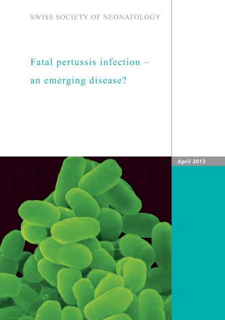
<strong>Fatal</strong> <strong>pertussis</strong> <strong>infection</strong> –<br />
an emerging disease?<br />
April 2013
Schlapbach LJ, Schibler A, Lourie R, Stocker C, Paediatric<br />
Critical Care Research Group (SLJ, SA, SC), Paediatric Intensive<br />
Care Unit, Mater Children’s Hospital, Department <strong>of</strong> Pathology<br />
(LR), Mater Health Services, Brisbane, Australia<br />
© <strong>Swiss</strong> <strong>Society</strong> <strong>of</strong> <strong>Neonatology</strong>, Thomas M Berger, Webmaster<br />
2
3<br />
Before the widespread implementation <strong>of</strong> vaccination<br />
strategies, <strong>pertussis</strong> caused large epidemics and was<br />
responsible for an estimated 10‘000 annual deaths in<br />
the United States (1). As a result <strong>of</strong> successful vaccina-<br />
tion programs, the public awareness <strong>of</strong> this disease in<br />
most developed countries at present is low, and most<br />
neonatologists will not get confronted with severe per-<br />
tussis <strong>infection</strong>s during their training. However, over<br />
the past decade, several countries including Australia<br />
have reported outbreaks <strong>of</strong> <strong>pertussis</strong> <strong>infection</strong>s, and<br />
there is concern about an increasing number <strong>of</strong> poten-<br />
tially preventable deaths, particularly in neonates and<br />
young infants (2). In Switzerland, approximately 50 in-<br />
fants with confirmed <strong>pertussis</strong> are hospitalized each<br />
year (3). We present the case <strong>of</strong> an infant with severe<br />
<strong>pertussis</strong> <strong>infection</strong> requiring extracorporeal membrane<br />
oxygenation (ECMO).<br />
INTRODUCTION
CASE REPORT<br />
A four-week-old boy was brought to the emergency<br />
department after his parents had observed a cyanotic<br />
episode at home that had responded to stimulation.<br />
The baby had been born through spontaneous vagi-<br />
nal delivery at 39 2/7 weeks, and the early neonatal<br />
period had been uneventful. The child and his family,<br />
including a two year old, fully vaccinated sibling, had<br />
all had cough and coryza for the past days.<br />
On arrival to the emergency department, the patient<br />
was saturating 100% in 2 L/min <strong>of</strong> oxygen, but sho-<br />
wed signs <strong>of</strong> respiratory distress with a respiratory<br />
rate <strong>of</strong> 66/min and moderate subcostal and tracheal<br />
recessions. The heart rate was 216/min with a capilla-<br />
ry refill time <strong>of</strong> 3 to 4 seconds. Pulses were palpable<br />
on all four limbs, the liver was <strong>of</strong> normal size, and no<br />
cardiac murmur was audible. The fontanel was s<strong>of</strong>t,<br />
but the child was irritable. Suspecting sepsis, a fluid<br />
bolus was given and intravenous antibiotic treatment<br />
with cefotaxime and gentamycin was started. A ve-<br />
nous blood gas showed a mixed acidosis with a pH<br />
<strong>of</strong> 7.19, pCO2 7.5 kPa (56 mmHg), a base excess <strong>of</strong><br />
-8.1 mmol/l and a lactate <strong>of</strong> 5.7 mmol/l. The full blood<br />
count revealed a severe leukocytosis with a white<br />
blood cell count (WCC) <strong>of</strong> 94.4 x10 9 /L, 44.3 x10 9 /L<br />
neutrophils, and 36.3 x10 9 /L lymphocytes. The hemo-<br />
globin level was 108 g/L, and platelets count was 758<br />
x10 9 /L. Suspecting <strong>pertussis</strong>, intravenous treatment<br />
with azithromycin was added.<br />
4
5<br />
Over the next two hours, the condition <strong>of</strong> the baby<br />
deteriorated despite high-flow nasal cannula oxygen<br />
support with persistent grunting and frequent desa-<br />
turations. A repeat venous blood gas now showed a<br />
severe respiratory acidosis with a pH <strong>of</strong> 7.03 and a<br />
pCO2 <strong>of</strong> 14.1 kPa (106 mmHg). The infant was there-<br />
fore intubated and retrieved to our tertiary pediatric<br />
intensive care unit (PICU). On arrival to the PICU, the<br />
chest X-ray showed bilateral consolidations with areas<br />
<strong>of</strong> hyperinflation (Fig. 1). Echocardiography showed<br />
a morphologically normal heart with normal func-<br />
tion but increased pulmonary pressures. Due to high<br />
peak ventilatory pressures and persistent hypercarbia,<br />
high-frequency oscillatory ventilation (Sensormedics<br />
3100A) was initiated, and a milrinone infusion was<br />
started. Inhaled nitric oxide was added at 20 ppm.<br />
Blood cultures were negative, but the polymerase<br />
chain reaction (PCR) in the nasopharyngeal aspirate<br />
tested positive for Bordatella <strong>pertussis</strong>. The WCC con-<br />
firmed hyperleukocytosis <strong>of</strong> 111 x10 9 /L. Given the cri-<br />
tical conditions <strong>of</strong> the infant with acute cardiorespira-<br />
tory failure, it was decided to perform a double blood<br />
volume exchange transfusion. Based on the protocol<br />
published by the Great Ormond Street Hospital (GOSH)<br />
(4), 200 ml/kg <strong>of</strong> blood were removed and continously<br />
replaced with packed red blood cells and 4% albumin<br />
over four hours. The procedure was hemodynamically<br />
well tolerated, and the WCC dropped to 24.8 x10 9 /L.
Fig. 1<br />
Chest X-ray on admission to the PICU: bilateral<br />
patchy infiltrates with partial consolidation <strong>of</strong> the<br />
right and left upper lobes alternating with<br />
hyperinflated areas.<br />
6
7<br />
Chest X-ray after cannulation for VA ECMO on HFOV:<br />
the pulmonary radiographic changes noted on admis-<br />
sion have progressed.<br />
Fig. 2
Fig. 3<br />
Chest CT on day 7 <strong>of</strong> ECMO: dense consolidations in<br />
both lungs with partial residual aeration <strong>of</strong> the right<br />
lung.<br />
8
9<br />
Chest X-ray on day 25 <strong>of</strong> ECMO: diffuse interstitial<br />
and alveolar opacifications in both lungs.<br />
Fig. 4
Fig. 5<br />
CT scan <strong>of</strong> the brain on day 25 <strong>of</strong> ECMO: several foci<br />
<strong>of</strong> intracerebral hemorrhages with associated edema;<br />
in addition, cerebral atrophy with widened extra-<br />
cerebral spaces is noted.<br />
10
11<br />
Twelve hours after admission to the PICU, the infant<br />
developed arterial hypotension requiring support with<br />
noradrenaline and dobutamine. Echocardiographic<br />
assessment now revealed suprasystemic pulmonary<br />
hypertension and a dilated failing right ventricle with<br />
bulging <strong>of</strong> the interventricular septum to the left.<br />
The decision to support the child with extracorporeal<br />
membrane oxygenation (ECMO) was made. Venoarterial<br />
(VA) ECMO was initiated through neck cannulation.<br />
While on VA ECMO, the patient was kept on HFOV<br />
(Fig. 2). Echocardiography showed significantly redu-<br />
ced right ventricular dimensions and a decompressed<br />
left atrium and ventricle. Since the WCC decreased to<br />
values between 5 and 20 x10 9 /L no additional leuko-<br />
depletion was performed.<br />
On day seven, given the absence <strong>of</strong> any improvement<br />
in pulmonary status, a thorough bronchoscopy was<br />
performed, and several mucous plugs were removed.<br />
A computed tomography (CT) <strong>of</strong> the head was normal,<br />
while the CT <strong>of</strong> the chest showed extensively conso-<br />
lidated lungs but no signs <strong>of</strong> necrotizing pneumonia<br />
(Fig. 3).<br />
Over the next two weeks, different strategies to recruit<br />
the consolidated lungs were applied, including repea-<br />
ted broncoscopies with DNAse washouts, and surfac-<br />
tant application. Lung function tests showed mini mal<br />
compliance, and chest x-ray showed persistent dif-<br />
fuse opacifications <strong>of</strong> the lungs (Fig. 4). Conversion
12<br />
<strong>of</strong> venoarterial to venovenous ECMO and weaning<br />
<strong>of</strong> ECMO was not possible since reduction <strong>of</strong> ECMO<br />
flows rapidly led to increased pulmonary pressures<br />
with right ventricular dilatation. At 25 days on ECMO,<br />
hyperechogenic changes were noted on cranial ultra-<br />
sound. A subsequent CT <strong>of</strong> the head confirmed the<br />
presence <strong>of</strong> several intracerebral bleedings and cere-<br />
bral atrophy with widened extracerebral spaces (Fig. 5).<br />
As there were no signs <strong>of</strong> pulmonary recovery, and<br />
given the evidence for permanent brain damage, con-<br />
tinuation <strong>of</strong> life support was considered futile by the<br />
team and the parents. The infant died upon cessation<br />
<strong>of</strong> ECMO.<br />
The postmortem MRI confirmed multiple hemorrhagic<br />
foci throughout the cerebral hemishperes and the basal<br />
ganglia. The postmortem pathology showed exten sive<br />
lung injury with totally consolidated lungs. In histopa-<br />
thology, highly inflamed alveoli with a mononuclear<br />
cellular infiltrate <strong>of</strong> the airspaces were found side-to-<br />
side with focally necrotizing pneumonia with fibrous<br />
scarring (Fig. 6). Multiple small arteries with thrombo-<br />
ses were noted (Fig. 7).
13<br />
Postmortem lung histology: acute necrotizing<br />
inflammatory infiltrates in a respiratory bronchiolus;<br />
the infiltrate is neutrophil-rich, and the background<br />
lung architecture is still intact in this area<br />
(HE stain, x20 objective).<br />
Fig. 6
Fig. 7<br />
14<br />
Postmortem lung histology: here, the lung architecture<br />
is mostly lost, and replaced by solid necrotic tissue and<br />
fibrin; a thrombosed vessel (arrow) with early calcifica-<br />
tion can be seen (HE stain, 10x objective).
15<br />
Bordatella <strong>pertussis</strong>, a gram-negative aerobic bacte-<br />
rium, causes airborne respiratory <strong>infection</strong>s in all age<br />
groups, ranging from coryza and persistent cough to<br />
apneas and life-threatening complications including<br />
pulmonary hypertension and acute respiratory distress<br />
syndrome (ARDS)-like presentations. Despite signi-<br />
ficant reduction in mortality over the past decades,<br />
<strong>pertussis</strong> is still ranking among the 10 leading causes<br />
<strong>of</strong> childhood mortality on a global scale with an esti-<br />
mated 195‘000 annual deaths (www.who.int/immuni-<br />
zation_monitoring/diseases/<strong>pertussis</strong>/en/index.html).<br />
Pathogenicity is related to different virulence factors<br />
including adhesins allowing binding to epithelial cells,<br />
cytotoxins which inhibit ciliary clearance, and Pertus-<br />
sis toxin. Pertussis toxin affects cellular signalling and<br />
causes lymphocytosis, which is pathognomonic for the<br />
disease (5). Traditional teaching on <strong>pertussis</strong> in text-<br />
books mainly refers to the paroxysmal coughing spells<br />
which may last up to several minutes and which can<br />
lead to intermittent hypoxemia, apneas, hypertensive<br />
encephalopathy and seizures.<br />
In 1993, Gouldin et al. reported refractory pulmonary<br />
hypertension in fatal <strong>pertussis</strong> (6). In fact, the majority<br />
<strong>of</strong> fatal <strong>pertussis</strong> cases that have been reported in re-<br />
cent series from developed countries were associated<br />
with hyperleukocytosis, severe pulmonary hypertensi-<br />
on and ARDS-like refractory respiratory failure (7, 8).<br />
The extreme leukocytosis may exceed 100 x10 9 /L and<br />
DISCUSSION
16<br />
results in a hyperviscosity syndrome which can lead to<br />
impaired microcirculation and organ failure. In addition<br />
to hyperviscosity, obstruction <strong>of</strong> pulmonary arterioles<br />
by leukocyte thrombi may result in severe pulmonary<br />
hypertension as a prominent feature (5). Furthermore,<br />
<strong>pertussis</strong> toxin can mediate an increase <strong>of</strong> cAMP levels<br />
which contributes to pulmonary vaso constriction. Fi-<br />
nally, aggregation and activation <strong>of</strong> lymphocytes and<br />
neutrophils in the pulmonary vascular bed may aggra-<br />
vate acute lung injury. Postmortem studies have found<br />
cilia loss in airway mucosa, necrotizing bronchiolitis,<br />
and focal or diffuse bronchopneumonia (5). Aggre-<br />
gates <strong>of</strong> leukocytes in pulmonary arterioles, small ar-<br />
teries and venules and occlusive thrombi are frequent<br />
postmortem findings, as observed in the presented<br />
case.<br />
The management <strong>of</strong> severe pulmonary hypertension in<br />
<strong>pertussis</strong> patients is extremely challenging, since clas-<br />
sic approaches at reducing pulmonary vascular resi-<br />
stance such as iNO may fail due to hyperviscosity and<br />
vascular obstruction. The observation <strong>of</strong> a high mor-<br />
tality in infants presenting with hyperleukocytosis has<br />
therefore lead to strategies for leukoreduction (4). The<br />
GOSH protocol recommends leuk<strong>of</strong>iltration in pati-<br />
ents requiring ECMO for cardiorespiratory failure with<br />
WCC above 50 x10 9 /L. In patients that do not require<br />
ECMO, urgent double-volume blood exchange trans-<br />
fusion is advocated if the WCC is above 100 x10 9 /L,<br />
or above 70 x10 9 /L and cardiorespiratory failure or se-
17<br />
vere pulmonary hypertension are present. Comparing<br />
a historical cohort <strong>of</strong> nine infants under 90 days <strong>of</strong><br />
age with severe <strong>pertussis</strong> managed without leukore-<br />
duction versus a recent group <strong>of</strong> ten infants treated<br />
according to this protocol, the group at GOSH obser-<br />
ved improved survival from 55% to 90%. To date, this<br />
small non-randomized study remains the only report<br />
on a significant treatment benefit for severe <strong>pertussis</strong>.<br />
ECMO is a valid treatment option in the setting <strong>of</strong><br />
refractory pulmonary hypertension due to a poten-<br />
tially reversible cause (such as meconium aspiration<br />
syndrome or sepsis), and has therefore been used in<br />
patients with <strong>pertussis</strong>-induced cardiorespiratory fai-<br />
lure (9). The Extracorporeal Life Support Organisati-<br />
on (ELSO) registry recorded 169 <strong>pertussis</strong> cases that<br />
were treated with ECMO between 1992 to 2009 (4).<br />
The prognosis <strong>of</strong> severe <strong>pertussis</strong> requiring ECMO was<br />
worse compared to other indications for respiratory<br />
ECMO (mortality <strong>of</strong> 70% compared to a mortality <strong>of</strong><br />
20% for respiratory syncytial virus bronchiolitis). The<br />
highest case fatality rate (84%) was observed in in-<br />
fants below 6 weeks <strong>of</strong> age. ECMO may be required<br />
for several weeks, which leads to a high risk <strong>of</strong> ECMO-<br />
related life-threatening complications such as sepsis,<br />
major bleeding or thromboembolic events.<br />
Recent serological follow-up data from vaccine cen-<br />
ters suggests that the protection after full immuniza-<br />
tion using the acellular <strong>pertussis</strong> vaccine wanes faster
18<br />
compared to whole-cell <strong>pertussis</strong> vaccines (1). In fact,<br />
adolescents and adults currently represent the main<br />
epidemic reservoir, leading to <strong>infection</strong> <strong>of</strong> non-vacci-<br />
nated neonates and infants (10). Therefore, the <strong>Swiss</strong><br />
Federal Office <strong>of</strong> Health (BAG) recently issued new<br />
recommendations that include a dTpa booster every<br />
ten years for all adolescents and adults with regular<br />
contact with infants less than 6 months <strong>of</strong> age (11).<br />
Obviously, decreasing adherence <strong>of</strong> parents to vacci-<br />
nation programs remains a major concern.<br />
In summary, the present case illustrates that <strong>pertussis</strong><br />
can lead to rapid cardiorespiratory failure and refrac-<br />
tory pulmonary hypertension in neonates and young<br />
infants. Hyperleukocytosis is associated with a high<br />
mortality, and rescue strategies including exchange<br />
transfusion for leukodepletion, or leukapheresis in pa-<br />
tients on ECMO should be considered early.
19<br />
1. Klein NP, Bartlett J, Rowhani-Rahbar A, et al. Waning protection<br />
after fifth dose <strong>of</strong> acellular <strong>pertussis</strong> vaccine in children.<br />
N Engl J Med 2012;367:1012-1019<br />
2. Cherry JD. The present and future control <strong>of</strong> <strong>pertussis</strong>.<br />
Clin Infect Dis 2010;51:663-667<br />
3. Posfay-Barbe KM. Infections in pediatrics: old and new diseases.<br />
<strong>Swiss</strong> Med Wkly 2012;142:w13654<br />
4. Rowlands HE, Goldman AP, Harrington K, et al. Impact <strong>of</strong> rapid<br />
leukodepletion on the outcome <strong>of</strong> severe clinical <strong>pertussis</strong> in<br />
young infants. Pediatrics 2010;126:e816-e827<br />
5. Paddock CD, Sanden GN, Cherry JC, et al. Pathology and patho-<br />
genesis <strong>of</strong> fatal Bordatella <strong>pertussis</strong> <strong>infection</strong> in infants.<br />
Clin Infect Dis 2008;47:328-338<br />
6. Goulin GD, Kaya KM, Bradley JS. Severe pulmonary hyperten-<br />
sion associated with shock and death in infants infected with<br />
Bordetella <strong>pertussis</strong>. Crit Care Med 1993;21:1791-1794<br />
7. Pierce C, Klein N, Peters M. Is leukocytosis a predictor <strong>of</strong><br />
mortality in severe <strong>pertussis</strong> <strong>infection</strong>? Intensive Care Med<br />
2000;26:1512-1514<br />
8. Wortis N, Strebel PM, Wharton M, et al. Pertussis deaths: report<br />
on 23 cases in the United States, 1992 and 1993. Pediatrics<br />
1996;97:607-612<br />
9. Halasa NB, Barr FE, Johnson JE, Edwards KM. <strong>Fatal</strong><br />
pulmonary hypertension associated with <strong>pertussis</strong> in infants:<br />
does extracorporeal membrane oxygenation have a role?<br />
Pediatrics 2003;112:1274-1278<br />
10. Wymann MN, Richard JL, Vidondo B, Heininger U. Prospec-<br />
tive <strong>pertussis</strong> surveillance in Switzerland, 1991-2006. Vaccine<br />
2011;29:2058-2065<br />
REFERENCES
11. Bundesamt für Gesundheit (BAG). Optimierung der<br />
Auffrischimpfungen gegen Diphtherie, Tetanus und Pertussis<br />
(dT/dTpa) bei Erwachsenen. Bull BAG 2011;51:1-11<br />
20
SUPPORTED BY<br />
CONTACT<br />
<strong>Swiss</strong> <strong>Society</strong> <strong>of</strong> <strong>Neonatology</strong><br />
www.neonet.ch<br />
webmaster@neonet.ch<br />
concept & design by mesch.ch


