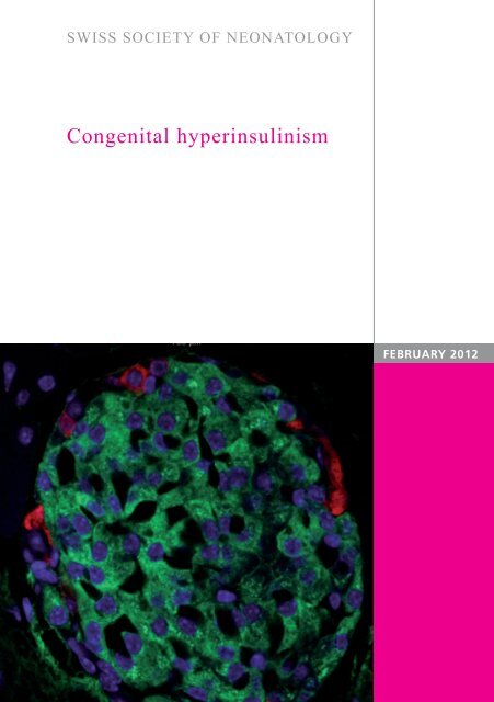
Congenital hyperinsulinism - Swiss Society of Neonatology
Congenital hyperinsulinism - Swiss Society of Neonatology
Congenital hyperinsulinism - Swiss Society of Neonatology

Create successful ePaper yourself
Turn your PDF publications into a flip-book with our unique Google optimized e-Paper software.
SWISS SOCIETY OF NEONATOLOGY<br />
<strong>Congenital</strong> <strong>hyperinsulinism</strong><br />
FEBRUARY 2012
Morgillo D, Berger TM, Caduff JH, Barthlen W, Mohnike K,<br />
Mohnike W, Neonatal and Pediatric Intensive Care Unit<br />
(MD, BTM), Department <strong>of</strong> Pediatric Radiology (CJH),<br />
Children‘s Hospital <strong>of</strong> Lucerne, Lucerne, Switzerland,<br />
Department <strong>of</strong> Pediatric Surgery, University Medicine<br />
Greifswald (BW), Greifswald, Germany, Department <strong>of</strong><br />
Pediatrics, University Hospital <strong>of</strong> Magdeburg (MK),<br />
Magdeburg, Germany, Diagnostic Therapeutic Centre<br />
Frankfurter Tor (MW), Berlin, Germany<br />
© <strong>Swiss</strong> <strong>Society</strong> <strong>of</strong> <strong>Neonatology</strong>, Thomas M Berger, Webmaster<br />
2
3<br />
<strong>Congenital</strong> <strong>hyperinsulinism</strong> (CHI) is characterized by<br />
inappropriate secretion <strong>of</strong> insulin by the ß cells <strong>of</strong> the<br />
islets <strong>of</strong> Langerhans and is an extremely heterogeneous<br />
condition in terms <strong>of</strong> clinical presentation, histological<br />
subgroups and underlying molecular biology.<br />
Histologically, CHI has been classified into two major<br />
subgroups: diffuse (affecting the whole pancreas)<br />
and focal (being localized to a single region <strong>of</strong> the<br />
pancreas) disease. Advances in molecular genetics,<br />
radiological imaging techniques (such as fluorine-18<br />
L-3,4-dihydroxyphenylalanine-PET-CT ( 18FDOPA-PET-CT) scanning) and surgical techniques have completely<br />
changed the clinical approach to infants with severe<br />
congenital forms <strong>of</strong> hyperinsulinemic hypoglycemia.<br />
This male infant was born to a healthy 35-year-old G3/P3<br />
by spontaneous vaginal delivery at 38 4/7 weeks. His<br />
birth weight was 3530 g (P 50-75), his head circumference<br />
was 35 cm (P 25) and his length was 50 cm<br />
(P 25-50). Postnatal adaptation was normal with an<br />
arterial cord pH <strong>of</strong> 7.28 and Apgar scores <strong>of</strong> 8, 9, and<br />
9 at 1, 5, and 10 minutes, respectively. Pregnancy had<br />
been uneventful without any evidence <strong>of</strong> gestational<br />
diabetes.<br />
On the second day <strong>of</strong> life, he was noted to have a<br />
grayish skin color and poor muscle tone. A POCT<br />
INTRODUCTION<br />
CASE REPORT
lood glucose measurement indicated a glucose con-<br />
centration <strong>of</strong> 0.1 mmol/l, which only increased to 0.4<br />
mmol/l after the administration <strong>of</strong> oral glucose solution.<br />
At that point, our neonatal transport team was<br />
called. On arrival, intravenous access was established,<br />
blood cultures were obtained and a bolus <strong>of</strong> 2 ml/kg<br />
<strong>of</strong> a 10% dextrose solution was given, followed by a<br />
continuous glucose infusion at a rate <strong>of</strong> 5 mg/kg/min.<br />
He was started on antibiotics and transferred to our<br />
neonatal intensive care unit.<br />
On transport, there was focal tonic-clonic seizure ac-<br />
tivity involving the right arm. Blood glucose concen-<br />
tration at that time was 3 mmol/l. Phenobarbital was<br />
started and the seizures did not reoccur. Antibiotics<br />
were discontinued after 72 hours. The further hospital<br />
course was remarkable for recurrent hypoglycemic episodes<br />
despite increasing rates <strong>of</strong> enteral and parenteral<br />
glucose administration (up to 18 mg/kg/min). High<br />
insulin concentrations were documented repetitively<br />
during episodes <strong>of</strong> hypoglycemia without concurrent<br />
increase in free fatty acids or ketone bodies. Cortisol<br />
and growth hormone responses, however, were adequate.<br />
Thus, a diagnosis <strong>of</strong> hyperinsulinemic hypoglycemia<br />
was made.<br />
At the age <strong>of</strong> one month, the patient was transferred<br />
to the University Children‘s Hospital <strong>of</strong> Zurich for further<br />
management. The patient did not respond to a<br />
trial with the potassium channel activator diazoxide.<br />
4
5<br />
A<br />
C<br />
Enhanced activity in the head <strong>of</strong> the pancreas<br />
(A, B: frontal view, C: coronal view); in addition,<br />
there is enhancement over the kidneys and bladder.<br />
B<br />
Fig. 1
One week later, he was started on octreotide, initi-<br />
ally by bolus injections but eventually by continuous<br />
subcutaneous infusion. At the age <strong>of</strong> seven weeks, he<br />
was discharged home fully breastfed on octreotide at<br />
a rate <strong>of</strong> 17 mcg/kg/day.<br />
At the age <strong>of</strong> 4 months, MR studies <strong>of</strong> the head and<br />
abdomen were normal. Shortly thereafter, an 18FDOPA- PET-CT scan was obtained. This study revealed increased<br />
focal activity in the region <strong>of</strong> the pancreatic head<br />
(Fig. 1, 2). At this time, a curative resection <strong>of</strong> the<br />
focal abnormality in the region <strong>of</strong> the pancreatic head<br />
was not scheduled because the parents were satisfied<br />
with the medical management and because <strong>of</strong> the<br />
considerable risks involved with a surgical approach.<br />
However, after several episodes <strong>of</strong> gastroenteritis<br />
with concurrent hypoglycemia, and after obtaining<br />
a second opinion at the University <strong>of</strong> Greifswald in<br />
collaboration with the University <strong>of</strong> Magdeburg and<br />
the Diagnostic Therapeutic Centre Frankfurter Tor in<br />
Berlin, Germany (a team that specializes in congenital<br />
<strong>hyperinsulinism</strong>) the parents opted for the operation.<br />
At the age <strong>of</strong> 16 months, a resection <strong>of</strong> the pancreatic<br />
head with preservation <strong>of</strong> the duodenum using<br />
a Roux-en-Y approach was performed (Fig. 3). Histology<br />
revealed hyperplasia <strong>of</strong> the islet cells without<br />
signs <strong>of</strong> malignancy (Fig. 4, 5).<br />
Following the intervention octreotide was no longer<br />
required. No further episodes <strong>of</strong> hypoglycemia were<br />
6
7<br />
noted and regular glucose measurements were no<br />
longer necessary. Today, at the age <strong>of</strong> three years, the<br />
patient is cured without neurological deficits (Fig.6).<br />
18F DOPA-PET-CT: Focus located in the head <strong>of</strong> the<br />
pancreas adjacent to the superior mesenteric vein<br />
(measuring 13.5 mm in diameter).<br />
Fig. 2
Fig. 3<br />
Schematic drawing <strong>of</strong> the surgical procedure with<br />
excision <strong>of</strong> the focal lesion and reconstruction using a<br />
Roux-en-Y loop (note: in our patient, the excision was<br />
located in the head <strong>of</strong> the pancreas).<br />
8
9<br />
Focal adenomatous hyperplasia with hyperplastic but<br />
normally structured islet and a peripheral rim <strong>of</strong><br />
non-ß cells (HE stain).<br />
Fig. 4
Fig. 5<br />
A B<br />
10<br />
Focal adenomatous hyperplasia: within the lesion, the ß<br />
cells are hyperactive, with enlarged cytoplasm and a large<br />
Golgi region full <strong>of</strong> proinsulin but relatively few insulin<br />
granules and little insulin labelling because the lesion<br />
hypersecretes but does not store insulin (A: HE stain, B:<br />
immunohistochemistry staining for proinsulin).
11<br />
Patient at the age <strong>of</strong> 3 years: normal psychomotor<br />
and cognitive development.<br />
Fig. 6
DISCUSSION<br />
12<br />
Hyperinsulinemic hypoglycemia (HH) occurs as a conse-<br />
quence <strong>of</strong> unregulated insulin secretion from pancreatic<br />
ß cells. This is the major cause <strong>of</strong> persistent and recurrent<br />
hypoglycemia in the neonatal and infancy period. Rapid<br />
diagnosis and appropriate management <strong>of</strong> these patients<br />
is essential to prevent brain injury, as HH is associated<br />
with a high risk <strong>of</strong> epilepsy, cerebral palsy and neurological<br />
handicap. Inappropriate insulin secretion drives glucose<br />
into insulin-sensitive tissues (such as skeletal muscle,<br />
adipose tissue and the liver) and simultaneously inhibits<br />
glucose production via glycolysis and gluconeogenesis,<br />
suppresses fatty acid release and ketone body synthesis<br />
(i.e., inhibition <strong>of</strong> lipolysis and ketogenesis). This metabolic<br />
„footprint“ <strong>of</strong> insulin action (hypoglycemia with inappropriately<br />
low fatty acid and ketone body formation)<br />
explains why patients with HH have an increased risk <strong>of</strong><br />
brain injury. The brain is not only deprived <strong>of</strong> its most important<br />
substrate (i.e., glucose) but also ketone bodies,<br />
which form an alternative source <strong>of</strong> fuel (1).<br />
HH may be congenital (CHI, congenital <strong>hyperinsulinism</strong>),<br />
secondary to certain risk factors (such as maternal diabetes,<br />
perinatal asphyxia or intrauterine growth restriction)<br />
or it can be associated with developmental syndromes<br />
(such as Beckwith-Wiedemann syndrome).<br />
CHI is a genetically heterogeneous disease with muta-<br />
tions having been described in 8 different genes (ABCC8,<br />
KCNJ11, GLUD1, GCK, HADH, HNF4A, UCP2 and SL-<br />
C16A1) (2, 3). Although dominant mutations have been
13<br />
reported in a number <strong>of</strong> these genes, recessively inheri-<br />
ted CHI is more common. The estimated incidence <strong>of</strong> CHI<br />
in the general population is 1:30‘000 to 1:50‘000 but it<br />
increases to 1:2500 in communities with high rates <strong>of</strong><br />
consanguinity (1). Mutations in the ABCC8 (ATP-binding<br />
cassette, sub-family C, member 8) and KCNJ11 (potassium<br />
inwardly rectifying channel, sub-family J, member<br />
11) genes that encode the ATP-sensitive potassium<br />
channels (KATP channels) in the pancreatic ß cells are by<br />
far the most common cause <strong>of</strong> CHI and are estimated<br />
to account for 40-45% <strong>of</strong> all cases, whereas mutations<br />
in the remaining 5 genes are identified in approximately<br />
5-10% <strong>of</strong> cases. The genetic etiology for the remaining<br />
45-55% <strong>of</strong> patients remains unknown (2).<br />
KATP channels play a central role in the regulation <strong>of</strong> in-<br />
sulin secretion in the pancreatic ß cells. The channels<br />
couple glucose metabolism to membrane electrical activity<br />
and insulin release. When glucose is metabolized<br />
by the ß cells the intracellular ratio <strong>of</strong> ATP/ADP increases<br />
and leads to closure <strong>of</strong> the channels; this results in cell<br />
membrane depolarization, Ca2+ influx via voltage-gated<br />
calcium channels and insulin exocytosis (1) (Fig. 7). CHI<br />
is associated with loss-<strong>of</strong>-function KATP channel mutations.<br />
There are two main histologic subtypes <strong>of</strong> CHI: diffuse<br />
(60-70% <strong>of</strong> patients) and focal (30-40% <strong>of</strong> patients)<br />
(Fig. 8). Focal pancreatic lesions appear as small regions<br />
<strong>of</strong> islet adenomatosis measuring 2-10 mm in diameter,
Fig. 7<br />
Pancreatic ß-cell metabolism in response to glucose<br />
uptake. After glucose has entered the cell via the<br />
GLUT2 transporter, a phosphate moiety is added to<br />
the glucose molecule which then is metabolized in<br />
the mitochondria. The ATP produced competes with<br />
MgADP and closes the KATP channel. The subsequent<br />
depolarization opens the voltage-gated Ca 2+ channels<br />
and entrance <strong>of</strong> Ca 2+ in turn triggers exocytosis <strong>of</strong><br />
the insulin secretory granules (from Ref.7).<br />
14
15<br />
which are characterized by ß cells with enlarged nuclei<br />
surrounded by normal tissue. In contrast, diffuse pancreatic<br />
disease affects all the ß cells within the islets <strong>of</strong><br />
Langerhans (1). The focal form <strong>of</strong> CHI exhibits a particular<br />
genetic pattern with a paternally inherited mutation<br />
on chromosome 11p15.1 and a loss <strong>of</strong> the maternal<br />
allele specifically in the cells <strong>of</strong> the focal lesion (4). The<br />
majority <strong>of</strong> patients with diffuse disease have homozygous<br />
or compound heterozygous mutations in ABCC8<br />
and KCNJ11.<br />
Advances in diagnostic imaging have revolutionized the<br />
ability to localize lesions in the pancreas by the introduction<br />
<strong>of</strong> integrated 18FDOPA-PET-CT that merges anatomical<br />
and functional data. L-DOPA is adsorbed by neuroendocrine<br />
and pancreas islet cells and metabolized into<br />
dopamine. Beta cells <strong>of</strong> the pancreas possess dopamine<br />
receptors. The uptake <strong>of</strong> 18FDOPA is considerably increased<br />
in foci with high insulin synthesis rates. It is not<br />
only possible to differentiate between diffuse and focal<br />
forms with high sensitivity and specificity, but localization<br />
<strong>of</strong> the focus can also be provided with a formerly<br />
unthinkable precision <strong>of</strong> up to a few millimetres (5).<br />
The goal <strong>of</strong> treatment in infants with CHI is to maintain<br />
plasma glucose levels > 4 mmol/l. Long-term treatment<br />
with diazoxide has dramatically reduced the need for<br />
extensive surgical procedures. Diazoxide acts by keeping<br />
the KATP channel open, thereby preventing depolarisation<br />
<strong>of</strong> the ß cell membrane and insulin secretion.
In patients unresponsive to diazoxide, it is essential to<br />
differentiate focal from diffuse disease, as the surgical<br />
approaches are radically different. In patients with focal<br />
disease, precise preoperative localization and limited<br />
surgical excision „cures“ the patient. In contrast, patients<br />
with diffuse disease may require a near-total pancreatectomy,<br />
which will have lifelong implications (high<br />
risk <strong>of</strong> diabetes mellitus and/or pancreatic exocrine insufficiency).<br />
Other medical treatments that can be used while awai-<br />
16<br />
ting surgical treatment include octreotide, glucagon,<br />
and continuous intragastric dextrose administration. Octreotide<br />
is the second line <strong>of</strong> medical therapy for infants<br />
with CHI who are unresponsive to diazoxide. Octreotide<br />
is a long-acting somatostatin analogue that inhibits insulin<br />
secretion by inducing hyperpolarization <strong>of</strong> ß cells<br />
and by direct inhibition <strong>of</strong> voltage-dependent calcium<br />
channels. Long-term medical management <strong>of</strong> diffuse<br />
disease with subcutaneous octreotide administration<br />
should not be taken lightly as it may impose a huge burden<br />
and is extremely stressful on the family. Glucagon<br />
can be given as a continuous intravenous infusion to<br />
help maintain euglycemia in infants who are awaiting<br />
surgery. Unfortunately, glucagon is too unstable in solution<br />
to be useful for chronic management (1, 6). Fig. 9<br />
outlines various treatment options in patients with CHI.
17<br />
A B<br />
Diffuse CHI (A) involves the entire pancreas while the<br />
focal form (B) is localized to a single region <strong>of</strong> the<br />
pancreas (from MediVisuals.inc).<br />
Fig. 8
Fig. 9<br />
Flow chart outlining the management cascade <strong>of</strong><br />
neonates with hyperinsulinemic hypoglycemia (CHI).<br />
Clinically, CHI can be classified into diazoxide-responsive<br />
and diazoxide-unresponsive disease. A 18FDOPA- PET-CT scan is currently only indicated in neonates<br />
who are unresponsive to diazoxide and do not have<br />
genetically confirmed diffuse disease.<br />
18
19<br />
1. Kapoor R, Flanagan S, Hussain K. et al. Hyperinsulinemic<br />
hypoglycemia. Arch Dis Child 2009;94:450-457<br />
2. Flanagan S, Kapoor R, Hussain K. Genetics <strong>of</strong> congenital<br />
hyperinsulinemic hypoglycemia. Semin Pediatr Surg<br />
2011;20:13-17<br />
3. Glaser B, Thornton P, Otonkoski T, et al. Genetics <strong>of</strong> neonatal<br />
<strong>hyperinsulinism</strong>. Arch Dis Child Fetal Neonat Ed 2000;<br />
82:F79-F86<br />
4. Rahier J, Guiot Y, Sempoux C. Morphologic analysis <strong>of</strong> focal<br />
and diffuse forms <strong>of</strong> congenital <strong>hyperinsulinism</strong>. Semin<br />
Pediatr Surg Ed 2011;20:3-12<br />
5. Mohnike W, Barthlen W, Mohnike K, Blankenstein O. Positron<br />
emission tomography and computed tomography diagnostics<br />
by means <strong>of</strong> fluorine-18-L-dihydroxy-phenylalanine in<br />
congenital <strong>hyperinsulinism</strong>. Semin Pediatr Surg 2011;20:23-27<br />
6. Palladino A, Stanley C. A specialized team approach to<br />
diagnosis and medical versus surgical treatment <strong>of</strong> infants<br />
with congenital <strong>hyperinsulinism</strong>. Semin Pediatr Surg<br />
2011;20:32-37<br />
7. Saint-Martin C, Arnoux JB, De Lonlay P, Bellanné-Chantelot<br />
C. KATP channel mutations in congenital <strong>hyperinsulinism</strong>.<br />
Semin Pediatr Surg 2011;20:18-22<br />
REFERENCES
SUPPORTED BY<br />
CONTACT<br />
<strong>Swiss</strong> <strong>Society</strong> <strong>of</strong> <strong>Neonatology</strong><br />
www.neonet.ch<br />
webmaster@neonet.ch<br />
concept & design by mesch.ch