Perinatal management of a large bronchogenic cyst - Swiss Society ...
Perinatal management of a large bronchogenic cyst - Swiss Society ...
Perinatal management of a large bronchogenic cyst - Swiss Society ...

You also want an ePaper? Increase the reach of your titles
YUMPU automatically turns print PDFs into web optimized ePapers that Google loves.
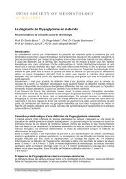
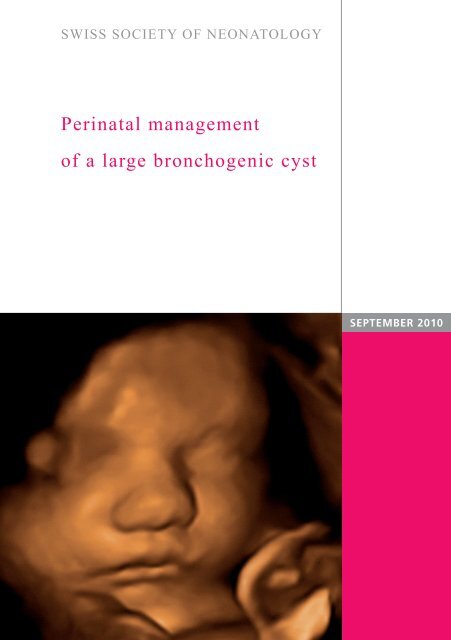
SWISS SOCIETY OF NEONATOLOGY<br />
<strong>Perinatal</strong> <strong>management</strong><br />
<strong>of</strong> a <strong>large</strong> <strong>bronchogenic</strong> <strong>cyst</strong><br />
SEPTEMBER 2010
Berger TM, Steurer MA, Winiker H, Caduff HJ, Zenklusen<br />
HR, Neonatal and Pediatric Intensive Care Unit (BTM,<br />
SMA), Department <strong>of</strong> Pediatric Surgery (WH),<br />
Department <strong>of</strong> Pediatric Radiology (CJH), Children‘s<br />
Hospital <strong>of</strong> Lucerne, Switzerland, Institute <strong>of</strong> Pathology<br />
(ZHR), Cantonal Hospital <strong>of</strong> Lucerne, Switzerland<br />
© <strong>Swiss</strong> <strong>Society</strong> <strong>of</strong> Neonatology, Thomas M Berger, Webmaster<br />
2
3<br />
This male infant was born to a 30-year-old G3/P3 by<br />
spontaneous vaginal delivery at 41 6/7 weeks <strong>of</strong> pregnancy.<br />
At 19 weeks, a right-sided <strong>cyst</strong>ic thoracic malformation<br />
had been detected on prenatal ultrasound<br />
examination (Fig. 1). Subsequently, the women was<br />
regularly followed to detect any fetal compromise,<br />
such as evidence <strong>of</strong> congestive heart failure or hydrops.<br />
At 39 0/7 weeks <strong>of</strong> gestation, the lesion measured<br />
3x3 cm on an axial view and displaced the mediastinal<br />
structures to the left (Fig. 2).<br />
The infant adapted without difficulties with Apgar<br />
scores <strong>of</strong> 9, 9 and 10 at 1, 5 and 10 minutes, respectively.<br />
There was no evidence <strong>of</strong> any respiratory<br />
distress and the baby was initially left with his parents<br />
on continuous pulse oxymetry monitoring. At<br />
the age <strong>of</strong> eight hours, ap and lateral X-ray views <strong>of</strong><br />
the chest were obtained and confirmed the presence<br />
<strong>of</strong> a <strong>cyst</strong>ic lesion in the right hemithorax. An air-fluid<br />
level was easily recognized suggesting that the lesion<br />
was connected either to the bronchial system or the<br />
esophagus (Fig. 3). On echocardiography, there was a<br />
structurally normal heart and mild pulmonary arterial<br />
hypertension which normalized one week later.<br />
Preoperatively, a CT scan <strong>of</strong> the chest was obtained.<br />
It demonstrated a <strong>cyst</strong>ic lesion in the right upper lobe<br />
with a connection to the bronchial system (Fig. 4, 5).<br />
On day <strong>of</strong> life 9, the infant was operated. The lesion<br />
was found to be a <strong>large</strong> <strong>cyst</strong> within the right upper<br />
CASE REPORT
lobe (Fig. 6, 7). Following aspiration <strong>of</strong> air, it was suc-<br />
cessfully separated from the intact lung tissue with<br />
the use <strong>of</strong> a hot knife. The postoperative course was<br />
uneventful.<br />
On histology, the <strong>cyst</strong> was lined with respiratory epi-<br />
thelium and its wall contained small areas <strong>of</strong> cartilage<br />
(Fig. 8-10). These findings are consistent with the clinical<br />
diagnosis <strong>of</strong> a <strong>bronchogenic</strong> <strong>cyst</strong>.<br />
4
5<br />
1 D 1.56cm<br />
2 D 1.65cm<br />
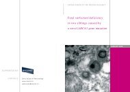
Prenatal US at 19 0/7 weeks <strong>of</strong> pregnancy<br />
demon strating a single right-sided thoracic <strong>cyst</strong>.<br />
Fig. 1
Fig. 2<br />
A B<br />
1 A D 3.46cm B<br />
2 D 3.26cm<br />
Prenatal US at 39 0/7 weeks <strong>of</strong> pregnancy<br />
demonstrating a single right-sided thoracic <strong>cyst</strong>.<br />
6
7<br />
CXR (ap and lateral views) on day 1 <strong>of</strong> life:<br />
right-sided dorsolateral <strong>cyst</strong>ic lesion with an air-fluid<br />
level.<br />
Fig. 3
Fig. 4<br />
A B<br />
A B<br />
CT scan with contrast on day <strong>of</strong> life 4: <strong>large</strong> (4x5x4<br />
cm) right-sided <strong>cyst</strong> with apparent connection to the<br />
bronchial system (arrow).<br />
8
9<br />
C<br />
CT scan with 3D reconstruction: the <strong>cyst</strong> appears hy-<br />
perlucent (C) and is easily separated from the partially<br />
compressed white lung tissue.<br />
Fig. 5
Fig. 6<br />
Appearance <strong>of</strong> the <strong>cyst</strong> following opening <strong>of</strong> the<br />
chest through the 4th intercostal space.<br />
10
11<br />
Cyst<br />
RUL<br />
Appearance <strong>of</strong> the <strong>cyst</strong> following aspiration <strong>of</strong> air:<br />
the <strong>cyst</strong> has collapsed and can now be delineated<br />
from the normally aerated part <strong>of</strong> the right upper<br />
lobe (RUL).<br />
Fig. 7
Fig. 8<br />
PS<br />
Histology <strong>of</strong> the <strong>cyst</strong> (H&E stain): compressed lung<br />
tissue between pleural surface (PS) and <strong>cyst</strong> (C).<br />
12<br />
C
13<br />
Ca<br />
Ca<br />
Ca<br />
Histology <strong>of</strong> the <strong>cyst</strong> (H&E stain): islets <strong>of</strong> primitive<br />
cartilage (Ca) within the <strong>cyst</strong> wall.<br />
Fig. 9
Fig. 10<br />
Histology <strong>of</strong> the <strong>cyst</strong> (H&E stain): higher magnifica-<br />
tion <strong>of</strong> the <strong>cyst</strong> wall with columnar respiratory epi-<br />
thelium (CL: lumen <strong>of</strong> <strong>cyst</strong>).<br />
CL<br />
14
15<br />
IES<br />
FOREGUT ABNORMALITIES<br />
BC<br />
BPS<br />
CLE CCAM<br />
BA<br />
Diagram <strong>of</strong> the spectrum <strong>of</strong> bronchopulmonary<br />
foregut malformations, including foregut, pulmonary,<br />
airway and vascular components (7) (BA: bronchial<br />
atresia; BC: <strong>bronchogenic</strong> <strong>cyst</strong>; BPS: bronchopulmonary<br />
sequestration; CCAM: congenital <strong>cyst</strong>ic<br />
adenomatoid malformation; CLE: congenital lobar<br />
emphysema).<br />
AIRWAY ABNORMALITIES PARENCHYMAL ABNORMALIT<br />
VASCULAR ABNORMALITIES<br />
Fig. 11
Table<br />
New nomenclature Old terms superseded<br />
Congenital hyperlucent lobe<br />
(CHL)<br />
Congenital thoracic<br />
malformation (CTM)<br />
Congenital lobar emphysema<br />
(CLE)<br />
Polyalveolar lobe<br />
Congenital <strong>cyst</strong>ic adenomatoid<br />
malformation (CCAM)<br />
Congenital pulmonary airway<br />
malformation<br />
Malinosculation<br />
Sequestration (intra- and<br />
extrapulmonary)<br />
Bronchogenic <strong>cyst</strong><br />
Reduplication <strong>cyst</strong><br />
Foregut <strong>cyst</strong><br />
Congenital small lung (CSL) Pulmonary hypoplasia<br />
Absent lung, absent trachea Agenesis <strong>of</strong> lung, tracheal<br />
aplasia<br />
Absent bronchus Bronchial atresia<br />
Comparison <strong>of</strong> new nomenclature with old terms (1).<br />
16
17<br />
Congenital lung lesions are increasingly being reco-<br />
gnized on prenatal ultrasound examinations. While<br />
they can be described as <strong>cyst</strong>ic, intermediate or solid,<br />
it is <strong>of</strong>ten not possible to make a more specific diagnosis.<br />
Some <strong>of</strong> these lesions disappear by the time<br />
the baby is born. Some infants are asymptomatic at<br />
birth, others have severe respiratory distress and may<br />
require emergency interventions. Postnatal imaging<br />
studies, including conventional X-ray, CT, MRI and ultrasound<br />
will help to better define the lesions and<br />
plan the appropriate surgical procedures. Finally, histological<br />
examination will allow a definitive description<br />
<strong>of</strong> the malformation.<br />
Recently, several authors have argued that many <strong>of</strong><br />
the terms used in the past to describe congenital<br />
lung malformations (e.g., congenital <strong>cyst</strong>ic adenomatoid<br />
malformation (CCAM), congenital lobar emphysema<br />
(CLE), bronchopulmonary sequestration (BPS),<br />
<strong>bronchogenic</strong> <strong>cyst</strong>) most likely describe extremes <strong>of</strong> a<br />
spectrum <strong>of</strong> malformations with the same underlying<br />
pathogenic mechanism (i.e., fetal airway obstruction)<br />
rather than separate disease entities (1, 2). In support<br />
<strong>of</strong> this concept, many authors have reported hybrid<br />
lesions with coexisting histological features <strong>of</strong> both<br />
CCAM and BPS (3, 4). Reviewing the final pathology<br />
reports <strong>of</strong> 25 patients who underwent surgical resection<br />
<strong>of</strong> prenatally diagnosed lung masses, Kunisaki et<br />
al. found that most congenital lung malformations<br />
were associated with an occult atretic bronchus (5).<br />
DISCUSSION
Their observation suggests that fetal airway obstruc-<br />
tion is likely to play a prominent role in the pathoge-<br />
nesis <strong>of</strong> congenital lung malformations (5).<br />
Langston has proposed a malformation sequence to<br />
explain the spectrum <strong>of</strong> pulmonary anomalies, depending<br />
on the level, timing, and degree <strong>of</strong> bronchial<br />
obstruction. For example, an atretic bronchus early<br />
in gestation might favor the formation <strong>of</strong> a CCAM<br />
or a <strong>bronchogenic</strong> <strong>cyst</strong>, whereas obstruction later in<br />
gestation (e.g., 16-18 weeks <strong>of</strong> gestation) might predispose<br />
the lung to develop BPS or CLE (Fig. 11) (6,<br />
7). Support for this theory also comes from experimental<br />
data that shows that <strong>cyst</strong>ic changes occur in<br />
fetal animal lungs after bronchial ligation. Newman<br />
has pointed out that this is also a common embryologic<br />
theme in other organ systems, such as the genitourinary<br />
system, with <strong>cyst</strong>ic renal dysplasia being<br />
a consequence <strong>of</strong> high-grade urinary obstruction (7).<br />
Bush (1) has made a plea to replace the old terms<br />
by a simplified new nomenclature (Table). He recommends<br />
to use the following principles to classify congenital<br />
lung disease:<br />
1. What is actually seen should be described,<br />
without indulgence in embryological speculation,<br />
which will almost certainly be proved wrong<br />
sooner or later.<br />
18
19<br />
2. The description should be in ordinary, everyday<br />
language.<br />
3. The lung and associated organs should be<br />
approached in a systematic manner, because ab-<br />
normalities are <strong>of</strong>ten multiple, and associated<br />
lesions will be missed unless carefully sought.<br />
4. Clinical and pathological descriptions should<br />
be kept separate; the same clinical appearance<br />
(e.g., a multi<strong>cyst</strong>ic mass) may have different<br />
pathological phenotypes.<br />
The final pathological diagnosis in our patient was a<br />
<strong>bronchogenic</strong> <strong>cyst</strong> (Fig. 8-10). Bronchogenic <strong>cyst</strong>s are<br />
thought to arise from abnormal buds from the primitive<br />
esophagus and tracheobronchial tree which do<br />
not extend to the site where alveolar differentiation<br />
occurs. Alternatively, the may be the consequence<br />
<strong>of</strong> bronchial obstruction early in gestation (5). In<br />
the majority <strong>of</strong> cases, they are located in the right<br />
paratracheal or carinal region, but intrapulmonary<br />
forms - as in our patient - have also been described<br />
(2). Depending on its location, a <strong>bronchogenic</strong> <strong>cyst</strong><br />
may cause airway compression resulting in cough,<br />
wheeze, dyspnea, or even respiratory distress. Secondary<br />
infection <strong>of</strong> the <strong>cyst</strong> is a frequent complication.<br />
Malignant transformation <strong>of</strong> epithelial cells <strong>of</strong> <strong>bronchogenic</strong><br />
<strong>cyst</strong>s has been reported repeatedly (2).<br />
Radiographic findings <strong>of</strong> <strong>bronchogenic</strong> <strong>cyst</strong>s are va-<br />
riable and range from a rounded mass, with uniform
20<br />
density similar to that <strong>of</strong> the cardiac shadow and pro-<br />
jecting from the mediastinum, to hyperinflation or ate-<br />
lectasis <strong>of</strong> a lobe or an entire lung. As illustrated by<br />
our patient (Fig. 3, 4), when the lesion communicates<br />
with the esophagus or the tracheobronchial tree, airfluid<br />
levels may be seen.<br />
Because <strong>of</strong> the risk <strong>of</strong> complications, such as infection,<br />
hemorrhage, pneumothorax, sudden respiratory compromise<br />
and malignant transformation, most authors<br />
recommend elective resection <strong>of</strong> <strong>bronchogenic</strong> <strong>cyst</strong>s.<br />
Histologically, the <strong>cyst</strong> wall has structural elements <strong>of</strong><br />
the air way, including cartilage (as an obligatory finding<br />
to allow differentiation from an enteric duplication<br />
<strong>cyst</strong>), smooth muscle, mucous glands and respiratory<br />
epithelium (1).
21<br />
1. Bush A. Congenital lung disease: a plea for clear thinking and<br />
clear nomenclature. Pediatr Pulmonol 2001;32:328-337<br />
2. Eber E. Antenatal diagnosis <strong>of</strong> congenital thoracic<br />
malformations: early surgery, late surgery, or no surgery? Sem<br />
Resp Crit Care Med 2007;28:355-366<br />
3. Samuel M, Burge DM. Management <strong>of</strong> antenatally diagnosed<br />
pulmonary sequestration associated with congenital <strong>cyst</strong>ic<br />
adenomatoid malformation. Thorax 1999;54:701-706<br />
4. Roggin KK, Breuer CK, Carr SR, et al. The unpredictable<br />
character <strong>of</strong> congenital <strong>cyst</strong>ic lung lesions. J Pediatr Surg<br />
2000;35:801-805<br />
5. Kunisaki SM, Fauza DO, Nemes LP, et al. Bronchial atresia: the<br />
hidden pathology within a spectrum <strong>of</strong> prenatally diagnosed<br />
lung masses. J Pediatr Surg 2006;41:61–65<br />
6. Langston C. New concepts in the pathology <strong>of</strong> congenital lung<br />
malformations. Semin Pediatr Surg 2003;12:17- 37<br />
7. Newman B. Congenital bronchopulmonary foregut<br />
malformations: concepts and controversies. Pediatr Radiol<br />
2006;36:773-791<br />
REFERENCES
SUPPORTED BY<br />
CONTACT<br />
<strong>Swiss</strong> <strong>Society</strong> <strong>of</strong> Neonatology<br />
www.neonet.ch<br />
webmaster@neonet.ch<br />
concept & design by mesch.ch