A Comparison Of Intensity-Modulated Radiotherapy And Dynamic ...
A Comparison Of Intensity-Modulated Radiotherapy And Dynamic ...
A Comparison Of Intensity-Modulated Radiotherapy And Dynamic ...

You also want an ePaper? Increase the reach of your titles
YUMPU automatically turns print PDFs into web optimized ePapers that Google loves.
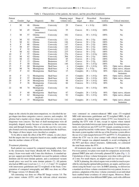
Patient<br />
no. Gender Age Diagnosis Site<br />
Table 1. Characteristics of the patients, the tumors, and the prescribed treatments<br />
Planning target<br />
volume (mL)<br />
shape on the criteria for plan intercomparison, we classified the target<br />
shapes into three categories: convex, concave, and complex. All<br />
gliomas had a regular convex shape and all but one convexity meningiomas<br />
were concave. The base of skull meningiomas were all<br />
irregularly shaped, mostly because of extensions in the cavernous<br />
sinus and through foramina. This was also the case for a very complex<br />
frontal convexity meningioma that extended into the skull base.<br />
The shapes of these targets were classified as complex.<br />
To be able to study the effect of the PTV volume, we also classified<br />
the targets into two categories based on size: smaller and larger<br />
than the median volume of 92 mL.<br />
Treatment planning<br />
Each patient was scanned by computed tomography while fixed<br />
in the stereotactic head frame (BrainLAB AG, Feldkirchen, Germany).<br />
Scans were made with 2-mm slice thickness. A standard upper<br />
jaw support provided by Brainlab was used for patients without<br />
dentition and for most dentate patients, and a customized vacuum<br />
mouth piece was used for some dentate patients (7). All patients<br />
also had an MRI scan (voxel size 1.1 1.1 1.3 mm 3 ; T1-weighted<br />
with intravenous gadolinium and T2-weighted) of the brain. Coregistration<br />
of computed tomography and MRI was done on i-Plan<br />
RT Image version 3.0 (BrainLAB).<br />
All contouring was done on i-Plan RT Image version 3.0. The<br />
gross tumor volume (GTV) and the relevant critical structures<br />
IMRT and DCA in intracranial tumors d R. G. J. WIGGENRAAD et al. 1019<br />
Shape of<br />
target<br />
Prescribed<br />
dose<br />
Prescription<br />
isodose Critical structures<br />
1 M 46 Glioma<br />
(recurrence)<br />
Convexity 29 Convex 4 8 Gy 100% No<br />
2 M 45 Glioma<br />
(recurrence)<br />
Convexity 55 Convex 30 1.8 Gy 100% No<br />
3 M 57 Glioma<br />
(recurrence)<br />
Convexity 102 Convex 30 1.8 Gy 100% No<br />
4 M 52 Glioma Convexity 121 Convex 30 2 Gy 100% No<br />
5 M 61 Glioma Convexity 146 Convex 30 2 Gy 100% No<br />
6 F 68 Glioma Convexity 147 Convex 30 2 Gy 100% No<br />
7 M 67 Glioma Convexity 148 Convex 30 2 Gy 100% No<br />
8 F 55 Glioma Convexity 152 Convex 30 2 Gy 100% No<br />
9 M 62 Glioma Convexity 235 Convex 30 2 Gy 100% No<br />
10 F 37 Glioma Convexity 276 Convex 30 1.8 Gy 100% No<br />
11 M 62 Glioma Convexity 304 Convex 30 2 Gy 100% No<br />
12 M 41 Glioma Brainstem 85 Convex 28 2 Gy 100% No<br />
13 F 23 Glioma Brainstem 97 Convex 30 2 Gy 100% No<br />
14 F 40 Glioma Brainstem 146 Convex 30 1.8 Gy 100% No<br />
15 F 60 Meningioma Skull base 6 Complex 28 1.8 Gy 80% Optic nerve, chiasm<br />
16 M 76 Meningioma Skull base 8 Complex 5 5 Gy 80% Optic nerve, chiasm<br />
17 F 42 Meningioma Skull base 15 Complex 28 1.8 Gy 80% Optic nerve, chiasm<br />
brainstem<br />
18 F 72 Meningioma Skull base 15 Complex 28 1.8 Gy 80% Optic nerve, chiasm<br />
19 F 57 Meningioma Skull base 21 Complex 28 1.8 Gy 80% Optic nerve, chiasm<br />
20 F 38 Meningioma Convexity 48 Concave 28 1.8 Gy 80% No<br />
21 F 36 Meningioma<br />
(atypical)<br />
Convexity 66 Concave 30 1.8 Gy 80% No<br />
22 M 74 Meningioma<br />
(atypical)<br />
Convexity 81 Concave 30 1.8 Gy 80% No<br />
23 F 68 Meningioma Skull base 87 Complex 28 1.8 Gy 80% Optic nerve, chiasm<br />
24 F 62 Meningioma<br />
(atypical)<br />
Convexity 92 Complex 30 1.8 Gy 80% Optic nerve, chiasm<br />
25 F 37 Meningioma Skull base 142 Complex 28 1.8 Gy 80% Optic nerve, chiasm<br />
brainstem<br />
were contoured on contrast-enhanced MRI scans (T1-weighted<br />
MRI with intravenous gadolinium and T2-weighted MRI). In glioma<br />
patients, the clinical target volume (CTV) was formed by expanding<br />
the GTV with 15 mm, except in regions where natural<br />
boundaries precluded microscopic tumor spread. In meningioma patients,<br />
we did not use any GTV to CTV margin, assuming no microscopic<br />
spread beyond the visible tumor. The positioning accuracy of<br />
the mask system together with the use of the Exactrac system allows<br />
the use of a 2-mm margin between CTV and PTV (7). For patients in<br />
whom the CTV was in close contact with critical structures, the<br />
CTV-PTV margin was adapted manually to avoid overlapping of<br />
the PTV and these critical structures. Additionally, we contoured<br />
the total brain minus the PTV.<br />
All treatment plans were made on Brainscan 5.31 (BrainLAB).<br />
For every patient, two plans were made for comparison: an inversely<br />
planned IMRT plan and a forwardly planned DCA plan (Fig. 1, 2).<br />
Calculations of dose–volume histograms were done with a grid size<br />
of 2 mm. For small objects, an adaptive grid size was used.<br />
In DCA planning, the critical organs were avoided as much as possible<br />
by selecting the optimal table positions, arc angles, and leave positions<br />
using the beam’s-eye view. In IMRT planning, we did not use<br />
a standard set of dose–volume constraints for every patient. In our experience,<br />
adaptation of the dose–volume constraints on an individual<br />
basis enabled better avoidance of critical organs. Moreover, Brainscan<br />
always produces four different IMRT plans, each with a different