A new penumbra generator for electron fields matching - UCSF ...
A new penumbra generator for electron fields matching - UCSF ...
A new penumbra generator for electron fields matching - UCSF ...

Create successful ePaper yourself
Turn your PDF publications into a flip-book with our unique Google optimized e-Paper software.
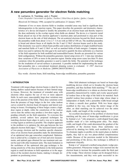
A <strong>new</strong> <strong>penumbra</strong> <strong>generator</strong> <strong>for</strong> <strong>electron</strong> <strong>fields</strong> <strong>matching</strong><br />
B. Lachance, D. Tremblay, and J. Pouliot<br />
Centre Hospitalier Universitaire de Québec, Pavillon L’Hôtel-Dieu de Québec, Québec, Canada<br />
Received 26 February 1996; accepted <strong>for</strong> publication 23 January 1997<br />
Abutment of two or more <strong>electron</strong> <strong>fields</strong> to irradiate extended areas may lead to significant dose<br />
inhomogeneities in the junction region. This paper describes the geometric and dosimetric characteristics<br />
of a device developed to modify the <strong>penumbra</strong> of an <strong>electron</strong> beam and thereby improve<br />
the dose uni<strong>for</strong>mity in the overlap region when <strong>fields</strong> are abutted. The device is a Lipowitz metal<br />
block placed on top of the <strong>electron</strong> applicator’s insertion plate and positioned to stop part of the<br />
<strong>electron</strong> beam on the side of field abutment. The air-scattered <strong>electron</strong>s beyond the block increase<br />
the <strong>penumbra</strong> width from about 1.4 to 2.7–3.4 cm with an SSD of 100 cm. The modified <strong>penumbra</strong><br />
is broad and almost linear at all depths <strong>for</strong> the 9 and 12 MeV <strong>electron</strong> beams used in this study.<br />
Film dosimetry was used to obtain beam profiles and isodose distributions of single modified beams<br />
and matched <strong>fields</strong> of 9 and 12 MeV as well as matched <strong>fields</strong> of both energies. Computer simulation<br />
was used to optimize the skin gap to be used and to quantify the dose uni<strong>for</strong>mity as a function<br />
of the field separation <strong>for</strong> both modified and nonmodified beams. Results are presented <strong>for</strong> various<br />
field configurations. Without the <strong>penumbra</strong> <strong>generator</strong>, lateral setup errors of 2–3 mm may introduce<br />
dose variations of 20% or more in the junction region. Similar setup errors cause less than 5% dose<br />
variations when the <strong>penumbra</strong> <strong>generator</strong> is used to match the <strong>fields</strong>. The potential of the technique<br />
<strong>for</strong> the irradiation of curved surfaces is presented. A possible method <strong>for</strong> implementing the modified<br />
<strong>penumbra</strong> into a conventional treatment planning system is evaluated. © 1997 American<br />
Association of Physicists in Medicine. S0094-24059701404-1<br />
Key words: <strong>electron</strong> beam, field <strong>matching</strong>, beam-edge modification, <strong>penumbra</strong> <strong>generator</strong><br />
I. INTRODUCTION<br />
Treatment with megavoltage <strong>electron</strong> beams is ideal <strong>for</strong> irradiating<br />
shallow seated tumors because of their limited range<br />
in tissues. However, the treatment of extended areas with<br />
<strong>electron</strong>s often requires the use of two or more adjacent<br />
<strong>fields</strong>. In such cases, unacceptably large dose variations may<br />
arise at the junction of the <strong>fields</strong>. These dose variations come<br />
from the presence of large bulges in the low value isodose<br />
curves created by <strong>electron</strong> beam divergence and lateral scattering<br />
in tissues. Overlapping of these bulges creates a highdose<br />
region at depth, while the constriction of the isodose<br />
curves near the surface may produce a low-dose region, depending<br />
critically on the field separation. To overcome this<br />
problem, several authors have proposed techniques <strong>for</strong><br />
<strong>matching</strong> <strong>electron</strong> beam edges in such a way as to make the<br />
overlap region as uni<strong>for</strong>m as possible. The simplest approach<br />
to the problem is to optimize the skin gap between the two<br />
adjacent <strong>electron</strong> field edges. 1–3 The increased lateral scatter<br />
of low-energy <strong>electron</strong>s and the machine specific characteristics<br />
of an <strong>electron</strong> beam <strong>penumbra</strong> make the determination<br />
of an optimized skin gap somewhat complicated. Optimization<br />
is achieved by a complete set of trial and error measurements,<br />
and optimal gaps obtained <strong>for</strong> one treatment unit cannot<br />
be used with other treatment units. The main limitation to<br />
the usefulness of the optimized skin gap technique is the<br />
strong sensitivity of the dose distribution in the field junction<br />
region to small deviations in field separation or in the angulation<br />
of the incident <strong>electron</strong> beams, making it strongly dependent<br />
on positioning errors.<br />
Other field abutment techniques are based on beam-edge<br />
modifying devices which act to broaden the <strong>electron</strong> beam<br />
<strong>penumbra</strong>, and thus facilitate field <strong>matching</strong>. 1,4,5 The aim of<br />
beam-edge modification is to obtain an <strong>electron</strong> beam with a<br />
<strong>penumbra</strong> characterized by widely and evenly spaced parallel<br />
isodose lines at the edge of the field to be matched. Such<br />
parallel isodose lines produce a beam edge whose shape is<br />
independent of depth, with a <strong>penumbra</strong> width broad enough<br />
to obtain a smooth dose gradient. With two beam edges<br />
modified in this way, one being the mirror image of the<br />
other, an ideal <strong>matching</strong> can be achieved over the entire<br />
depth, width, and length of the overlap region.<br />
An excellent description of the problem of <strong>electron</strong> beam<br />
<strong>matching</strong> and the aims of beam-edge modification is given<br />
by Kalend et al., 4 who proposed a comb-shaped beam-edge<br />
modifier made of a low melting-point alloy. The shape,<br />
width, and length of the comb determined the gradient of the<br />
<strong>penumbra</strong> of the <strong>electron</strong> beam. Dose uni<strong>for</strong>mity was<br />
achieved by abutting two <strong>fields</strong>, each modified by a comb.<br />
To eliminate ripples in profiles in the match plane at shallow<br />
depths, they had to use two phase-shifted complementary<br />
combs. Because of the precision needed in the fabrication<br />
and positioning of the device, this technique is difficult to<br />
implement clinically.<br />
Kurup et al. 5 proposed a method using plastic wedge <strong>penumbra</strong><br />
<strong>generator</strong>s. Polystyrene wedges were inserted into the<br />
<strong>electron</strong> beam to increase the <strong>penumbra</strong> under the thick part<br />
of the wedge. These <strong>electron</strong> wedges can be designed from a<br />
few measurements, and they were quite successful in im-<br />
485 Med. Phys. 24 (4), April 1997 0094-2405/97/24(4)/485/11/$10.00 © 1997 Am. Assoc. Phys. Med.<br />
485
486 Lachance, Tremblay, and Pouliot: New <strong>penumbra</strong> generation 486<br />
proving the dosimetry of the junction region; however, the<br />
presence of the wedges causes a slight degradation of the<br />
beam energy and penetration. Furthermore, the <strong>penumbra</strong><br />
broadening occurs at all field edges in the thick part of the<br />
wedge and is not limited to the region of abutment.<br />
Another approach is to match <strong>electron</strong> beams without the<br />
secondary collimation. 6,7 Such beams have profiles close to<br />
Gaussian and are there<strong>for</strong>e easy to match at 50% intensity<br />
points to produce a homogeneous dose distribution across a<br />
large field. This method is successfully used by a few centers<br />
as an <strong>electron</strong> beam pseudoarc technique 7–11 <strong>for</strong> the treatment<br />
of large superficial volumes which follow curved surfaces;<br />
but the dosimetric gain comes at the expense of the<br />
setup time in the treatment room because of the necessary<br />
lead shielding on the patient to provide a sharp dose cutoff at<br />
the edges of the composite treatment field.<br />
In this paper, we describe a <strong>new</strong> and simple method to<br />
modify an <strong>electron</strong> beam to produce a wide <strong>penumbra</strong> and<br />
yield an excellent dose uni<strong>for</strong>mity at the junction between<br />
adjacent <strong>electron</strong> <strong>fields</strong>. The <strong>new</strong> <strong>penumbra</strong> <strong>generator</strong> consists<br />
of a metal block made of Lipowitz metal and placed on<br />
the applicator insertion plate to stop part of the <strong>electron</strong><br />
beam on the side of the field abutment. In this configuration,<br />
the <strong>electron</strong>s of the unblocked part of the beam have a large<br />
distance to be scattered in air under the block be<strong>for</strong>e reaching<br />
the patient, and with little disturbance from the <strong>electron</strong> applicator.<br />
The result is a wide and smooth <strong>penumbra</strong>, ideal <strong>for</strong><br />
field <strong>matching</strong>. The <strong>penumbra</strong> broadening occurs only at the<br />
edge of abutment, while the remaining field boundaries are<br />
defined near the patient in the usual way with the <strong>electron</strong><br />
cone.<br />
The objectives of this study are to present a systematic<br />
evaluation of modified <strong>electron</strong> field <strong>matching</strong> on a flat surface<br />
to better understand the behavior and physical characteristics<br />
of the <strong>penumbra</strong> <strong>generator</strong>, and to investigate the<br />
feasibility of using this technique <strong>for</strong> <strong>matching</strong> <strong>electron</strong><br />
<strong>fields</strong> on curved surfaces in order to irradiate the thoracic<br />
region. The first objective involves obtaining a quantitative<br />
indicator of the dose uni<strong>for</strong>mity in the junction region and<br />
optimizing the field separation. Because of the large amount<br />
of data required, we limit our discussion to the dosimetry of<br />
9 and 12 MeV <strong>electron</strong> beams used more frequently at our<br />
institution. These <strong>electron</strong> beam energies are widely documented<br />
<strong>for</strong> the treatment of extensive chest-wall<br />
recurrence. 5,6,12–14<br />
II. METHODS AND MATERIALS<br />
A. Adjacent <strong>electron</strong> beams on a flat surface<br />
The <strong>penumbra</strong> <strong>generator</strong> and the setup used <strong>for</strong> measurements<br />
are depicted in Fig. 1. The device is a Cerrobend block<br />
placed on top of the applicator’s insertion plate see Fig.<br />
1a to stop part of the <strong>electron</strong> beam on the side of the field<br />
abutment. Cerrobend is a trade name <strong>for</strong> Lipowitz metal:<br />
50% bismuth, 27% lead, 13% cadmium, and 10% tin. The<br />
insertion plate of the applicator and the <strong>penumbra</strong> <strong>generator</strong><br />
are situated 44 cm above the surface of a phantom placed at<br />
the nominal source-surface distance SSD of 100 cm. The<br />
Medical Physics, Vol. 24, No. 4, April 1997<br />
FIG. 1. Experimental setup showing: a The Siemens <strong>electron</strong> applicator<br />
with the <strong>penumbra</strong> <strong>generator</strong> placed on the insertion plate. The measurement<br />
point of the distance X is indicated. SI indicates the base of the applicator<br />
where tertiary collimation is provided by the insertion of Shields insert<br />
defining the field boundaries. b The two matched <strong>fields</strong> light <strong>fields</strong> are<br />
pictured with the gap measured at the phantom surface.<br />
block is aligned parallel to the edge of the beam to be<br />
matched. Its relative position with respect to the center of the<br />
field the distance X, is defined as the distance between the<br />
central axis of the beam and the position of the edge of the<br />
light field under the block Fig. 1a. This distance is measured<br />
on top of the custom-made cutouts insert shields insert<br />
SI in Fig. 1a, which is approximately 6 cm above the<br />
phantom patient surface at the nominal SSD of 100 cm.<br />
This measurement point was chosen <strong>for</strong> convenience, and the<br />
corresponding distance at the surface of the phantom can be<br />
found from a simple scaling factor. The gap Fig. 1b between<br />
adjacent modified <strong>electron</strong> beams is measured between<br />
the light <strong>fields</strong> at the surface of the phantom patient.<br />
Comparative measurements were carried out to evaluate<br />
how the modified <strong>penumbra</strong> is affected by either varying the<br />
thickness of the block or accounting <strong>for</strong> beam divergence.<br />
Tests were per<strong>for</strong>med with a 9-mm-thick block and with 20-
487 Lachance, Tremblay, and Pouliot: New <strong>penumbra</strong> generation 487<br />
mm-thick blocks having either a divergent edge <strong>for</strong> various<br />
distance X or a straight edge. These tests showed that no<br />
measurable change in the modified <strong>penumbra</strong> all other parameters<br />
being the same is visible when using either of the<br />
blocks. Thus, provided that the thickness of the block is<br />
larger than the <strong>electron</strong> range in the material, changing the<br />
thickness of the block has little effect on this technique.<br />
Also, accounting <strong>for</strong> beam divergence would only be an additional<br />
complication providing no dosimetric gain. All the<br />
data reported here were collected with a <strong>penumbra</strong> <strong>generator</strong><br />
having a straight edge and a thickness of 2 cm. According to<br />
transmission measurements per<strong>for</strong>med by Purdy and Choi, 15<br />
this thickness of Cerrobend is required to attenuate the 12<br />
MeV open <strong>electron</strong> beam dose to the 97% level with a<br />
2525 cm 2 applicator. The thickness used ensures that <strong>for</strong><br />
all distances X, only a weak bremsstrahlung contamination is<br />
transmitted through the block so that this is not of a major<br />
concern and that the <strong>penumbra</strong> <strong>generator</strong> provides sufficient<br />
attenuation to allow a simple extension of the technique to<br />
higher <strong>electron</strong> beam energies.<br />
For clinical applications, a frame made of thin aluminum<br />
plates is used to support the Cerrobend blocks. The size of<br />
the frame is sufficient to completely clear the opening in the<br />
insertion plate of the <strong>electron</strong> applicator. One such frame is<br />
used <strong>for</strong> every modified <strong>electron</strong> field. Four holes are drilled<br />
in the insertion plate, and the aluminum frame is screwed to<br />
the applicator. The <strong>penumbra</strong> <strong>generator</strong> is firmly taped in its<br />
appropriate position on the aluminum frame during the first<br />
treatment fraction. For subsequent fractions, one simply has<br />
to screw back in place the whole assembly consisting of the<br />
block taped on its aluminum support. The screw holes placed<br />
in the aluminum frame are 2 cm slots which provide the<br />
needed flexibility to obtain a perfect lateral alignment of the<br />
<strong>penumbra</strong> <strong>generator</strong> from day to day.<br />
Film dosimetry was used in this study to obtain isodose<br />
distributions and beam profiles at various depths in phantom.<br />
Ready-pack Kodak XV-2 films were placed in phantom in a<br />
plane parallel to the central axis of the beam. The films were<br />
aligned in the crossplane direction at the center of the field.<br />
A30cm30 cm30 cm polystyrene phantom consisting of<br />
2.5 cm slabs stacked together was used. The setup was designed<br />
to minimize the effects of potential film artifacts. The<br />
film jacket was punctured at each corner and the entire assembly,<br />
consisting of the phantom slabs and the aligned film,<br />
was tightly clamped together with a large carpenter clamp to<br />
avoid any perturbation to the dose distributions caused by air<br />
trapped in the film jacket. The films were kept in their jacket<br />
during irradiation to exclude light and Čerenkov emissions.<br />
The film edge facing the source was carefully aligned with<br />
the phantom surface. The excess wrapper extending beyond<br />
the film edge was folded over to one side and taped to the<br />
phantom.<br />
The sensitometric curve was obtained <strong>for</strong> each batch of<br />
films used in this study, and our Kodak XV-2 films showed a<br />
linear response to dose up to about 40 cGy. For this linear<br />
portion of the curve, the net optical density can be used <strong>for</strong><br />
relative dose measurements without any corrections. Hence,<br />
unless otherwise specified, 40 monitor units MU 38 cGy<br />
Medical Physics, Vol. 24, No. 4, April 1997<br />
at the depth of maximum dose in polystyrene were used to<br />
irradiate films, ensuring that no absolute dose larger than the<br />
upper limit of the linear portion of the sensitometric curve is<br />
recorded on film and that isodensity curves can be considered<br />
as isodose curves.<br />
All measurements were made using the 9 and 12 MeV<br />
<strong>electron</strong> beams from a Siemens Mevatron KD-2 linear accelerator.<br />
Film dosimetry was per<strong>for</strong>med with a Wellhöfer dosimetry<br />
system WP600. Experiments were per<strong>for</strong>med <strong>for</strong><br />
the following clinically useful <strong>electron</strong> cones: 1010, 15<br />
15, and 2020 cm 2 . All the isodose distributions presented<br />
in this paper were obtained with the phantom surface placed<br />
at the nominal SSD of 100 cm. The effect of extended SSDs<br />
105 and 110 cm on the modified <strong>penumbra</strong> width and on<br />
the gaps to be used <strong>for</strong> field abutment was investigated by<br />
beam profile measurements per<strong>for</strong>med in water with the<br />
scanning ionization chamber of the Wellhöfer.<br />
The initial data were taken by irradiating films with a<br />
single modified beam <strong>for</strong> various distances X and <strong>for</strong> all the<br />
<strong>electron</strong> cones used. Beam profiles at seven different depths<br />
were extracted from these films and saved in an ASCII <strong>for</strong>mat<br />
from the Wellhöfer. The depth of the saved profiles was<br />
chosen to uni<strong>for</strong>mly cover the dose distribution down to the<br />
50% isodose line, thus covering the range of depths of clinical<br />
interest. The depths chosen were 0.8, 1,3, 1.8, 2.0, 2.7,<br />
3.2, and 3.6 cm <strong>for</strong> the 9 MeV <strong>electron</strong> beams and 0.8, 1.5,<br />
2.0, 2.5, 3.3, 3.8, and 4.5 cm <strong>for</strong> the 12 MeV beams. These<br />
profiles were then fed into a custom-made computer program<br />
which generates a mirror image of the profiles and fictitiously<br />
combines them with various gaps between the two<br />
<strong>fields</strong>. The resulting combined profiles were used to find an<br />
optimal gap <strong>for</strong> various distances X, <strong>for</strong> the 9 and 12 MeV<br />
beams, and <strong>for</strong> the three <strong>electron</strong> cones used. This procedure<br />
was also per<strong>for</strong>med with regular <strong>electron</strong> beams <strong>for</strong> comparison<br />
with the results obtained <strong>for</strong> modified beams. On-line<br />
visualization of the composite profiles and quantification of<br />
the uni<strong>for</strong>mity of the resulting dose distribution <strong>for</strong> different<br />
gaps between the <strong>fields</strong> was provided by the program.<br />
Bagne 2 has defined the depth dose ratio DDR to analyze<br />
the composite dose distribution of adjacent <strong>fields</strong>. The DDR<br />
is defined as the ratio of the dose at a depth d on the midseparation<br />
axis to the dose at the same depth on the central<br />
axis of a single field,<br />
PDD d at midseparation axis<br />
DDR d .<br />
PDD d at central axis<br />
The shortcoming of the DDR method of evaluating abutment<br />
of <strong>electron</strong> beams is that only doses along the junction line<br />
are evaluated. This interesting concept fails to account <strong>for</strong><br />
possible hot or cold spots outside of the midseparation plane<br />
and there is no specification of the volume of the possible<br />
high or low dose regions. For these reasons, another indicator<br />
has been defined. In this study, the parameter chosen to<br />
describe the goodness of the composite dose distribution <strong>for</strong><br />
a particular field separation is the average field flatness<br />
AFF, defined as the average value of the flatness <strong>for</strong> the<br />
five shallowest composite profiles generated by our custom-
488 Lachance, Tremblay, and Pouliot: New <strong>penumbra</strong> generation 488<br />
made program. Here, the flatness definition used <strong>for</strong> a given<br />
composite profile at a depth d is the largest absolute value<br />
between<br />
maximum dose dcentral dose d<br />
central dose <br />
d<br />
and<br />
central dose dminimum dose d<br />
central dose ,<br />
d<br />
where the central dose is the dose value at midseparation<br />
axis, and the maximum and minimum dose values are taken<br />
from within 80% of the composite field width. This AFF is a<br />
good indicator of the dose uni<strong>for</strong>mity throughout the volume<br />
of clinical interest, and it also provides a good basis <strong>for</strong><br />
quantitative comparison with results obtained when <strong>matching</strong><br />
unmodified beams. The optimal gap <strong>for</strong> a given set of<br />
parameters beam energy, distance X, and <strong>electron</strong> cone size<br />
is the one which minimizes the AFF.<br />
The precision of the actual off-axis distances fed into our<br />
custom-made program was limited by the method used to<br />
identify the beam central axis position on the films irradiated<br />
with a single modified beam. Any misalignment with respect<br />
to the beam central axis was reflected in the values of optimal<br />
gaps obtained with our program. To account <strong>for</strong> this,<br />
verification films were doubly exposed with the modified<br />
beams matched with the field separation prescribed by the<br />
output of the program. Profiles extracted from these verification<br />
films were reproduced by our custom-made program<br />
with the beam profiles extracted from single modified beam<br />
exposures the initial data. This reconstruction of matched<br />
beam profiles provided the correction needed on the previously<br />
obtained optimal gaps. With this iterative procedure,<br />
we obtained a 0.5 mm precision on the optimal gaps to be<br />
used with a given set of conditions, namely, beam energy,<br />
distance X, and <strong>electron</strong> cone size.<br />
Isodose measurements were made by irradiating films<br />
with two modified beams matched with the optimal gaps.<br />
Results are presented <strong>for</strong> 9 and 12 MeV beams and <strong>for</strong><br />
matched beams of both energies. The beam’s-eye-view composite<br />
dose distribution was measured <strong>for</strong> selected geometries<br />
to verify that this technique also works well <strong>for</strong> the<br />
off-axis planes.<br />
The output factor of the modified <strong>electron</strong> beams must be<br />
considered <strong>for</strong> clinical applications. Determination of the<br />
output factor was achieved by comparison of ionization measurements<br />
per<strong>for</strong>med in water with a Farmer chamber<br />
Nuclear Enterprises, NE 2581. Readings were taken at a<br />
depth of d max at the center of both the modified <strong>electron</strong><br />
beams and the same energy beam with the unmodified <strong>electron</strong><br />
applicators. In the case of the modified beams, the center<br />
of the field was defined as the center of the light field at<br />
the phantom surface <strong>for</strong> an SSD of 100 cm. This field center<br />
does not correspond to the usual central axis of the open<br />
field. The isodose distributions <strong>for</strong> <strong>electron</strong> beams incident<br />
on flat surfaces presented in this paper were normalized to<br />
Medical Physics, Vol. 24, No. 4, April 1997<br />
FIG. 2. Experimental setup <strong>for</strong> irradiation of the Rando phantom with three<br />
9 MeV modified beams. Two <strong>penumbra</strong> <strong>generator</strong>s are used <strong>for</strong> the central<br />
field. The light <strong>fields</strong> are pictured.<br />
100% at the usual depth of d max <strong>for</strong> an open field and at the<br />
center of a single modified field defined above.<br />
To facilitate the clinical application of this technique, it is<br />
desirable to implement the generated <strong>penumbra</strong> into computerized<br />
treatment planning. A possible approach to this problem<br />
was evaluated with our commercial treatment planning<br />
system. THERAPLAN Plus, Theratronics International<br />
Limited, Kanata, Ontario, Canada. The solution proposed<br />
uses the ability of the system to allow beam modifiers <strong>for</strong><br />
<strong>electron</strong> beams. Indeed, with Theraplan Plus, the algorithm<br />
used to compute <strong>electron</strong> dose distributions is almost the<br />
same as the one used <strong>for</strong> photon beams. Although the algorithm<br />
is somewhat artificial <strong>for</strong> <strong>electron</strong> beams, it provides<br />
good results and has the advantage of allowing beam modifiers.<br />
The modified <strong>penumbra</strong> is obtained simply by optimizing<br />
the modifier shape in order to match the measured data.<br />
An example of computed modified <strong>penumbra</strong> is presented<br />
see Fig. 10.<br />
B. Application to irradiation of curved surfaces<br />
In order to apply the technique on curved surfaces, a few<br />
tests were per<strong>for</strong>med with an anthropomorphic phantom<br />
Rando phantom. A piece of film was cut in the dark room<br />
to con<strong>for</strong>m to the shape of a Rando slice and sandwiched,<br />
light proof, between two slices. A few slices were then added<br />
on both sides of the ‘‘sandwich’’ and the whole assembly<br />
was tightly clamped together. A given arc along the film<br />
edge was chosen as a target area, and this was irradiated with<br />
three 9 MeV modified beams. The tests were per<strong>for</strong>med with<br />
the 1515 cm 2 <strong>electron</strong> applicator. The technique is similar<br />
to the three-field wraparound technique described by<br />
Norris, 13 except that the dosimetry of the field junctions is<br />
improved by the <strong>penumbra</strong> <strong>generator</strong>s. Here, the central field<br />
had to be modified at both edges to ensure good <strong>matching</strong><br />
with the two lateral <strong>fields</strong> see Fig. 2.
489 Lachance, Tremblay, and Pouliot: New <strong>penumbra</strong> generation 489<br />
FIG. 3. Electron beam profiles modified by the <strong>penumbra</strong> <strong>generator</strong>. Data are<br />
obtained with a 9 MeV beam and the 1515 cm 2 applicator with a distance<br />
X of 3 cm.<br />
For a given target area, the gantry angles and the field<br />
sizes distances X are determined in the following way.<br />
First, the gantry angles are chosen so that each beam covers<br />
its intended area without too much obliquity, so that the isodose<br />
curves are not excessively shifted toward the surface.<br />
Then, the matchline positions are chosen to minimize the<br />
incidence angle at the edges of abutment in order <strong>for</strong> the<br />
overlapping of the modified field edges evaluated <strong>for</strong> flat<br />
surfaces to be approximately valid. The distances X to be<br />
used <strong>for</strong> each beam are automatically determined once the<br />
gantry angles and the matchline positions are chosen, since<br />
all beams are kept at the nominal SSD. Successive film measurements<br />
per<strong>for</strong>med in the Rando phantom are then used to<br />
empirically derive the ideal weight <strong>for</strong> the central field.<br />
III. RESULTS AND DISCUSSION<br />
A. Adjacent <strong>electron</strong> beams of the same energy: Flat<br />
surfaces<br />
Figure 3 shows a typical set of beam profiles modified by<br />
the <strong>penumbra</strong> <strong>generator</strong>. They are <strong>for</strong> the 9 MeV <strong>electron</strong><br />
beam with the 1515 cm 2 applicator and a distance X of 3<br />
cm. The profiles show a <strong>penumbra</strong> which is smooth and linear<br />
over a broad width at all depths. Such a linear dose<br />
gradient around the 50% intensity point is ideal <strong>for</strong> obtaining<br />
a perfect match. Beam profiles measured at d max and at<br />
nominal SSD showed that the <strong>penumbra</strong> width measured<br />
between the 80% and 20% dose values was increased from<br />
about 1.4 to 3–3.4 cm depending on the distance X and the<br />
<strong>electron</strong> cone size <strong>for</strong> 9 MeV <strong>electron</strong>s. The 12 MeV <strong>penumbra</strong><br />
was increased from about 1.3 to 2.7–2.9 cm under the<br />
same conditions. These <strong>penumbra</strong> widths are comparable to<br />
those obtained by Kurup et al. 5 It was found that the <strong>penumbra</strong><br />
width eventually decreases past a certain distance X. This<br />
corresponds to the point where the edge of the shields insert<br />
SI on Fig. 1a begins to interfere with the air-scattered<br />
<strong>electron</strong>s. For both energies this effect becomes clearly observable<br />
at about X4, 6, and 8 cm <strong>for</strong> the 1010, 1515,<br />
and 2020 cm 2 applicators, respectively. The resulting con-<br />
Medical Physics, Vol. 24, No. 4, April 1997<br />
FIG. 4. Electron isodose curves <strong>for</strong> beams with <strong>penumbra</strong> modified by the<br />
<strong>penumbra</strong> <strong>generator</strong>: a 9 MeV, 1515 cm 2 applicator and distance X3<br />
cm; b 12 MeV, 1515 cm 2 applicator and distance X3 cm. The arrows<br />
indicate the position of the light field edge at the surface of the phantom.<br />
striction of the low value isodose curves near the surface<br />
results in a degraded uni<strong>for</strong>mity of the dose distribution in<br />
the junction region, which is reflected as an increased AFF<br />
<strong>for</strong> large distances X see Table I.<br />
Figure 4 shows isodose distributions <strong>for</strong> modified beams<br />
at a 9 MeV and b 12 MeV. Both distributions were obtained<br />
with the 1515 cm 2 applicator and a distance X of 3<br />
cm. The modified edges correspond in all essential respects<br />
to the ideal <strong>penumbra</strong> described earlier. Namely, the iso-<br />
FIG. 5. Electron isodose curves <strong>for</strong> adjacent <strong>fields</strong> matched using the <strong>penumbra</strong><br />
<strong>generator</strong>: a both <strong>fields</strong> are 9 MeV beams with the 1010 cm 2<br />
applicator and distance X of 2 cm; b both <strong>fields</strong> are 12 MeV beams with<br />
the 1515 cm 2 applicator and distance X of 4 cm.
490 Lachance, Tremblay, and Pouliot: New <strong>penumbra</strong> generation 490<br />
FIG. 6. Beam’s-eye-view composite dose distributions <strong>for</strong> adjacent <strong>fields</strong><br />
matched using the <strong>penumbra</strong> <strong>generator</strong>: a both <strong>fields</strong> are 12 MeV beams<br />
with the 1515 cm 2 applicator and distance X of4cmdose distribution<br />
recorded at the depth of maximum dose of 2.5 cm; b matched <strong>fields</strong> of 9<br />
and 12 MeV with the 1515 cm 2 applicator and distance X of 3 cm, 40 MU<br />
37.2 cGy at the depth of d max in polystyrene <strong>for</strong> the 9 MeV beam and 39<br />
MU 36.7 cGy at the depth of d max in polystyrene <strong>for</strong> the 12 MeV beam<br />
dose distribution recorded at the depth of maximum dose <strong>for</strong> the 9 MeV<br />
beam: 2 cm.<br />
doses are widely and evenly spaced, and they are quite parallel<br />
to the beam axis. The arrows indicate the position of the<br />
light field at the surface of the phantom. For a given energy,<br />
the shape of the isodose distribution in the <strong>penumbra</strong> region<br />
was found to be slightly influenced by the distance X. This is<br />
true up to the point where the effect of the <strong>electron</strong> cone wall<br />
becomes important, as discussed previously.<br />
Examples of isodose distributions <strong>for</strong> abutted <strong>electron</strong><br />
beams are presented in Fig. 5 <strong>for</strong>: a 9 MeV beams with the<br />
1010 cm 2 applicator and distance X of 2 cm, and b 12<br />
MeV beams with the 1515 cm 2 applicator and distance X<br />
of 4 cm. The curves indicate small less than 2% variations<br />
in the match region <strong>for</strong> both energies. The beam’s-eye-view<br />
composite dose distribution <strong>for</strong> the example presented in Fig.<br />
5b is shown in Fig. 6a. This dose distribution was recorded<br />
in a polystyrene phantom at the depth of maximum<br />
dose 2.5 cm. The beam’s-eye-view composite dose distribution<br />
shows that the technique works well <strong>for</strong> the off-axis<br />
planes since a good dose uni<strong>for</strong>mity is achieved over the<br />
Medical Physics, Vol. 24, No. 4, April 1997<br />
entire match plane. The field sizes presented in Fig. 5 could<br />
be obtained more simply on a flat surface by using a larger<br />
<strong>electron</strong> applicator with secondary shields defining the field<br />
boundaries near the patient’s skin. The examples of Fig. 5<br />
are presented to illustrate the dose uni<strong>for</strong>mity achievable. In<br />
practice, field sizes of up to 3620 cm 2 with the 2020<br />
cm 2 applicator can be obtained using this technique. AFF<br />
values ranging from 1.7% to 5.6% <strong>for</strong> optimal field separations<br />
were obtained <strong>for</strong> distances X where the effect of the<br />
<strong>electron</strong> cone wall was not a problem.<br />
The optimal gaps measured see Fig. 7, and the corresponding<br />
calculated AFF values <strong>for</strong> various distances X are<br />
given in Table I. The measured output factors <strong>for</strong> the modified<br />
<strong>electron</strong> beams are also given in Table I. Figure 7 reveals<br />
that the optimal gaps mm to be used as a function of<br />
the distance X cm can be well approximated by linear functions.<br />
Only the slopes of the linear fits are needed to obtain<br />
the gap to be used <strong>for</strong> a given distance X. Slopes close to 1<br />
were obtained <strong>for</strong> the 1515 and 2020 cm 2 cones, while<br />
<strong>for</strong> the 1010 cm 2 cone the slopes were close to 1.5. This<br />
dependence of the optimal gap on the distance X is probably<br />
the result of the combined effect of the increased beam divergence<br />
under the <strong>penumbra</strong> <strong>generator</strong> <strong>for</strong> larger values of<br />
X and the virtual source distance being shorter than the light<br />
source distance the nominal source position. Because of<br />
this increased divergence the 50% isodose is displaced away<br />
from the light field edge as the distance X is made larger.<br />
There<strong>for</strong>e, it is necessary to increase the field separation in<br />
order to match the 50% isodoses of both <strong>fields</strong> as the distance<br />
X is made larger.<br />
The above assumption is strongly supported by first-order<br />
trigonometric calculations. The distance measured at the<br />
phantom surface between the light field projection under the<br />
<strong>penumbra</strong> <strong>generator</strong> and the ray line emanating from the virtual<br />
source position was connected analytically to the actual<br />
distance X. This distance should correspond to half of the<br />
skin gap necessary to obtain an optimal dosimetric match<br />
with a given distance X. The virtual source position was<br />
measured 16 <strong>for</strong> both 9 and 12 MeV beams with the full width<br />
at half-maximum method. For both energies, the values obtained<br />
are 95 cm <strong>for</strong> large field sizes 1515 and 2020<br />
cm 2 and 93 cm <strong>for</strong> the 1010 cm 2 cone. Using these values,<br />
the ratio of the optimal gap over the distance X predicted by<br />
trigonometric calculations at nominal SSD are 0.092 <strong>for</strong><br />
large field sizes 1515 and 2020 cm 2 and 0.134 <strong>for</strong> the<br />
1010 cm 2 cone. These values are well correlated with the<br />
ones determined experimentally Fig. 7. This geometric approach<br />
predicts a small dependence of optimal gap on the<br />
applied SSD <strong>for</strong> large virtual source distances. For example,<br />
with a virtual source distance of 95 cm, the ratio of the<br />
optimal gap over the distance X should change from 0.092<br />
<strong>for</strong> an SSD of 100 cm to 0.113 <strong>for</strong> an SSD of 110 cm. This<br />
small dependence on SSD was confirmed by modified beam<br />
profile measurements per<strong>for</strong>med in water <strong>for</strong> extended SSDs<br />
105 and 110 cm. In these experiments, the measured 50%<br />
intensity position in the modified <strong>penumbra</strong> was compared to<br />
the known position of the light field projection at the phantom<br />
surface <strong>for</strong> various distance X. Results indicate that, <strong>for</strong>
491 Lachance, Tremblay, and Pouliot: New <strong>penumbra</strong> generation 491<br />
TABLE I. Measured optimal gaps, calculated average field flatness and output factors as a function of the<br />
distance X.<br />
Distance X<br />
cm0.1<br />
Optimal gap<br />
mm0.5<br />
extended SSDs, the optimal gaps to be used are very similar<br />
to those presented in Fig. 7. Thus, <strong>for</strong> large virtual source<br />
distances, the only important effect of extended SSD is to<br />
further spread the modified <strong>penumbra</strong>. However, the reader<br />
must be aware that the dependence of optimal gap on the<br />
applied SSD should be more pronounced with shorter virtual<br />
source distances, such that the proposed technique should not<br />
be applied clinically without experimental verifications of<br />
the optimal gaps to be used.<br />
Experiments were per<strong>for</strong>med with standard unmodified<br />
<strong>electron</strong> beams in order to quantify clearly the gain in dose<br />
uni<strong>for</strong>mity provided by the use of the <strong>penumbra</strong> <strong>generator</strong> to<br />
match adjacent <strong>fields</strong>. The dosimetry procedure described in<br />
Sec. II A was applied to unmodified <strong>electron</strong> beams in order<br />
to see how their AFFs would compare to the ones obtained<br />
when using the <strong>penumbra</strong> <strong>generator</strong>. We found that <strong>for</strong> optimal<br />
field separations the AFFs of matched regular 9 MeV<br />
beams were 8.6%, 9.6%, and 12.9% <strong>for</strong> the 1010, 1515,<br />
and 2020 cm 2 <strong>electron</strong> cones, respectively. For the 12 MeV<br />
beam, these values are 9.2%, 12.3%, and 16.2%. In addition<br />
to providing a better dose uni<strong>for</strong>mity in the junction region,<br />
the <strong>penumbra</strong> <strong>generator</strong> makes the dose uni<strong>for</strong>mity less sensitive<br />
to positioning errors. This is shown <strong>for</strong> 9 MeV <strong>electron</strong><br />
beams in Fig. 8, where AFF is plotted as a function of field<br />
separation <strong>for</strong> a typical modified beam square symbols and<br />
<strong>for</strong> a regular 1515 cm 2 field diamond symbols. Lateral<br />
setup errors at the field junction of the order of 2 mm might<br />
cause only about 3.5% variation in the AFF, compared to<br />
Medical Physics, Vol. 24, No. 4, April 1997<br />
9 MeV 12 MeV<br />
AFF<br />
%<br />
Output factor<br />
0.002<br />
Optimal gap<br />
mm0.5<br />
AFF<br />
%<br />
Output factor<br />
0.002<br />
10 cm10 cm applicator 10 cm10 cm applicator<br />
0.0 0.5 2.49 0.869 0.5 3.57 0.904<br />
1.0 ••• ••• 0.923 2.0 3.02 0.945<br />
2.0 3.0 2.18 0.950 2.5 2.34 0.961<br />
3.0 4.5 4.14 0.966 4.5 3.74 0.973<br />
4.0 5.5 13.08 0.978 6.5 10.48 0.980<br />
15 cm15 cm applicator 15 cm15 cm applicator<br />
0.0 0.0 1.80 0.953 0.5 1.85 0.970<br />
1.0 ••• ••• 0.971 ••• ••• 0.982<br />
2.0 ••• ••• 0.981 1.5 2.16 0.985<br />
3.0 2.0 1.92 0.986 2.5 2.53 0.988<br />
4.0 5.0 3.25 0.990 4.5 3.61 0.989<br />
5.0 5.5 4.37 0.992 5.0 4.86 0.990<br />
6.0 7.5 10.30 0.993 7.0 9.02 0.991<br />
6.5 7.0 15.79 0.993 7.0 12.80 0.991<br />
20 cm20 cm applicator 20 cm20 cm applicator<br />
0.0 1.0 2.70 0.986 0.5 1.71 0.996<br />
1.0 ••• ••• 0.992 ••• ••• 0.998<br />
2.0 1.0 1.85 0.994 1.0 1.99 0.997<br />
3.0 2.0 2.28 0.996 2.5 2.48 0.997<br />
4.0 ••• ••• 0.998 3.5 2.93 0.998<br />
5.0 5.0 3.91 0.997 ••• ••• 0.998<br />
6.0 6.0 3.69 0.999 5.0 4.64 0.997<br />
7.0 7.0 5.19 0.998 6.0 5.59 0.997<br />
8.0 8.0 9.75 0.999 ••• ••• 0.996<br />
more than 12% in the absence of the <strong>penumbra</strong> <strong>generator</strong>.<br />
Results with other X values at 9 and 12 MeV are similar.<br />
The percent depth dose PDD at beam central axis <strong>for</strong> the<br />
standard <strong>electron</strong> beams and at midseparation axis <strong>for</strong><br />
matched modified beams are compared in Fig. 9. Here, the<br />
midseparation axis is the one passing through the <strong>fields</strong><br />
matchline position, as it is used in the definition of the DDR.<br />
The PDDs at midseparation axis are <strong>for</strong> the composite dose<br />
distributions presented in Fig. 5, while the PDDs at central<br />
axis are <strong>for</strong> the corresponding standard <strong>electron</strong> cones. The 9<br />
MeV curves are almost identical and the DDR values are<br />
close to 1 <strong>for</strong> the entire range of depths. For the 12 MeV<br />
curves, deviation between the central axis and midseparation<br />
axis PDDs is only significant very close to the surface, with<br />
a surface dose about 6% lower at midseparation axis. These<br />
examples are representative of what is obtained when <strong>matching</strong><br />
modified beams at optimal field separation or with a<br />
lateral deviation from optimal gap of up to 3 mm. There<strong>for</strong>e,<br />
neither the surface dose nor the bremsstrahlung tail are<br />
significantly affected by the placement of the <strong>penumbra</strong> <strong>generator</strong><br />
in the field. The photon contamination is increased by<br />
about 1% <strong>for</strong> 9 MeV <strong>electron</strong>s, while this value is about 2%<br />
<strong>for</strong> 12 MeV <strong>electron</strong> beams.<br />
The results of using our treatment planning system to<br />
model the modified 9 MeV beam presented in Fig. 4a are<br />
shown by the dotted lines in Fig. 10. The modified <strong>penumbra</strong><br />
was obtained by applying a beam modifier whose shape has<br />
been manually optimized to match the measured data. The
492 Lachance, Tremblay, and Pouliot: New <strong>penumbra</strong> generation 492<br />
FIG. 7. Optimal gaps as a function of the distance X <strong>for</strong> the 9 and 12 MeV modified <strong>electron</strong> beams and the 1010, 1515, and 2020 cm 2 applicators. The<br />
straight lines are linear fits to the data.<br />
agreement between the measured and calculated data is<br />
clearly satisfactory <strong>for</strong> illustrative purposes.<br />
B. Adjacent <strong>electron</strong> beams of different energies: Flat<br />
surfaces<br />
Abutted <strong>fields</strong> of different energies are shown in Fig. 11.<br />
They are <strong>for</strong> a the 1010 cm 2 <strong>electron</strong> cone with a distance<br />
X of 2 cm, and b the 1515 cm 2 <strong>electron</strong> cone with a<br />
distance X of 3 cm. Unequal weights had to be used <strong>for</strong> the<br />
9 and 12 MeV modified beams to account <strong>for</strong> their different<br />
output factors and the slight energy dependence of the film<br />
response. The isodose distributions presented were normalized<br />
to 100% at a depth of d max 2 cmand at the center as<br />
defined in Sec. II A of the 9 MeV modified beam. The composite<br />
dose distributions present small variations 3%<br />
caused by the unequal <strong>penumbra</strong> width of the 9 and 12 MeV<br />
beams. Being slightly wider, the 9 MeV <strong>penumbra</strong> contributes<br />
more on the 12 MeV side than the 12 MeV <strong>penumbra</strong><br />
does on the 9 MeV side. As a result, beam profiles at a depth<br />
of about 2 cm exhibit a hump on the 12 MeV side and a<br />
small hollow on the 9 MeV side. However, these variations<br />
are acceptably small, and the 90% and 80% isodose surfaces<br />
are found to be very smooth. This is further illustrated in Fig.<br />
6b, which shows the beam’s-eye-view composite dose dis-<br />
Medical Physics, Vol. 24, No. 4, April 1997<br />
tribution <strong>for</strong> the example presented in Fig. 11b. This dose<br />
distribution was recorded in a polystyrene phantom at the<br />
depth of maximum dose <strong>for</strong> the 9 MeV <strong>electron</strong> beam 2<br />
cm. There<strong>for</strong>e beams of different energies can be matched<br />
with good dose uni<strong>for</strong>mity by using the same optical gaps<br />
presented in Table I. The optimal gaps found <strong>for</strong> 9 and 12<br />
MeV beams are equal within the experimental uncertainty<br />
<strong>for</strong> a given distance X. Thus, taking the optimal gap value <strong>for</strong><br />
9 or 12 MeV will ensure good dose uni<strong>for</strong>mity when <strong>matching</strong><br />
beams of either energy.<br />
C. Adjacent <strong>electron</strong> beams on curved surfaces<br />
An example of achievable dose distributions with three 9<br />
MeV <strong>electron</strong> <strong>fields</strong> is presented in Fig. 12. This dose distribution<br />
was obtained in the Rando phantom with the following<br />
field parameters: Field A refer to Fig. 11 is an AP field<br />
0° gantry angle with a distance X of 2.8 cm, field B has a<br />
gantry angle of 39° and distances X of 2.35 cm on field A<br />
and field C sides, field C has a gantry angle of 78° and a<br />
distance X of 0.65 cm. All <strong>fields</strong> were positioned at nominal<br />
SSD and the monitor units used were: 40, 32, and 40 <strong>for</strong><br />
<strong>fields</strong> A, B, and C, respectively. These number of monitor<br />
units correspond to doses at d max and at the center of each<br />
field as defined in Sec. II A of 39.4, 24.3, and 38.6 cGy,
493 Lachance, Tremblay, and Pouliot: New <strong>penumbra</strong> generation 493<br />
FIG. 8. Average field flatness AFF as a function of field separation <strong>for</strong><br />
adjacent <strong>electron</strong> beams: squares are <strong>for</strong> 9 MeV beams modified by the<br />
<strong>penumbra</strong> <strong>generator</strong>, with the 1515 cm 2 applicator and a distance X of 4<br />
cm; diamonds are <strong>for</strong> standard unmodified 9 MeV beams with the 1515<br />
cm 2 applicator.<br />
respectively. The dose distribution presented in Fig. 12 was<br />
normalized to 100% at the depth of maximum dose along the<br />
beam central axis of the central field. The skin gaps used are<br />
simple averages of the optimal gaps Table I obtained previously<br />
on flat surfaces <strong>for</strong> the different distances X to be<br />
matched. The curves indicate no important variations near<br />
the field junctions. The dose in the central field region is<br />
slightly higher about 2%, but the uni<strong>for</strong>mity of the dose<br />
distribution in the whole target volume is excellent. The iso-<br />
FIG. 9. Percent depth doses PDDs at beam central axis <strong>for</strong> the standard<br />
<strong>electron</strong> beams and at midseparation axis <strong>for</strong> matched modified beams. The<br />
PDDs at midseparation axis are <strong>for</strong> 9 MeV beams with the 1010 cm 2<br />
applicator and distance X of2cmopen circles; and <strong>for</strong> 12 MeV beams<br />
with the 1515 cm 2 applicator and distance X of4cmdiamonds. PDDs at<br />
central axis are <strong>for</strong> the 9 MeV beam with the standard 1010 cm 2 <strong>electron</strong><br />
applicator dashed curve; and <strong>for</strong> the 12 MeV beam with the standard<br />
1515 cm 2 <strong>electron</strong> applicator solid curve.<br />
Medical Physics, Vol. 24, No. 4, April 1997<br />
FIG. 10. An enlargement of the modified <strong>penumbra</strong> of Fig. 4a <strong>for</strong> the 9<br />
MeV beam with a distance X of 3 cm. The dashed lines represent computer<br />
calculations obtained with our treatment planning system.<br />
doses smoothly follow the phantom surface and the 90% and<br />
80% isodose curves uni<strong>for</strong>mly cover the selected target area.<br />
Such uni<strong>for</strong>m distributions should be obtainable <strong>for</strong> a wide<br />
range of curvatures and target volumes with appropriate positioning<br />
of the <strong>fields</strong> and adequate selection of the beam<br />
weights.<br />
In the example presented in Fig. 12 a relatively small<br />
central field was used. In this case, an important part of the<br />
dose imparted in the central field region is contributed by the<br />
modified field edges of the two lateral <strong>fields</strong>. This contribu-<br />
FIG. 11. Electron isodose curves <strong>for</strong> adjacent <strong>fields</strong> matched using the <strong>penumbra</strong><br />
<strong>generator</strong>. Both distributions are <strong>for</strong> matched <strong>fields</strong> of 9 and 12 MeV<br />
with: a the 1010 cm 2 <strong>electron</strong> applicator and distance X of 2 cm, 40 MU<br />
36 cGy at the depth of d max in polystyrene <strong>for</strong> the 9 MeV beam and 38 MU<br />
35.1 cGy at the depth of d max in polystyrene <strong>for</strong> the 12 MeV beam; b the<br />
1515 cm 2 applicator and distance X of 3 cm, 40 MU 37.2 cGy at the<br />
depth of d max in polystyrene <strong>for</strong> the 9 MeV beam and 39 MU 36.7 cGy at<br />
the depth of d max in polystyrene <strong>for</strong> the 12 MeV beam.
494 Lachance, Tremblay, and Pouliot: New <strong>penumbra</strong> generation 494<br />
FIG. 12. An experimental isodose distribution obtained in the Rando phantom.<br />
The three <strong>fields</strong> used are indicated and the arrows indicate the field<br />
junctions. The tick marks on the beam central axis lines indicate the centimeter<br />
scale. See the text <strong>for</strong> experimental details.<br />
tion from the lateral <strong>fields</strong> has to be quantified clearly in<br />
order to correctly choose the weight to be used <strong>for</strong> the central<br />
field. Excellent dose distributions can be obtained by trial<br />
and error, but the problem of the central field weight will<br />
require further investigations.<br />
In these experiments, the rationale <strong>for</strong> using a small central<br />
field was to minimize the angle of incidence at the abutting<br />
field edges and to prevent large additional air gaps produced<br />
by the extended SSD. When the radius of curvature is<br />
not too small in the central field region, another approach<br />
would be to use a larger central field, thus minimizing the<br />
problem associated with the contribution from the lateral<br />
<strong>fields</strong>. In this case, the central field weight would no longer<br />
be a problem since only the output factor of the modified<br />
beam is needed. This will have to be further investigated to<br />
come up with a systematic approach <strong>for</strong> curved surfaces irradiation.<br />
A detailed study of the changes in the modified<br />
<strong>penumbra</strong> under oblique incidence will be required.<br />
For the tests per<strong>for</strong>med with the Rando phantom we successfully<br />
used the same optimal gaps Table I obtained previously<br />
on flat surfaces <strong>for</strong> the different distances X to be<br />
matched. The technique is quite insensitive to lateral setup<br />
precision and using the skin gaps obtained by experiments<br />
with flat surfaces was found to provide adequate dose uni<strong>for</strong>mity<br />
in the junction regions when <strong>matching</strong> <strong>fields</strong> on<br />
curved surfaces. This tends to confirm the slight dependence<br />
of optimal gap on SSD discussed previously.<br />
The tests described in Sec. II B and the example presented<br />
above were designed to tackle the somewhat complicated<br />
clinical situations involving large arc angles and necessitating<br />
the use of multiple <strong>fields</strong>. However, even with a two<br />
field technique, the <strong>penumbra</strong> <strong>generator</strong> can find interesting<br />
applications when the curvature of the region to be treated<br />
prohibits the use of a single field. In such clinical situations,<br />
the technique can be implemented in a relatively straight<strong>for</strong>ward<br />
manner since there is no problem related to the beam<br />
Medical Physics, Vol. 24, No. 4, April 1997<br />
weights. The first step is to establish the <strong>fields</strong> needed to<br />
obtain a good dosimetric coverage of the intended target<br />
area, based on the principles described in Sec. II B. Once the<br />
<strong>fields</strong> are chosen, the output factor of each modified beam is<br />
measured with both the custom-made cutout and the <strong>penumbra</strong><br />
<strong>generator</strong> in place. Finally, the skin gaps to be used can<br />
be verified or established by measurements in phantom. A<br />
refined implementation of the modified <strong>penumbra</strong> into the<br />
treatment planning system will allow one to easily tackle a<br />
wider range of clinical situations without the need <strong>for</strong> measurements<br />
in phantom.<br />
This technique has the advantage that the percent depth<br />
dose characteristics of the resulting composite dose distribution<br />
are essentially the same as that <strong>for</strong> the standard beam of<br />
the same energy incident on a flat phantom. This allows <strong>for</strong><br />
an easy manual treatment planning and dose prescription<br />
procedure. Also, the field boundaries can be defined in the<br />
simple usual way with secondary shields custom-made cutouts<br />
inserted into the standard <strong>electron</strong> cone near the patient’s<br />
skin at SI on Fig. 1a, while the field <strong>matching</strong> zone<br />
is defined by the <strong>penumbra</strong> <strong>generator</strong>. No lead shielding on<br />
the patient is necessary. When using the <strong>penumbra</strong> <strong>generator</strong><br />
with shields, the intended dosimetric junction line position<br />
must be chosen such that the shape of the composite treatment<br />
field in the junction region allows a complementary<br />
<strong>matching</strong> of both modified field edges throughout the volume<br />
of the junction. Also, the cutouts must be opened wide at the<br />
field junctions beyond the dosimetric junction line to avoid<br />
interference with the air-scattered <strong>electron</strong>s under the block.<br />
The <strong>penumbra</strong> <strong>generator</strong> technique is limited to a single<br />
slice optimization. However, the dose dependence on longitudinal<br />
variations in SSD is less critical with this approach<br />
since it basically uses regular stationary <strong>fields</strong> than with<br />
<strong>electron</strong> arc. Thus, the technique proposed could prove to be<br />
an interesting alternative <strong>for</strong> a wide range of clinical situations,<br />
even though it is clear that <strong>electron</strong> arc is still the<br />
method of choice to treat large superficial volumes which<br />
follow curved surfaces.<br />
IV. SUMMARY<br />
A <strong>new</strong> approach <strong>for</strong> the modification of an <strong>electron</strong> beam<br />
<strong>penumbra</strong> to improve field abutment has been presented.<br />
Beam-edge modification by means of a low melting point<br />
alloy block placed on the <strong>electron</strong> applicator’s insertion plate<br />
was found to yield a beam <strong>penumbra</strong> sufficiently wide and<br />
linear to ensure good dose uni<strong>for</strong>mity at the field junction.<br />
The dose distribution in the junction region was shown to be<br />
much less sensitive to positioning errors then when regular<br />
<strong>fields</strong> are matched. Since the device acts mainly by complete<br />
absorption of part of the <strong>electron</strong> beam, it modifies the beam<br />
without degrading its energy. Clinical application demonstrated<br />
that the technique is easy to implement and can be<br />
introduced into the daily routine of a radiotherapy department.<br />
Furthermore, the device is simple and inexpensive, and<br />
no special measurement is needed <strong>for</strong> its design. The block<br />
has simply to be of a thickness larger than the maximum
495 Lachance, Tremblay, and Pouliot: New <strong>penumbra</strong> generation 495<br />
<strong>electron</strong> range in the material, and its length and width have<br />
to be correctly chosen to cover the part of the beam to be<br />
blocked.<br />
A wide range of field sizes can be obtained with the existing<br />
square <strong>electron</strong> applicators by varying the distances X<br />
used. This provides the needed flexibility to irradiate different<br />
target areas. Furthermore, the technique was shown to be<br />
useful <strong>for</strong> <strong>matching</strong> <strong>electron</strong> beams of different energy.<br />
These two characteristics can prove to be useful when faced<br />
with a target volume of a complicated shape.<br />
The potential of the technique <strong>for</strong> the irradiation of curved<br />
surfaces was presented. It is universally accepted that the<br />
modality of choice <strong>for</strong> treatment of large superficial tumors<br />
which follow curved surfaces is the <strong>electron</strong> arc. Un<strong>for</strong>tunately,<br />
this technique is complex and time consuming in<br />
planning, and not all the centers can implement it. The technique<br />
proposed here could prove to be a simple and acceptable<br />
alternative to <strong>electron</strong> arcs <strong>for</strong> chest-wall and other<br />
curved surfaces irradiation when the off-central axis contours<br />
do not differ too much from the central axis contour such<br />
that the target volumes can be approximated by cylindrical<br />
geometries.<br />
ACKNOWLEDGMENT<br />
We would like to thank Ervin B. Podgorsak of the Department<br />
of Medical Physics, McGill University, Montreal<br />
General Hospital, <strong>for</strong> his constructive input and discussions.<br />
1 P. R. Almond, Clinical Applications of Electron Beams, edited by N. duv<br />
Tapley Wiley, New York, 1976.<br />
2 F. Bagne, ‘‘Adjacent <strong>fields</strong> of high-energy x-rays and <strong>electron</strong>s: Flat surfaces,’’<br />
Phys. Med. Biol. 23, 1186–1191 1978.<br />
3 W. B. Harms and J. A. Purdy, ‘‘Abutment of high energy <strong>electron</strong><br />
Medical Physics, Vol. 24, No. 4, April 1997<br />
<strong>fields</strong>,’’ Int. J. Radiat. Oncol. Biol. Phys. 20, 853–858 1991.<br />
4 K. M. Kalend, R. D. Zwicker, A. Wu, and E. S. Sternick, ‘‘A beam edge<br />
modifier <strong>for</strong> abutting <strong>electron</strong> <strong>fields</strong>,’’ Med. Phys. 12, 793–799 1985.<br />
5 R. G. Kurup, S. Wang, and G. P. Glasgow, ‘‘Field <strong>matching</strong> of <strong>electron</strong><br />
beams using plastic wedge <strong>penumbra</strong> <strong>generator</strong>s,’’ Phys. Med. Biol. 37,<br />
145–153 1992.<br />
6 V. Feygelman, Y. Mandelzweig, and E. Baral, ‘‘Matching <strong>electron</strong> beams<br />
without secondary collimation <strong>for</strong> treatment of extensive recurrent chestwall<br />
carcinoma,’’ Med. Dosimetry 19, 23–27 1994.<br />
7 A. L. Boyer, G. D. Fullerton, and J. G. Mira, ‘‘An <strong>electron</strong> beam<br />
pseudoarc technique <strong>for</strong> irradiation of large areas of chest wall and other<br />
curved surfaces,’’ Int. J. Radiat. Oncol. Biol. Phys. 8, 1969–1974 1982.<br />
8 M. Pla, E. B. Podgorsak, C. Pla, and C. R. Freeman, ‘‘Determination of<br />
secondary collimator shape in <strong>electron</strong> arc therapy,’’ Phys. Med. Biol. 38,<br />
999–1006 1993.<br />
9 M. Pla, E. B. Podgorsak, C. Pla, C. R. Freeman, L. Souhami, and J.<br />
Guerra, ‘‘Physical aspects of the angle- concept in <strong>electron</strong> arc<br />
therapy,’’ Int. J. Radiat. Oncol. Biol. Phys. 20, 1331–1339 1991.<br />
10 M. Pla, E. B. Podgorsak, and C. Pla, ‘‘Electron dose rate and photon<br />
contamination in <strong>electron</strong> arc therapy,’’ Med. Phys. 16, 692–697 1989.<br />
11 M. Pla, C. Pla, and E. B. Podgorsak, ‘‘The influence of beam parameters<br />
on percentage depth dose in <strong>electron</strong> arc therapy,’’ Med. Phys. 15, 49–55<br />
1988.<br />
12 M. Norris, ‘‘Implementation of the three-field <strong>electron</strong> wraparound technique<br />
<strong>for</strong> extensive recurrent chest-wall carcinoma: Dosimetric and clinical<br />
considerations,’’ Med. Dosimetry 16, 153–158 1991.<br />
13 J. J. Spunberg, N. D. Kessaris, and C. H. Chang, ‘‘Film dosimetry of<br />
multiple <strong>electron</strong> beam ports with wedges,’’ Int. J. Radiat. Oncol. Biol.<br />
Phys. 6, 379–380 1980.<br />
14 R. D. Pezner, J. A. Lipsett, B. Forell, N. L. Vora, K. R. Desai, J. Y.<br />
Wong, and K. H. Luk, ‘‘The reverse hockey stick technique: Postmastectomy<br />
radiation therapy <strong>for</strong> breast cancer patients with locally advanced<br />
tumor presentation or extensive loco-regional recurrence,’’ Int. J. Radiat.<br />
Oncol. Biol. Phys. 17, 191–197 1989.<br />
15 J. A. Purdy and M. C. Choi, ‘‘Lipowitz metal shielding thickness <strong>for</strong> dose<br />
reduction of 6–20 MeV <strong>electron</strong>s,’’ Med. Phys. 7, 251–253 1980.<br />
16 J. A. Meyer, J. R. Palta, and K. R. Hogstrom, ‘‘Determination of relatively<br />
<strong>new</strong> <strong>electron</strong> dosimetry measurement techniques on Mevatron 80,’’<br />
Med. Phys. 11, 670 1984.