Lecture Craniofacial Limoudei Hemsech
Lecture Craniofacial Limoudei Hemsech
Lecture Craniofacial Limoudei Hemsech

Create successful ePaper yourself
Turn your PDF publications into a flip-book with our unique Google optimized e-Paper software.
1<br />
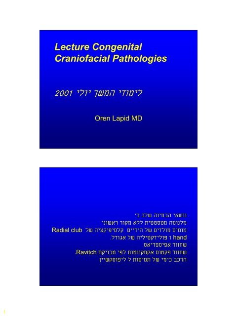
<strong>Lecture</strong> Congenital<br />
<strong>Craniofacial</strong> Pathologies<br />
2001 ילוי ךשמה ידומיל<br />
Radial club<br />
Oren Lapid MD<br />
'ב<br />
בלש הניחבה יאשונ<br />
ינושאר רוקמ אלל תיטטסטמ המונלמ<br />
לש היצקיפיסלק םיידיה לש םידלומ םימומ<br />
. לדוגא לש היליטקדילופ ו hand<br />
. Ravitch<br />
סאידפסיפא רוזחש<br />
תקינכט יפל םוטווקסקא סוטקפ רוזחש<br />
ןיישקסופיל ל תוסימת לש ימיכ בכרה
2<br />
<strong>Craniofacial</strong> surgery<br />
• Plastic surgery of the cephalic extremity<br />
– skull, face, Orbit<br />
– Congenital<br />
– Post traumatic<br />
– Tumors<br />
Craniosynostosis:<br />
• A general term encompassing a variety of<br />
developmental disorders characterized by<br />
inadequate capacity of the cranium to<br />
accommodate the growing brain, which<br />
results in compensatory deformities. (Less<br />
accurate for the minor forms where there is<br />
deformity with out problems of capacity)
3<br />
Simple: involves the cranial sutures,<br />
although there often is an extension into<br />
the base of the skull.<br />
Syndromatic: involves also the face with<br />
possible involvement of the limbs.<br />
Impact<br />
Aesthetic disfigurement<br />
Functional disorders: Increased ICP,<br />
hydrocephalus, visual impairment.
4<br />
Epidemiology<br />
• Detecteble abnormalities due to<br />
craniosynostosis are seen in<br />
1:1700-1900 births.<br />
• Most common types<br />
• Sagital schaphocephaly<br />
• Metopic trignocephaly<br />
• Unilateral coronal Plagiocephaly<br />
1:10,000<br />
• Bilateral coronal Brachicephaly<br />
Pathogenesis mechanisms to<br />
be completed
6<br />
ICP<br />
• Increased ICP is seen in cases of<br />
craniosynostosis more in cases where<br />
several sutures are involved.<br />
• Surgery decreases ICP.<br />
• A small skull doesn’t imply ICP elevation but<br />
ICP elevations usually seen in small skulls.<br />
Not all authors agree.<br />
• Signs of increased ICP<br />
• Papiledema, headaches, vomiting, lethargy,<br />
anorexia, beaten silver appearance on X-ray.
7<br />
Hydrocephalus<br />
• Hydrocephalus<br />
• Rare, seen more in the syndromatic<br />
cases.<br />
• Enlarged ventricles may be seen as part<br />
of the syndrome as in Apert’s. Changes<br />
in ventricle size are more accurate for<br />
diagnosis.<br />
Mental retardation<br />
• It is not clear how many of these children<br />
really have mental retardation. It is not clear if<br />
it is related to the problem or independent<br />
• Some just look retarded - environmental<br />
influences.<br />
• In children with non syndromatic<br />
craniosynostosis there was no increase in IQ<br />
after surgery. Other studies claim there was<br />
an improvmentt.
8<br />
Visual abnormalities<br />
• Optic atrophy and papilledema,<br />
stretching of optic nerve, pressure on<br />
the nerve. Damage from increased ICP.<br />
Morphology
9<br />
Acrocephaly ( also tower skull or<br />
oxycephaly of Virchow)<br />
Cranium in which the anterior part is<br />
higher than the posterior part, slanting<br />
in a front to back direction.
10<br />
Brachycephaly<br />
AP shortening, forehead and suprorbital<br />
bar are retropositioned.<br />
Results from bi-coronal stenosis. Typical<br />
of Apert’s and Crouzon.<br />
Oxycephaly<br />
Extreme upward growth with loss of AP &<br />
Lat diameters. Obtuse or absent<br />
nasofrontal angle. Usually a result of<br />
closure of the coronal suture.
11<br />
Turricephaly<br />
Exaggerated upward growth caused by<br />
frontoparietal suture closure. Seen in<br />
Apert’s<br />
Scaphocephaly<br />
Hull shaped cranium,reduced width with<br />
increased length, seen with premature<br />
sagital suture closure. The most<br />
common type. M:F 4:1
12<br />
Trigonocephaly<br />
Narrow and sharp forehead caused by<br />
premature closure of<br />
metopic suture. May be accompanied by<br />
hypotelorism.<br />
Clover leaf (Kleebattscaädel)<br />
Multiple sutural fusions, constriction ring<br />
in the lamboid-squamosal zone bulging<br />
of frontal and temporal lobes
13<br />
Plagiocephaly<br />
• Cranial asymmetry (various sutures)<br />
Plagiocephaly<br />
• Frontal<br />
– Ipsilateral coronal and frontosphenoidal sutures, flat<br />
forehead, backward displacement of orbit, bulging of<br />
contarlateral parietal area. Mandibular<br />
asymmetry.”harlequin” orbit.<br />
• Occipital<br />
– Flattening Lamboid suture must be distinguished from<br />
positional plagiocephaly.
14<br />
Plagiocephaly<br />
•Positional caused by extrinsic<br />
forces<br />
•Positional plagiocephaly does not<br />
require surgical treatment.<br />
Plagiocephaly
15<br />
Syndromes:<br />
• Different sutures may be involved in<br />
different patients with the same<br />
syndrome.<br />
• The difference may be in other parts of<br />
body.<br />
• Mental retardation found to some extent<br />
in all syndromes.
16<br />
Crouzon (<strong>Craniofacial</strong><br />
dysostosis)<br />
(Acrocephalosyndactyly, type<br />
II)<br />
• 1:25,000<br />
• Autosomal dominant (Most cases sporadic)<br />
Chromosome 10<br />
• Craniosynostosis, midfacial hypoplasia,<br />
exopthalmos.<br />
Crouzon cont<br />
• Various dformations of skull- usually brachy.<br />
• Midfacial retrusion – class III bite and shallow<br />
orbits. May cause damage to eyes. Beaked<br />
nose, High arched palate<br />
• Conductive hearing loss is common.<br />
• Synostosis develops during 1 st year of life<br />
usually complete by 3 rd .<br />
• Increased ICP common.
17<br />
Apert<br />
(Acrocephalosyndactyly,<br />
type I)<br />
• 1:160,000<br />
• Autosomal dominant (Most cases sporadic)<br />
• High cranial vault, flat posteriorly while<br />
bulging in the front. Brachycephalic,<br />
Turribracephalic.<br />
• Syndactyly of all four extremities- symmetrical<br />
middle three digits or more.
18<br />
Apert cont<br />
• Mild telorbitism, midfacial retrusion,<br />
deficient maxilla, abnormal dentition,<br />
Cleft palate 11-30%, low hair line,<br />
hypertrichosis of the eye brows, ptosis<br />
(mainly lateral), well developed<br />
protruding tongue.<br />
• Mental reardation seen in most.
19<br />
Pfeiffer<br />
(Acrocephalosyndactyly,<br />
type V)<br />
• Autosomal dominant (Most cases sporadic)<br />
Chromosome 8<br />
• Brachy-turricephalic, broad thumbs and great<br />
toes, occasional minor syndactyly,maxillary<br />
retrusion, possible high arched palate and<br />
submucous cleft.<br />
• Intelligence normal to mild mental retardation.
20<br />
Sathre-Chotzen<br />
(Acrocephalosyndactyly,<br />
type III)<br />
• Autosomal dominant (Most cases sporadic)<br />
chromosome 7<br />
• Brachycephalic cranium, various degrees of<br />
craniosynostosis, facial asymmetry, shallow<br />
orbits, telecantus, nasal septal deviation, low<br />
hair line minor syndactyly. Maxillary<br />
hypoplasia.<br />
• Intelligence normal to mild mental retardation.
21<br />
Carpenter<br />
• Autosomal recesive (Most cases sporadic)<br />
• Various cranial malformations, flat nasal<br />
canthi, dystrophic orbits, (possible anomalies<br />
of globe), low set ears<br />
• Hand (obligatory, syndactyly, brachydactyly,<br />
polydactyly)<br />
• Obesity, hypogonadism.<br />
• Mental retardation.<br />
Clefts<br />
• Major facial clefts 1.5-4.5/100,000 (CL<br />
CLP 1:750-1000)<br />
• Various degrees of such defects.
22<br />
Classification of Tessier<br />
• The orbit is the point of reference as it<br />
belongs both to cranial and facial clefts.<br />
• Counter clockwise, South to North<br />
• There may be both north and south<br />
components<br />
• Clefts always have hypoplasia (which may be<br />
minimal)<br />
• Pictures clefts 0-14
23<br />
Cleft 0-14 Cleft 1-13 bilateral<br />
Cleft 3 Cleft 4<br />
Cleft 5 bilateral<br />
Cleft 10<br />
Cleft 6 bilateral
24<br />
Clasification of Van der<br />
Mulen<br />
• The facial skeleton develops along an<br />
“S”<br />
• Focal fetal dysplasia,
25<br />
Telorbitism-Hypertelorism<br />
• Distance between<br />
anterior lacrimal<br />
crests<br />
• Men 19.5-<br />
30.7 (28) mm<br />
• Women 18.5-29.5<br />
(25) mm<br />
• 1 0 30-35 mm<br />
• 2 0 35-39 mm<br />
• 3 0 >40 mm<br />
Telorbitism-Hypertelorism<br />
• Up to 40 mm no problem<br />
• Tessier claims it is secondary to clefts or<br />
craniostenosis while Van-der-Mulen attributes<br />
it to arrest during 5 th -8 th weeks of<br />
embryogenesis.<br />
• Repair is undertaken to try and restore<br />
binocular vision and to improve the esthetic<br />
results.
26<br />
Frontoethmoidal<br />
Encephalomeningoceles<br />
• Failure of closure of the neural tube with<br />
herniation of CNS tissues from the<br />
forehead until the sacrum.<br />
• Nasofrontal, Naasoethmoidal and<br />
Nasoorbital<br />
• Rare seen mostly in Far East in<br />
Thailand 1:6000<br />
• Clefts 0,14, 1,13<br />
Oculomotor disturbances in<br />
craniofacial malformation.<br />
• Abnormal vectors of extraocular<br />
muscles or absence of muscles.<br />
• Altered intraocular distance and angle<br />
• Involvement of cranial nerves
27<br />
Laterofacial Microsomias<br />
• Represent groups of clefts 6,7,8<br />
Treacher Collins Complex<br />
(Franceschetti-Zwahlen-<br />
Klein Mandibulofacial<br />
dysostosis)<br />
• 1:25,000-50,000<br />
• Autosomal dominant. Chromosome 5<br />
• Variable penetration
28<br />
Treacher Collins<br />
• Bilateral and symetric<br />
• Convex profile, prominent nasal dorsum<br />
over a retrusive jaw and chin. Typical<br />
eyes, toungue shaped sideburns.<br />
Maldeveloped ears.<br />
• Complete form absence of the malar<br />
bone and the zygomatic arch.
29<br />
Treacher Collins<br />
• EYES<br />
– Eye<br />
• Strabismus, amblyopia<br />
• Lower eyelid<br />
Notch between lateral and<br />
middle third, absent eye lashes<br />
– Upper eyelid Microform coloboma<br />
– Eyebrow Notch<br />
– Lacrimal Absence of lower punctum<br />
– Lateral CantusNo insertion, free, small<br />
palpabrale fissure.<br />
– Orbit Open laterally because of the<br />
absent structures. The<br />
orbital floor is inclined<br />
outwards 45 0 .<br />
• Nose<br />
– Narrow, deviated, hooked, possible cohanal atresia.<br />
• Cheek<br />
– Sclerodermic furrow from the lower eye lid notch to the mandibular angle.<br />
• Oral comissure<br />
– Frequently macromastia<br />
• Alveola Palate<br />
– Narrow dental arch, high palate,<br />
• Maxilla& sinus<br />
– Under developed and small<br />
• Temporalis<br />
– The muscle is usually atrophic the masseter is not absent.<br />
• TMJ<br />
– Frequently hypoplastic<br />
• Mandible<br />
– Short ascending ramus. Lower body is hyoplastic.<br />
• Chin<br />
– Retrusion and increase in height.<br />
• Ear<br />
– Microtia and Cryptotia, Middle ear abnormalities<br />
• Cleft palate<br />
– May be present
30<br />
Nager syndrome (Acrofacial<br />
Dysostosis)<br />
• Similar to Treacher Collins but rarer<br />
• Autosomal recesive<br />
Binder’s Syndrome<br />
(Maxillonasal Dysplasia)<br />
• Short nose with flat bridge short<br />
columella
31<br />
Pierre Robin Sequence<br />
• Retrognathia, glosoptosis, airway obstruction.<br />
Cleft of soft and occasionally hard palate<br />
seen in 50%.<br />
• Etiology- late straightening of the fetus or late<br />
descent of the tongue.<br />
• Children have FTT de to difficulty breathing<br />
and feeding. Vicious cycle<br />
• Treatment- prone position, tongue adhesion.<br />
Hemifacial microsomia<br />
First and second branchiall<br />
arch syndrome<br />
Otomandibular dystosis<br />
<strong>Craniofacial</strong> microsomia<br />
Lateral facial dysplasia<br />
Otomandibular syndrome
33<br />
Hemifacial microsomia<br />
• No genetic background 1:3500-5600 births<br />
• Unilateral microtia, macrostomia, and failure of<br />
formation of the mandibular ramus and condyle.<br />
• Orbit, zygoma, temporal bone, maxilla and nose may<br />
be involved.<br />
• The defect may seem minor at birth but progresses<br />
with growth.<br />
• Soft tissue deficits range from normal to major<br />
deficiency of sub cutaneous tissue and muscle, the<br />
parotid may also be affected.<br />
• CNS Abnormalities: agenesis the corpus callosum,<br />
hydrocephalus, cranial nerve anomalies facial palsy)<br />
• Tongue depressor test.<br />
Pruzansky’s clasification by<br />
grades<br />
I Minimal hypoplasia<br />
II Deformed TMJ, distortion of<br />
condyle, ramusand sigmoid notch.<br />
•III complete absence of the ramus<br />
and glenoid fossa, the mandibular<br />
body ends in the molar area.
34<br />
Goldenhar Syndrome<br />
• Similar to Treacher Collins<br />
• Asymetrical skull, prominent frontal<br />
bossing, low hair line, mandibular<br />
hypoplasia, low set ears, colobomas of<br />
upper eyelids, epibulbar dermoids,<br />
accessory ear lobes, vertebral<br />
anomalies and a change in the clefting<br />
pattern.
35<br />
Romberg’s disease<br />
(Progresive facial<br />
hemiatrophy)<br />
• First or second decade of life. F:M 1.5:1<br />
• Unilateral in 95% of cases<br />
Etiology?<br />
• Infection, scleroderma,cervical<br />
sympathetic loss.<br />
• Progresive hemifacial atrophy of skin,<br />
soft tissue and bone.<br />
• Coup de sabre sign in 50%.
36<br />
Reconstruction<br />
• Reconstruction is under taken at least a<br />
year after the disease is static.<br />
• Reconstruction by soft tissue flaps and<br />
grafts and bone. Fat injection is also an<br />
option.<br />
Surgical repair
37<br />
Pre operative evaluation<br />
• History<br />
– Family, pregnancy..<br />
• Physical<br />
– Observation, palpation of sutures,<br />
fontanels, neurological exam.<br />
• Other abnormalities<br />
– search for other abnormalities<br />
Pre operative evaluation<br />
• Imaging<br />
– X-ray (including C-spine)<br />
– Cephalometris<br />
– CT 3DCT<br />
– MRI<br />
• Imaging has a major importance in the<br />
management of these cases with the<br />
possibility of performing mock surgery
38<br />
Timing of surgery<br />
• The consensus is before one year.<br />
• Earlier<br />
– Better growth, early relief of ICP.<br />
• Later<br />
– Child stands surgery better, nearer to adult<br />
size.<br />
• Facial osteotomies<br />
– are performed at a later age.<br />
Principles of craniofacial<br />
surgery<br />
• Work is on a dynamic structure<br />
Surgery is performed on a growing skull,<br />
growth can alter the results and at the same<br />
time may change the growth patterns.<br />
• Patients positioned according to the operative<br />
plan and area.<br />
• Wide dissection of affected areas, Bicoronal<br />
incisions.<br />
• Combined intra and extracranial approaches.
39<br />
• Extensive use of bone grafts<br />
• Bone grafts, free flaps, Distraction<br />
osteogenesis.<br />
• Strip craniotomies.<br />
• Total craniectomy (abandoned)<br />
• Dead space is replaced by brain growth.<br />
• Elevation of scalp flaps and then periostal<br />
flaps.<br />
• Via burr holes the dura is separated from the<br />
skull.<br />
• Starting at the burr holes the bones are<br />
sawed in-situ and also shaped and modified<br />
outside the patient.<br />
• The older the child the more brittle the bones<br />
– less easy to work.<br />
• Methods of fixation. Wires, plates, sutures.<br />
• Absorbable vs. permanent plates, Plate<br />
migration.
40<br />
Correction of simple sutural<br />
synostosis<br />
• Aim: correct functional and esthetic<br />
deficits<br />
• Strip craniotomies<br />
– Not suficent for an esthablished deformity<br />
but may be used in infancy, Bone growth<br />
and regeneration fill the defects.<br />
Correction of simple sutural<br />
synostosis<br />
• Plagiocephaly<br />
– Frontoorbital remodeling<br />
• Brachycephaly<br />
– Frontoorbital remodeling<br />
– The orbital bar is remodeled and repositioned in<br />
an appropriate location<br />
• Metopic<br />
– Deformed frontal bone Remodeled or<br />
replaced by parietal bone
43<br />
Correction of syndromatic<br />
sutural synostosis<br />
• Advancement of forehead and midface<br />
with monoblock frontal osteotomy.<br />
• Le fort osteotomies.<br />
• Facial bipartition for telorbitism.<br />
• Mid face deformities<br />
• Le Fort III, Le Fort III+I, Monoblock<br />
advancment
44<br />
Le Fort III<br />
Facial<br />
Bipartition
45<br />
Forehead Advancement<br />
Forehead Advancement Floating
46<br />
Forehead Advancement
47<br />
Forehead Advancement for Trigonocephaly
48<br />
Correction of Laterofacial<br />
Microsomias<br />
• Dental- orthodontic treatment can improve<br />
results and direct growth.<br />
• Distraction osteogenesis<br />
• Mandibular osteotomies<br />
• Bone grafts transfers for reconstruction of the<br />
mandibular ramus TMJ<br />
• Onlay bone grafts for contour restoration.<br />
Complications<br />
• Death 1.5-2%<br />
• Hemorhagic complications. There is<br />
significantand continous blood loss in<br />
these operations. Replacment required<br />
• Air emboli.
49<br />
Complications<br />
• Infection of soft tissues and bones more when<br />
there is a communication of bone with the<br />
respiratory system. Related to the length of<br />
surgery. Prophylactic antibiotics are given.<br />
• Infection (meningitis)<br />
• CSF leak<br />
• Ophthalmic complications- blindness.<br />
• Cerebral edema<br />
• Respiratory obstruction<br />
• Sagital sinus thrombosis<br />
• Seizures<br />
Thanks<br />
When will you fix me ?