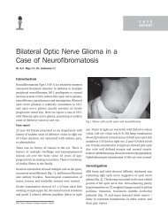
Management of Subluxated Lenses - KSOS
Management of Subluxated Lenses - KSOS
Management of Subluxated Lenses - KSOS

Create successful ePaper yourself
Turn your PDF publications into a flip-book with our unique Google optimized e-Paper software.
166 Kerala Journal <strong>of</strong> Ophthalmology Vol. XXI, No. 2<br />
shallowing <strong>of</strong> anterior chamber, a negative<br />
pressure in AC and further vitreous prolapse as<br />
the anterior segment is less pressurised than the<br />
posterior segment.<br />
12. Divide and or chop technique are preferred in eyes<br />
with zonular weakness. This technique minimises<br />
zonular stress during phacoemulsification if<br />
surgeon is careful to apply equal forces in opposing<br />
directions to avoid displacing the nucleus.<br />
13. “Visco dissect” nuclear halves / quadrants in areas<br />
<strong>of</strong> zonular weakness. The viscoelastic should be<br />
injected below the nuclear fragment and the<br />
capsular bag- lifting the nuclear fragment as well<br />
as expanding and stabilizing the capsular bag.<br />
Additional cortical removal by visco dissection<br />
will limit stress on the remaining zonules during<br />
aspiration <strong>of</strong> cortex.<br />
14. Automated Irrigation and Aspiration device is not<br />
preferred for cortex removal as it can hydrate<br />
vitreous and increase vit prolapse. Manually<br />
aspirate with a 24/ 27 G canula striping cortex in<br />
a tangential manner instead <strong>of</strong> radially to limit<br />
stress on zonules. A ‘J’ cannula can be used for<br />
sub incisional cortex .Ensure removal <strong>of</strong> all<br />
vitreous from the anterior chamber if it is present.<br />
Use ‘Dry vitrectomy’ with automated vitrector<br />
after filling anterior chamber with viscoelastics.<br />
For significant vitreous loss a bimanual vitrectomy<br />
should be performed.<br />
IOL placement options<br />
1. The surgeon should decide if it is safe to use an<br />
ACIOL or PCIOL.<br />
2. If an ACIOL is used the remnants <strong>of</strong> the capsular<br />
bag should be removed to prevent contraction and<br />
opacification.<br />
3. If the surgeon uses a PCIOL it should be either<br />
a. Sutured to the scleral wall or<br />
b. Placed in the capsular bag<br />
Ciliary sulcus placement <strong>of</strong> PCIOL without suture<br />
fixation in an eye with significant zonular compromise<br />
is not recommended.<br />
Placement <strong>of</strong> PCIOL into the capsular bag<br />
1. Placement <strong>of</strong> PCIOL into the capsular bag is<br />
challenging when there is significant zonular<br />
weakness as one must achieve IOL centration, and<br />
maximize long term stability.<br />
2. Use <strong>of</strong> 6 mm optic diametre IOL decreases the<br />
chances <strong>of</strong> undesirable edge - glare symptoms<br />
should lens decentration occur post operatively.<br />
Haptic configuration designed for broad contact<br />
with equatorial capsular bag increases the chances<br />
<strong>of</strong> long term centration. Use <strong>of</strong> silicone plate haptic<br />
IOL. should be avoided in the presence <strong>of</strong> zonular<br />
dialysis as there is greater chance <strong>of</strong> capsular<br />
contraction and decentration.<br />
3. Insertion <strong>of</strong> CTR to provide 360 0 capsular bag<br />
expansion and greater stabilization.<br />
4. If the ZD is located at the incision site, lens<br />
placement is more difficult.<br />
a. One Option is to first place the entire lens into<br />
the AC. Then using a two handed technique, the<br />
superior haptic is inserted into the capsular bag<br />
followed by a similar maneuvre for the inferior<br />
haptic.<br />
5. Orientation <strong>of</strong> the IOL: There are 2 schools <strong>of</strong><br />
thought.<br />
a. Orienting the IOL in a plane parallel to the zonular<br />
dialysis (ZD) in order to take advantage <strong>of</strong> the<br />
remaining intact zonules. This orientation will<br />
provide optimum support but may induce ovaling<br />
<strong>of</strong> the capsular bag and an increased risk <strong>of</strong><br />
postoperative decentration.<br />
b. Placing one haptic in area <strong>of</strong> ZD will ensure<br />
stretching <strong>of</strong> the bag and decrease ovaling.<br />
However it should be borne in mind that only one<br />
haptic is adequately supported.<br />
It is recommended to orient the haptics in whichever<br />
axis that provides the best centration intraoperatively.<br />
This is accomplished by careful rotation <strong>of</strong> PCIOL.<br />
Capsular Tension Ring (CTRs)<br />
Drs Witschel and Legler (1993) from Germany<br />
demonstrated that CTRs could provide both<br />
intraoperative and postoperative stabilization <strong>of</strong>