Macular Hole Surgery Sans Gas - KSOS
Macular Hole Surgery Sans Gas - KSOS
Macular Hole Surgery Sans Gas - KSOS

Create successful ePaper yourself
Turn your PDF publications into a flip-book with our unique Google optimized e-Paper software.
402 Kerala Journal of Ophthalmology Vol. XXI, No. 4<br />
ORIGINAL<br />
ARTICLE<br />
<strong>Macular</strong> <strong>Hole</strong> <strong>Surgery</strong> <strong>Sans</strong> <strong>Gas</strong><br />
Dr. Sonia Rani John DNB, Dr. Meena Chakrabarti MS DO DNB, Dr. Arup Chakrabarti MS DO<br />
Introduction<br />
<strong>Macular</strong> holes affect one in every 1,000 individuals,<br />
72 % of them are women between 60 and 70 years of<br />
age 1 with a bilaterality ranging between 6 and 22 %.<br />
Eyes with idiopathic macular hole have reduced vision,<br />
secondarily to the loss of tissue, cystic retinal changes,<br />
detachment of the retinal ring surrounding the hole<br />
and photoreception degeneration 2 . In 1988, <strong>Gas</strong>s<br />
described the pathogenesis of idiopathic macular hole,<br />
considering tangential vitreoretinal traction as the<br />
cause 3 . Also in 1988, Smiddy et al performed this<br />
surgery without being able to define the usefulness of<br />
vitrectomy and due to the progression of lens sclerosis<br />
and potential retinal complications; concluded that<br />
conservative management in this stage would be a<br />
better option 4 . Subsequently, studies were made in<br />
patients with stage 3 or 4 idiopathic macular hole in<br />
which pars plana vitrectomy with removal of the<br />
posterior vitreous and tamponade with expandable<br />
gas induced a healing of the macular hole in 58 - 97 %<br />
of cases, with visual improvement between 42 and<br />
85 % 2,5,6 .<br />
The first report of successful macular hole surgery was<br />
published by Kelly and Wendel in 1991 1 In recent years,<br />
a number of authors have attributed increased success<br />
rate of surgery to the use of healing adjuvants<br />
(autologous serum, transforming growth factor â,<br />
platelets or thrombin), the initial stage and duration<br />
of macular hole 2-8<br />
The surgical technique necessitates post operative face<br />
down posturing in order to achieve effective tamponade<br />
Chakrabarti Eye Care Centre, Kochulloor, Trivandrum 695 011 E-mail:<br />
tvm_meenarup@sancharnet.in<br />
of the macular hole. Indeed, there is evidence to suggest<br />
that longer duration of intraocular gas tamponade may<br />
have a favorable effect on the outcome of macular hole<br />
surgery 9,10 .<br />
Although 2 studies have reported comparable results<br />
with no face-down posture 11 and four days of<br />
posture 12 postoperatively, most studies advocate strict<br />
face-down posturing for at least one week after surgery<br />
as this is believed to be an important factors in closure<br />
of the hole. Because of this, macular hole surgery has<br />
been restricted to patients who are able to comply with<br />
the postoperative face-down posturing.<br />
A number of patients however are unable to maintain<br />
posture because of positioning difficulties due to neck,<br />
back, spine, chest, other diseases or social reasons. This<br />
study reviews our experience of macular hole surgery<br />
without gas tamponade in a consecutive series of<br />
25 patients and presents the results of a comparative<br />
analysis of patients undergoing macular hole surgery<br />
with and without postoperative gas tamponade.<br />
Clinical Objective<br />
To study whether gas tamponade was necessary to<br />
improve anatomic and functional outcomes in macular<br />
hole surgery.<br />
Materials and Methods<br />
This was a comparative case control study in which a<br />
retrospective analysis of 50 patients who underwent<br />
macular hole surgery at our centre between 2007 and<br />
2009 June . They were divided into 2 groups. Group I<br />
which included 25 patients in whom surgery was
December 2009 Sonia Rani John et al. - <strong>Macular</strong> hole surgery sans gas 403<br />
performed with intra operative gas tamponade and<br />
Group II which included 25 patients without gas<br />
tamponade. In both groups ILM peeling had been<br />
performed intraoperatively.<br />
The inclusion criteria comprised patients between<br />
50 and 80 years of age, males and females with macular<br />
hole diagnosed and evaluated based on clinical features,<br />
digital fluorescein angiogram and Optical Coherence<br />
Tomography (OCT) scan of stage 3 and 4 with a<br />
duration of greater than 6 months.<br />
The exclusion criteria were vitreous or retinal pathology,<br />
aphakia, uveitis, glaucoma, corneal pathology, high<br />
myopia and previous vitreoretinal surgery. In all cases,<br />
a detailed evaluation was made, including assessment<br />
of visual acuity, slit lamp evaluation, assessment of<br />
lenticular changes, non contact tonometry and indirect<br />
ophthalmoscopy. All patients also underwent digital<br />
fluorescein angiography, fundus imaging and OCT Scan.<br />
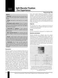
Fig. 1. Serial OCT pictures of a patient in Group 2 showing<br />
progression of macular hole over a 9 month period :<br />
(a) Vitreomacular traction (d) Full thickness macular<br />
hole (e) Postoperative OCT<br />
Surgical Technique<br />
Three port pars plana vitrectomy was performed by<br />
the same vitreoretinal surgeon (Dr. M C), with<br />
separation and removal of the posterior cortical vitreous<br />
after staining and demarcating it with triamcinolone<br />
acetonide. The internal limiting membrane was lifted,<br />
torn from the retinal surface by blunt dissection<br />
employing the technique of maculorhexis and peeled<br />
with a forceps. In patients whom gas tamponade was<br />
performed, a fluid / air exchange was done with<br />
infusion of air-perfluoropropane gas (C 3 F 8 ) mixture at<br />
a non-expansible concentration of 17 %. A thorough<br />
examination of 360 0 of the peripheral retina was<br />
performed with indirect ophthalmoscope and scleral<br />
indentation. The sclerotomies were then closed.<br />
Post operative management<br />
The post operative visits were scheduled on day 1,<br />
weekly for the 1 st 1 month and then monthly for the<br />
second, fourth, sixth and 12 th months. The post<br />
operative evaluation comprised of best corrected visual<br />
acuity ,non contact tonometry, slit lamp evaluation<br />
(to evaluate the condition of the cornea, lens, macular<br />
hole), indirect ophthalmoscopy and presence of<br />
complications, if any. OCT scans were performed on<br />
the 1 st , 6 th and 12 th month after the procedure.<br />
Results<br />
The study included 50 eyes of 50 patients, 17 males<br />
and 33 females, between 52 and 80 years of age with<br />
an average of 61.4 ± 11.9 years. (Table: 1)<br />
The pretreatment best corrected visual acuity ranged<br />
from 6/9 to counting fingers at half meter distance.<br />
The post operative visual acuity ranged from 6/9 to<br />
hand movements.<br />
Distribution of the sample patients according to<br />
duration of the macular hole is shown in table 2. 32 %<br />
of the patients had a duration of < 5 months while<br />
42 % between 5-8 months and 26 % of the patients<br />
had macular holes of more than 9 months duration.<br />
The average duration of holes prior to surgery was<br />
6.9 m ± 5.6 m.<br />
58 % of the patients had a macular hole size of > 400<br />
micrometre while 42 % had macular hole sized < 400<br />
micrometers (Table 3)<br />
Associated findings included cystoid macular edema<br />
(CME) 6 %, epiretinal membranes (ERM) 22 %,<br />
subretinal fluid (SRF) 4 % and Berlin’s edema 2 %.<br />
56 % of the cases with macular hole did not have any<br />
other associated findings. One patient had an associated<br />
peripheral hole which was lasered intraoperatively.<br />
Retinal tears and postoperative retinal detachments<br />
are the most serious posterior segment complications<br />
and they occurred in 12 % of cases (Table 4) 88 %
404 Kerala Journal of Ophthalmology Vol. XXI, No. 4<br />
Table 1. Distribution of the sample patients according to age<br />
Age Without gas With gas Total χ2 ρ<br />
Count Percent Count Percent Count Percent<br />
< 50 2 8.0 3 12.0 5 10.0 0.86 0.834<br />
50-59 5 20.0 4 16.0 9 18.0 60-69 14<br />
56.0 12 48.0 26 52.0 70-79 4 16.0 6<br />
24.0 10 20.0 Average 61.5 ± 11.9 61.3 ± 12.2 61.4 ± 11.9<br />
Table 2. Distribution of the sample patients according to duration<br />
Duration without gas With gas Total χ 2 p<br />
Count Percent Count Percent Count Percent<br />
400 11 44.0 18 72.0 29 58.0 4.02* 0.045<br />
< 400 14 56.0 7 28.0 21 42.0<br />
Table 4. Distribution of the sample patients based on intraoperative complications encountered.<br />
Intra OP Problem Without gas With gas<br />
Count Percent Count Percent<br />
Nil 22 88.0 21 84.0<br />
Retinal Tear 2 8.0 3 4.0<br />
Peripheral <strong>Hole</strong> 0 0.0 1 4.0<br />
of patients did not encounter any intra operative<br />
problems.<br />
36 % of the patients underwent staining of the internal<br />
limiting membrane (ILM) with indocyanine green dye<br />
while 4 % of the ILM was stained with brilliant blue<br />
green (BBG) and 60 % with trypan blue dye.<br />
50 % of the patients underwent perfluoropropane (C 3 F 8 )<br />
injection following ILM peeling while the remaining<br />
50 % did not received any gas.<br />
Patients who received C 3 F 8 gas underwent post<br />
operative positioning for a period of 3 weeks. (6 hrs /<br />
day X 1 week ; 4 hrs / day X 2 nd week ; and 2 hrs / day<br />
Table 5. Distribution of the sample patients according to closure.<br />
in the 3 rd week). They were instructed to maintain facedown<br />
posture for the prescribed duration and to sleep<br />
on either sides avoiding supine position.<br />
In 80 % of the patients in both the groups, the macular<br />
hole closed completely while the hole remained open<br />
in 20 % of the cases in both groups (Table 5)<br />
Post operative complications included cataract in<br />
40 % of patients who underwent macular hole<br />
surgery with gas and only 8% in those without gas.<br />
Glaucoma was encountered in 4 % of patients with gas<br />
while none of the patients in the other group developed<br />
glaucoma.<br />
Closure Without gas With gas Total χ2 p<br />
Count Percent Count Percent Count Percent<br />
closed 20 80.0 20 80.0 40 80.0 0 1.000<br />
open 5 20.0 5 20.0 10 20.0
December 2009 Sonia Rani John et al. - <strong>Macular</strong> hole surgery sans gas 405<br />
Discussion<br />
The rate of closure of idiopathic macular hole depends<br />
on many factors like size of the hole (measured by OCT<br />
scan), duration of symptoms, ILM peeling, type of<br />
tamponade used (air, SF6, C 3 F 8 , silicone oil) and length<br />
of face-down positioning (2 weeks, 1 week, 5 days,<br />
1 day or more).<br />
Majority of the surgeons use gas tamponade. A 2006<br />
survey by Mittra and Pollack showed that 63 % of surgeons<br />
use C3F8 while 33 % use SF6 as 6 weeks of visual<br />
disturbances with C3F8 is disabling for most patients.<br />
The patient compliance to postoperative positioning is<br />
also poor as indicated in a patient survey conducted by<br />
the AAO (Pollack J.S,Packo.J 2000). Only 3 %(4 weeks),<br />
8 % (3 weeks), 63 % (2 weeks) and 25 % (1week)<br />
compiled with the postoperative positioning.<br />
So if buoyancy of the gas is small and the patients are<br />
generally unable to comply with the positioning, then<br />
what are we positioning for ? A metanalysis of relevant<br />
studies on shortened post operative positioning is given<br />
below (Table 6).<br />
Park in 1999 conducted a study in 58 patients with<br />
macular hole and concluded that 91 % of the macular<br />
holes closed with gas tamponade and positioning for<br />
4 days.<br />
Isomac in 2002 studied the effect of C 3 F 8 tamponade<br />
and 1 day post operative positioning in 21 eyes with<br />
recent onset macular holes which also had a hole<br />
closure rate of 91 %.<br />
Table 6. Metanalysis of published articles on MHS with shortened duration of posturing.<br />
Sato in 2003 also attained 91% hole closure rate with<br />
air tamponade and 1 day face down positioning in a<br />
case series of 23 patients with small macular holes.<br />
Krohn in 2005 studied 24 eyes with full thickness<br />
macular hole who underwent pars plana vitrectomy<br />
along with C 3 F 8 tamponade and 3 days post operative<br />
face down positioning and showed that 87.5 % of the<br />
macular holes closed well while Wockens in 2006<br />
achieved 95 % closure rate (21 eyes case series) with<br />
C 3 F 8 gas tamponade and 3 days face down positioning.<br />
Toruambe in 1997 achieved 79 % closure rate with C 3 F 8<br />
tamponade alone without any face down positioning,<br />
while Simcock in 2001 with C 3 F 8 tamponade alone<br />
achieved 90 % closure rate.<br />
Tranos and Merkur in 2007 used C 3 F 8 alone for<br />
tamponade and had a hole closure rate of 88 % and<br />
92 % respectively.<br />
Thus smaller, recent holes will likely close with ILM<br />
peeling and minimal or no positioning (as long as<br />
patient avoids the supine position). Most holes will close<br />
with ILM peeling, SF 6 gas and 1 day of positioning. For<br />
chronic or larger holes or if there are doubts regarding<br />
the patients ability to position, 3 or more days of face<br />
down positioning or C 3 F 8 gas can be considered.<br />
Clinical studies have shown that small holes that are<br />
of recent onset will have high success rates using almost<br />
any technique. Tadayoni et al (Br J Ophthalmol 2006)<br />
has shown that 100 % hole closure can be obtained for<br />
holes < 400 micrometre, with or without ILM peel.<br />
STUDY PATIENT SIZE/DURATION GAS POSITION CLOSURE<br />
PARK 1999 58 ALL AIR 4 DAYS 91%<br />
ISOMAC 2002 21 RECENT C3F8 1 DAY 91%<br />
SATO 2003 23 SMALL AIR 1 DAY 91%<br />
KROHNS 2005 24 ALL C3F8 3 DAYS 87.5 %<br />
WICKENS 2006 21 ALL C3F8 3 DAYS 95 %<br />
Table 7. <strong>Hole</strong> Closure Rates for macular holes without positioning<br />
STUDY PATIENT SIZE/DURATION ILM GAS POSITION CLOSURE<br />
TORNABE 33 20 % CHRONIC - C3F8 NONE 79 %<br />
SIMCOCK 20 STAGE II/III - C2F6 NONE 90 %<br />
TRANOS 16 ALL + C3F8 NONE 88 %<br />
MERKUR 72 RECENT + C3F8 NONE 92 %
406 Kerala Journal of Ophthalmology Vol. XXI, No. 4<br />
100 % closure could be obtained with ILM peel in<br />
macular holes > 400 micrometre while only 73 % of<br />
the macular holes closed without ILM peel.<br />
Although ILM peel remains controversial, most<br />
surgeons peel ILM for idiopathic holes. ILM peeling<br />
allows for complete removal of perifoveal vitreous<br />
traction.<br />
Most surgeons use C 3 F 8 for gas tamponade. However<br />
6 weeks of visual disturbance with C 3 F 8 is disabling for<br />
most patients.<br />
The buoyant pressure and interfacial tension of the gas<br />
tamponade reattaches retina. C 3 F 8 allows for 2-3 weeks<br />
of contact between hole and gas if supine position is<br />
avoided. Longer acting gas allows for more leeway in<br />
positioning. Shorter acting gas requires more<br />
positioning to achieve hole / gas contact as bubble<br />
dissipates. Positioning may actually be more critical as<br />
the bubble dissipates.<br />
<strong>Hole</strong> closure rates for macular holes without positioning<br />
has been studied by several workers and the results<br />
show that it does not affect closure rate or functional<br />
outcomes (Table: 7)<br />
The anatomic and functional outcomes in macular hole<br />
surgery are similar irrespective of whether<br />
intraoperative gas tamponade was used or not in the<br />
present study. Face down positioning is burdensome<br />
for the patient with reported side effects like ulnar<br />
neuropathy.<br />
We decided to conduct our study without gas to<br />
tamponade the macular hole in 20 consecutive cases<br />
of idiopathic 3 and 4 macular holes and compared our<br />
results with our own series where C3F8 was used for<br />
postoperative tamponade. The rate of hole closure was<br />
similar in both groups, with the added advantage of<br />
fewer complications in the group where gas was not<br />
used. The patients were also saved from the discomfort<br />
of postoperative positioning, lesser progression of<br />
cataract and negligible postoperative glaucoma.<br />
However for repeat procedures and large chronic holes,<br />
tamponade should still be considered.<br />
References<br />
(1) Aakerg TM, Blair CI, <strong>Gas</strong>s JD <strong>Macular</strong> holes Am J<br />
Ophthalmol 1970; 69; 555-562.<br />
(2) Wendel RT, Patel AC, Kelly NE, Salzane TC, Wells JW,<br />
Novak GD. Vitreous surgery for macular holes.<br />
Ophthalmology 1993; 100: 1671-1676.<br />
(3) <strong>Gas</strong>s JD. Idiopathic senile macular hole. Its early stages<br />
& pathogenesis. Arch Ophthalmol 1988; 106: 629-639.<br />
(4) Smiddy WE, Michels RG, Glaser BM, de Bustros S.<br />
Vitrectomy for impending idiopathic macular holes. Am<br />
J Ophthalmol 1988; 105:371-376.<br />
(5) Kutchins RK. Complications of macular hole surgery.<br />
Ophthalmology 1998 105:762.<br />
(6) Sjaarda RN, Glaser BM, Thompson JT, Murphy RP,<br />
Hanham A. Distribution of iatrogenic retinal breaks in<br />
macular hole surgery. Ophthalmology 1995; 102:1387-<br />
1392.<br />
(7) Chen CJ. Glaucoma after macular hole surgery.<br />
Ophthalmology 1998; 105:94-100.<br />
(8) Torrambe PE, Poliner LS, Grote K. <strong>Macular</strong> hole surgery<br />
without face-down positioning. A pilot study-Retina<br />
1997: 17:179-185.<br />
(9) Thompson JT, Sjaarda RN, Lansing MB. The results of<br />
vitreous surgery for chronic macular holes. Retina 1997:<br />
17:493-501.<br />
(10) Kelly NE, Wendel. R, T. Vitreous surgery for idiopathic<br />
macular holes; results of a pilot study. Arch Ophthalmol<br />
1991; 109:654-659.<br />
(11) Willis AW, Garcia-Cosio JF. <strong>Macular</strong> hole surgery:<br />
comparison of long standing versus recent macular<br />
holes. Ophthalmology 1996; 103:1811-1814.<br />
(12) Thompson JT, Smiddy WE, Glasor BM et al. Intraocular<br />
tamponade duration and success of macular hole<br />
surgery. Retina 1996; 16:373-382.