Ocular Surface Disorders - Current Concepts and ... - KSOS
Ocular Surface Disorders - Current Concepts and ... - KSOS
Ocular Surface Disorders - Current Concepts and ... - KSOS

Create successful ePaper yourself
Turn your PDF publications into a flip-book with our unique Google optimized e-Paper software.
66 Kerala Journal of Ophthalmology Vol. XX, No. 1<br />
CURRENT<br />
CONCEPTS<br />
<strong>Ocular</strong> <strong>Surface</strong> <strong>Disorders</strong> - <strong>Current</strong><br />
<strong>Concepts</strong> <strong>and</strong> Management<br />
Dr. Jayasree Menon P. MS, Dr. C.V. Anthrayose MS, Dr. Alex Joseph MS<br />
<strong>Ocular</strong> <strong>Surface</strong> <strong>Disorders</strong><br />
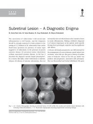
<strong>Ocular</strong> surface disorders are a group of disorders 1 of<br />
diverse pathogenesis in which, the disease results in failure<br />
of mechanisms that maintain a healthy ocular surface.<br />
(Fig. 1)<br />
Fig. 1. OSD with unhealthy ocular surface<br />
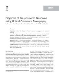
<strong>Ocular</strong> <strong>Surface</strong><br />
The ocular surface refers to the<br />
mucosal lining between upper <strong>and</strong><br />
lower eyelids. The functional unit<br />
is comprised of tear film,<br />
conjunctival epithelium, limbal<br />
epithelium, corneal epithelium,<br />
lacrimal gl<strong>and</strong> <strong>and</strong> meibomian<br />
gl<strong>and</strong> 2 (Fig. 2).<br />
The 2 components essential for<br />
maintaining ocular surface<br />
health 3 Fig. 2. Lacrimal gl<strong>and</strong><br />
<strong>and</strong> drainage<br />
apparatus<br />
are<br />
(1) Healthy ocular surface epithelium <strong>and</strong><br />
(2) Normal, stable preocular tear film<br />
These act as a vicious cycle.<br />
Jubilee Mission Medical College, Thrissur<br />
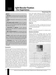
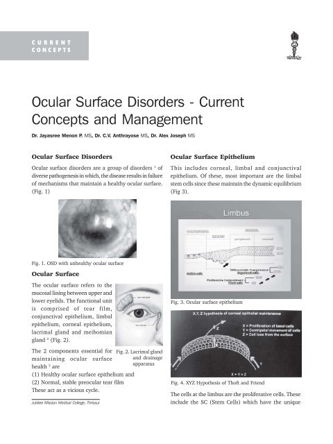
<strong>Ocular</strong> <strong>Surface</strong> Epithelium<br />
This includes corneal, limbal <strong>and</strong> conjunctival<br />
epithelium. Of these, most important are the limbal<br />
stem cells since these maintain the dynamic equilibrium<br />
(Fig 3).<br />
Fig. 3. <strong>Ocular</strong> surface epithelium<br />
Fig. 4. XYZ Hypothesis of Thoft <strong>and</strong> Friend<br />
The cells at the limbus are the proliferative cells. These<br />
include the SC (Stem Cells) which have the unique
March 2008 Jayasree Menon et al. - OSD 67<br />
property of self renewal <strong>and</strong> the TAC (Transiently<br />
Amplifying Cells) that are derived from mitotic division<br />
of stem cells <strong>and</strong> amplify by undergoing a few rounds<br />
of cell division. Cells in the non proliferative compartment<br />
are the PMC (Post Mitotic Cells) which are in the<br />
different stages of maturation by differentiation into<br />
the TDC (Terminally Differentiated Cells) 3<br />
This follows the limbal stem cell X,Y,Z hypothesis<br />
proposed by Thoft & Friend 4 in 1983 (Fig. 4).<br />
<strong>Ocular</strong> <strong>Surface</strong> Defence<br />
The 2 factors essential for the ocular surface defence 3<br />
are<br />
(1) Normal, adequate <strong>and</strong> stable tear film <strong>and</strong><br />
(2) Normally functioning hydrodynamic factors<br />
The tear film consists of<br />
mucin layer, aqueous<br />
layer <strong>and</strong> lipid layer<br />
(Fig. 5).<br />
Hydrodynamic factors<br />
include periodic,<br />
adequate <strong>and</strong> complete<br />
lid blinking to<br />
distribute an even tear<br />
film over the ocular<br />
surface <strong>and</strong> proper tear<br />
clearance to ensure adequate turnover <strong>and</strong> refreshment 3 Fig. 5. Layers of tear film<br />
.<br />
Thus, the eyelids, the external adnexal gl<strong>and</strong>s <strong>and</strong> the<br />
ocular surface epithelia all play a major role in<br />
maintaining a normal tear film <strong>and</strong> ocular surface<br />
(Fig. 6).<br />
Two neuronal reflex arcs function in this process. For<br />
both the arcs, the 1 st branch of trigeminal nerve controls<br />
the ocular sensitivity as the afferent sensory input <strong>and</strong><br />
the parasympathetic branch <strong>and</strong> the motor branch of<br />
the facial nerve are the afferent output 3 (Fig. 7).<br />
Fig. 6. Normal ocular surface breaks in the tear film<br />
Fig. 7. Neoronal feed back loop on which normal tearing<br />
depends<br />
These 2 components are interrelated. An alteration in<br />
the quantity or quality of any of the elements of the<br />
tear film can lead to an unstable tear film <strong>and</strong> secondary<br />
changes in the epithelium. Vice versa, primary changes<br />
of the ocular surface epithelium as part of ocular surface<br />
failure can lead to a secondary dry eye. Thus, an<br />
intimate relationship exists between the two <strong>and</strong> any<br />
change in this can lead to the occurrence of various<br />
ocular surface disorders.<br />
<strong>Ocular</strong> <strong>Surface</strong> Failure - Etiopathogenesis<br />
2 major types of ocular surface failure have been<br />
identified based on the epithelial phenotype in<br />
impression cytology 5 .<br />
(1) With intact limbal stem cells<br />
(2) With limbal stem cell deficiency<br />
With intact limbal stem cells<br />
Here, the normal nonkeratinized ocular surface<br />
epithelium undergoes squamous metaplasia into<br />
keratinized epithelium 5,6 . This is also associated with<br />
loss of goblet cells <strong>and</strong> mucin expression. This altered<br />
epithelial differentiation renders the ocular surface<br />
epithelium non wettable. This leads to an unstable tear<br />
film, the hallmark of various dry eye disorders.<br />
This is usually due to poor ocular surface defense <strong>and</strong><br />
dry eye may be secondary to 7,8,9 :<br />
(a) Aqueous tear deficiency<br />
Idiopathic – Age related, Hormonal<br />
Hyponutritional – Vit.A deficiency
68 Kerala Journal of Ophthalmology Vol. XX, No. 1<br />
Sensory denervation – After surgery or keratitis<br />
Collagen vascular diseases – Rheumatoid arthritis,<br />
Wegeners, SLE<br />
Sjogren’s syndrome <strong>and</strong> other autoimmune<br />
disorders<br />
Drugs – Oral Contraceptives, Antidepressants,<br />
Antihistamines, Beta blockers<br />
Lacrimal gl<strong>and</strong> infiltration – Amyloidosis,<br />
Sarcoidosis, Tumors<br />
Lacrimal gl<strong>and</strong> fibrosis – Radiation<br />
Contact lens use<br />
(b) Lipid tear layer deficiency<br />
Blepharitis<br />
Acne rosacea<br />
Contact lens<br />
(c) Mucin tear layer deficiency<br />
Vit.A deficiency<br />
Trachoma<br />
Mucocutaneous disorders<br />
Contact lens<br />
Conjunctival scarring – <strong>Ocular</strong> pemphigoid,<br />
Steven Johnson Syndrome, Chemical burns<br />
Topical medications<br />
(d) Increased tear film evaporation<br />
Lagophthalmos<br />
Ectropion<br />
Computer vision syndrome<br />
Lid retraction<br />
Exophthalmos<br />
Defective sensation<br />
(e) Delayed tear clearance<br />
Obstruction of tear outflow<br />
Ineffective lacrimal pump<br />
With limbal stem cell deficiency<br />
Here, the normal corneal epithelium is replaced by<br />
conjunctival epithelium. The salient features here are<br />
conjunctivalisation, vascularisation, chronic<br />
inflammation, poor epithelial integrity manifested as<br />
an irregular surface, recurrent erosion <strong>and</strong> persistent<br />
ulcer, destruction of basement membrane <strong>and</strong> fibrous<br />
ingrowth 3 . Conjunctivalization is the hallmark of limbal<br />
stem cell deficiency. Conditions with limbal stem cell<br />
deficiency can be classified in terms of the following.<br />
(a) Primary limbal stem cell deficiency<br />
These patients exhibit a gradual loss of limbal stem cell<br />
function over time. Seen in association with Peripheral<br />
keratitis<br />
Pterygium/pseudopterygium<br />
Aniridia<br />
Neurotrophic keratopathy<br />
(b) Secondary limbal stem cell deficiency<br />
These patients have a clear pathogenic cause that is<br />
responsible for destruction of limbal stem cells.<br />
Chemical / Thermal burns<br />
Steven Johnson Syndrome<br />
<strong>Ocular</strong> rosaceae<br />
<strong>Ocular</strong> pemphigoid<br />
Contact lens wear<br />
Multiple surgeries/Cryotherapies<br />
Antimetabolite (5 FU) toxicity<br />
Etiology – Common factors<br />
Aging<br />
Hormonal - Post menopausal females<br />
Excessive computer use – reduced blinking<br />
Excessive use of contact lens<br />
Eye surgeries, injuries<br />
Drugs<br />
Keratoconjunctivitis sicca<br />
Classification<br />
The Madrid Triple Classification of Dry Eye 10<br />
Dry eye classified depending upon 3 factors –<br />
Etiopathogenesis, Anatomo-pathologic <strong>and</strong> Severity<br />
The features are:<br />
A. Classification according to etiopathogenesis<br />
The etiologic factors are divided into 10 groups:<br />
1. Age related – With aging, all cellular structures of<br />
body undergo a progressive apoptosis including<br />
lacrimal gl<strong>and</strong>s. The lacrimal secretion begins to<br />
diminish from the age of 30 years <strong>and</strong> becomes<br />
insufficient for the necessities by 60 years.<br />
2. Hormonal – Lacrimal secretion is affected by some<br />
endocrine gl<strong>and</strong> activity, the most important of which<br />
are <strong>and</strong>rogens, estrogens <strong>and</strong> prolactin. Aqueoserous<br />
<strong>and</strong> lipid secretions are the most affected.
March 2008 Jayasree Menon et al. - OSD 69<br />
3. Pharmacologic – Systemic- Antidepressants<br />
(Fluoxetine, Imipramine), Anxiolytics<br />
(Bromazepam, Diazepam, Clorazepate),<br />
Antiparkinsons (Bipiredin, Benztropine), Diuretics<br />
(Chlorthalidone, Frusemide), Antihypertensives<br />
(Clonidine, Chlorothiazide), Anticholinergics<br />
(Atropine, Metoclopramide), Antihistaminics<br />
(Dexchlorpheniramine, Cetrizine), Antiarrythmics<br />
(Disopyramide, Mexiletine)<br />
Topical – Preservatives (Benzalkonium chloride,<br />
Thiomersal, Chlorobutanol, EDTA), Anasthetics<br />
(Tetracaine, Proparacaine, Lidocaine)<br />
4. Immunopathic 11 – Autoimmune disorders –<br />
(1) Primary Sjogren’s syndrome – those<br />
preferentially affecting gl<strong>and</strong>s – where<br />
vasculitis by immune complex deposits,<br />
pseudolymphomas <strong>and</strong> lymphomas are<br />
sometimes associated.<br />
(2) Secondary Sjogren’s syndrome which<br />
includes Rheumatoid Arthritis, Systemic<br />
Lupus erythematosis, Dermatomyositis,<br />
Scleroderma etc.<br />
(3) Autoimmune attack of other tissues <strong>and</strong><br />
secondary destruction of gl<strong>and</strong>s as in Steven<br />
Johnsons Syndrome, <strong>Ocular</strong> pemphigoid etc.<br />
(4) Affecting other tissues – Thyroid <strong>and</strong> adrenal<br />
insufficiency<br />
5. Hyponutritional- Vit.A deficiency, Omega 3 FA<br />
deficiency, Vit. B2, B12 <strong>and</strong> C deficiency<br />
6. Dysgenetic – Genetic <strong>and</strong> congenital diseases that<br />
affect one or several types of dacryogl<strong>and</strong>s –<br />
Aqueoserous (Alacrima, Dysplasia ectodermica<br />
anhidrotica), Lipid (Blepharophimosis syndrome,<br />
Keratopathy- icthyosis- deafness syndrome, First<br />
branchial arc syndrome), Mucin (Aniridia, Bietti<br />
syndrome), <strong>Ocular</strong> surface epithelium (Meesmann<br />
dystrophy, Cogan microcystic dystrophy)<br />
7. Inflammatory/Infectious – Dacryoadenitis,<br />
Blepharitis, Trachoma, H.simplex, H.zoster<br />
8. Traumatic – Surgical, Chemical, Radiation<br />
induced, Accidental<br />
9. Neurologic – Lacrimal secretion is very dependent<br />
on nervous stimulation.<br />
(1) Hypothalamic <strong>and</strong> limbic influences –<br />
Circadian rhythm of tear production that is<br />
maximum at morning <strong>and</strong> noon <strong>and</strong><br />
minimum at night. Limbic influences such as<br />
anxiety, tiredness, psychic influences <strong>and</strong><br />
somnolescence diminish the basal tear<br />
secretion.<br />
(2) Afferent neurodeprivation – Any condition<br />
causing ocular surface anesthesia diminishes<br />
lacrimal secretion.<br />
(3) Efferent neurodeprivation – Trauma, Tumors,<br />
Botulinum toxin injection.<br />
10. Tantalic – These patients despite having enough<br />
tears, have a dry ocular surface. There are 3 types<br />
of tantalic dry eyes 12 :<br />
(1)Lid-eye incongruency – Lid cannot create,<br />
maintain <strong>and</strong> reshape the tear film onto the<br />
ocular surface as in lid palsy, ectropion, lagophthalmos,<br />
lid coloboma, exophthalmos, local<br />
protrusion of pterygium or dermoid cyst etc.<br />
(2)Epitheliopathic – Epithelial dystrophies, limbal<br />
deficiency, Corneal conjunctivalisation,<br />
Endothelial decompensation etc. makes<br />
corneal epithelium less wettable.<br />
(3)Evaporation – Environmental conditions like<br />
hot dry climates, excessive air conditioning,<br />
open car window etc.<br />
Most of the dry eye conditions are multifactorial<br />
B. Classification according to damaged<br />
gl<strong>and</strong>s <strong>and</strong> tissues<br />
The affected parts of the lacrimal basin may be<br />
summarized in this histopathologic classification with<br />
the acronym ALMEN;<br />
A – Aqueoserous deficiency<br />
L – Lipid deficiency<br />
M – Mucin deficiency<br />
E – Epithelial deficiency<br />
N – Nondacryologic exocrine deficiencies<br />
C. Classification according to severity<br />
In the initial Madrid classification severity of dry eye<br />
was divided into 5 grades:
70 Kerala Journal of Ophthalmology Vol. XX, No. 1<br />
Subclinical – Symptoms only when overexposure<br />
Mild – Habitual symptoms<br />
Moderate – Symptoms plus reversible signs<br />
Severe – Symptoms plus permanent signs<br />
Disabling – All of the above plus visual incapacity<br />
Recent Triple Classification of dry eye for<br />
practical clinical use<br />
In 8 th congress of the International Society of<br />
Dacryology <strong>and</strong> Dry eye (Madrid, April, 2005), the<br />
previous Triple classification of dry eye approved in<br />
the XIV congress of European Society of Ophthalmology<br />
(Madrid, June, 2003) was modified.<br />
Here, classification according to etiopathogenesis <strong>and</strong><br />
affected gl<strong>and</strong>s <strong>and</strong> tissues are retained. Classification<br />
depending on severity was modified into 3 grades for<br />
practical use<br />
(1) Grade 1 or Mild – Symptoms without slit lamp<br />
signs<br />
(2) Grade 2 or Moderate – Symptoms with reversible<br />
signs<br />
(3) Grade 3 or Severe – Symptoms with permanent<br />
signs.<br />
Goals of Therapy<br />
Major goals include:<br />
1. Supplementation of a deficient tear film<br />
2. Preservation of the available tear by reestablishment<br />
of lid motility with normal lidcorneal<br />
congruity.<br />
3. Supplementation of limbal tissue containing<br />
epithelial stem cells for the management of<br />
epithelial disease of cornea.<br />
4. Improvement or supplementation of a basement<br />
membrane substrate.<br />
5. Restoration of clear visual axis.<br />
Treatment Guidelines<br />
A. Medical<br />
Along with tear substitutes, it is important to treat coexisting<br />
lid disease like blepharitis, trichiasis <strong>and</strong><br />
meibomian gl<strong>and</strong> dysfunction as well as to preserve<br />
the available tear by punctual occlusion.<br />
1. Tear substitutes <strong>and</strong> Lubricants<br />
Lubricants act as a physical means of protecting<br />
already compromised ocular surface from desiccation<br />
<strong>and</strong> irritation, but the preservatives like benzalkonium<br />
chloride may counteract the benefit 12 . Hence<br />
preservative free lubricants are preferable although<br />
more expensive.<br />
Guide to various types of therapy for specific tear film<br />
abnormalities 13<br />
Abnormality Forms of therapy<br />
Aqueous deficiency Artificial tears, Lubricants<br />
Punctal occlusion, Tarsorraphy,<br />
Moist chamber spectacles<br />
Mucin deficiency Artificial tears, Lubricants<br />
Acetylcysteine<br />
Immunosuppressants, Vit. A<br />
Lipid deficiency Lid hygiene, Warm compress<br />
Oral Tetracycline<br />
Tear spreading/ Artificial tears/ Lubricants<br />
Lid problems Taping lids/ Tarsorraphy/ Other lid<br />
surgeries<br />
Tear base (Epithelial) Artificial tears/ Lubricants<br />
Therapeutic soft CL<br />
Anterior stromal puncture/<br />
Excimer Photo Therapeutic<br />
Keratoplasty<br />
2. Nutritional Supplements<br />
Boerner et al 14 found 98 % patients reported improvement<br />
in the symptoms with omega 3 supplementation. Omega<br />
3 fatty acids produces anti-inflammatory eicosanoid<br />
acid which suppress inflammation by blocking the gene<br />
expression of pro inflammatory cytokines.<br />
3. Tear Stimulants (Secretagogues) 15<br />
(a) Diquafosol tetrasodium<br />
This molecule acts as a uridine nucleotide analogue<br />
that acts as a agonist of P2Y2 receptor present on<br />
ocular surface. Diquafosol increases water transport via<br />
chloride channel activation <strong>and</strong> enhances nongl<strong>and</strong>ular<br />
secretion of tear fluid. The drug is under clinical trial.<br />
(b) Pilocarpine<br />
Pilocarpine is a cholinergic parasympathomimetic that<br />
binds to muscarinic M3 receptors <strong>and</strong> stimulates<br />
salivary <strong>and</strong> lacrimal gl<strong>and</strong>s.<br />
(c) Cevimeline<br />
Cevimeline is an acetylcholine analogue <strong>and</strong> has high
March 2008 Jayasree Menon et al. - OSD 71<br />
affinity for muscarinic M3 receptors of salivary <strong>and</strong><br />
lacrimal gl<strong>and</strong>s<br />
(d) Eledoisin<br />
Eledoisin is an endecapeptide which when applied<br />
locally has secretostimulant effect.<br />
(e) Mucinous stimulators<br />
Bromhexine <strong>and</strong> N-acetylcysteine are stimulants of<br />
mucin production. Topical medicines such as geranyl<br />
farnesylacetate <strong>and</strong> hydroxyeicosatetraenoic acids have<br />
been introduced which improve the epithelium <strong>and</strong><br />
stimulate the goblet cells.<br />
B. Suppression of Inflammation<br />
(1) Corticosteroids<br />
Topical corticosteroids are beneficial for ocular surface<br />
defects associated with intense inflammation as in<br />
Sjogrens syndrome, Pemphigoid, Steven Johnsons<br />
Syndrome. The anti-inflammatory effect of corticosteroids<br />
are mediated through stabilization of the cytoplasmic<br />
<strong>and</strong> lysosomal membranes, thereby reducing the release<br />
of inflammatory mediators <strong>and</strong> inhibiting chemotaxis.<br />
However, careful monitoring of these patients is<br />
essential to watch out for steroid related complications.<br />
(2) Non steroidal anti-inflammatory agents<br />
Antiinflammatory agents like salicylates, indomethacin,<br />
flurbiprofen <strong>and</strong> progestational steroids reduce<br />
inflammation without suppressing wound repair.<br />
(3) Immunosuppressives<br />
Immunosuppressants like antimetabolites have been<br />
effective in treating ocular surface disorders of<br />
autoimmune origin like rheumatoid arthritis, Systemic<br />
Lupus Erythematosus <strong>and</strong> ocular cicatricial pemphigoid<br />
C. Limitation of Tissue Destruction<br />
1. Tissue Adhesives<br />
Tissue adhesives like isobutyl cyanoacrylate have been<br />
used as an adjuant in the management of corneal ulcers<br />
<strong>and</strong> small perforations 16 . It gives structural support<br />
<strong>and</strong> can arrest further stromal loss. Early application<br />
of tissue adhesive in the management of stromal melts,<br />
can postpone or reduce the need for keratoplasty or<br />
conjunctival flaps.<br />
2. Mechanical protection of corneal epithelium<br />
Methods:<br />
(a) Taping of lids<br />
(b) Tarsorraphy<br />
(c) Botulinum induced ptosis<br />
(d) Therapeutic soft contact lens<br />
This is useful to protect the loosely adherent remaining<br />
transient amplifying cells or regenerating epithelium<br />
from the action of blinking eyelids has significantly<br />
improved the management of persisting epithelial<br />
defects 17 . Soft contact lenses are undesirable in dry<br />
eye patients because of the high risk of infection. Only<br />
silicone provides adequate oxygen transmission for<br />
continuous wear, however Omafilcon A (proclear), a<br />
novel biomimetic, 59 % water content hydrogel soft<br />
contact lenses for daily wear has been found to give<br />
better comfort.<br />
3. Promotion of epithelial wound healing <strong>and</strong><br />
differentiation<br />
(a) Topical autologous serum<br />
Topical autologous serum not only moistens the ocular<br />
surface, but also provides necessary tear proteins such<br />
as epidermal growth factor (EGF), vitamin A,<br />
transforming growth factor-B (TGF-B), fibronectin <strong>and</strong><br />
other cytokines. The effect of EGF is primarily on<br />
epithelial wound healing whereas fibronectin appears<br />
to be involved in stromal healing.<br />
(b) Topical retinoids<br />
These are essential for epithelial growth <strong>and</strong><br />
differentiation. All trans retinoic acid 0.05 % in Vaseline<br />
BD has shown to increase the epithelial healing rate 18 .<br />
Retinol palmitate ophthalmic solution has also shown<br />
an increase in goblet cells <strong>and</strong> non keratinized cells.<br />
(c) Topical trisodium citrate 10 % <strong>and</strong> sodium<br />
ascorbate 10 %<br />
These have been found to reduce the incidence of<br />
ulceration <strong>and</strong> perforation in the immediate treatment<br />
of alkali burns.<br />
(d) Topical Cyclosporin A 0.05 % & 0.1 %<br />
Its efficacy may be due to an immunomodulatory <strong>and</strong><br />
anti-inflammatory effect on the ocular surface, thus
72 Kerala Journal of Ophthalmology Vol. XX, No. 1<br />
facilitating ocular surface healing. This has been used<br />
in severe forms of dry eye where long term use of<br />
corticosteroids are required.<br />
D. Preservation of natural tears<br />
(a) Punctal occlusion<br />
Punctal <strong>and</strong> canalicular closure increases mainly the<br />
aqueous component of natural tears but also has<br />
secondary beneficial effects on goblet cell density, tear<br />
film stability, <strong>and</strong> tear osmolality 20 . This also increases<br />
the retension of artificial tears.<br />
(1) Thermal occlusion<br />
(i) The hot cautery method utilizes the direct<br />
transmission of heat from a hot probe to produce<br />
a controlled burn injury to the punctual opening.<br />
It is important to treat not only the surface of<br />
punctum, but also to insert the tip of cautery<br />
gently into the proximal lumen to achieve a more<br />
effective <strong>and</strong> permanent closure.<br />
(ii) Diathermy utilizes 455 kHz to 100 mHz<br />
radiofrequency energy to heat the tissues in the<br />
area of punctual opening <strong>and</strong> proximal lumen. A<br />
fine needle electrode is introduced into the<br />
canaliculus through the punctum <strong>and</strong> the<br />
electromagnetic current is activated until the<br />
surrounding tissues blanch <strong>and</strong> contract.<br />
(iii) Argon laser photocoagulation 21, 22 has a shorter<br />
duration of effect compared to thermal cautery 22, 23 .<br />
Here, the punctual opening is first encircled with<br />
laser spots <strong>and</strong> then additional spots are delivered<br />
into the punctum itself.<br />
(b) Punctal obstruction<br />
Lacrimal punctum <strong>and</strong> canaliculi may be occluded<br />
temporarily or permanently with tissue glue or implanted<br />
foreign bodies. Temporary obstructive procedures are<br />
useful in assessing the beneficial effects of lacrimal<br />
obstruction prior to restoring to permanent occlusion.<br />
(i) Glue<br />
Cyanoacrylate tissue adhesive or the more recent fibrin<br />
surgical glue may be applied to the punctal opening or<br />
proximal canaliculus using 25-27 G canula or needle.<br />
Occlusion with glue lasts for only several days to week<br />
since the epithelial cells lining the lumen slough during<br />
the natural cell turnover cycle.<br />
(ii) Absorbable implants<br />
Collagen implants are the widely used absorbable<br />
implants. They degrade over 3-7 days, although<br />
total degradation takes upto 14 days. Catgut (2-0) or<br />
chromic catgut (4-0) sutures are alternative absorbable<br />
implants.<br />
(iii) Non absorbable implants <strong>and</strong> plugs<br />
Non absorbable implant materials include polyethylene,<br />
silicone <strong>and</strong> acrylic (Fig. 8).<br />
Newer one is hydrophobic acrylic which is heat<br />
responsive <strong>and</strong> its physical dimensions undergo<br />
Fig. 8. Punctal plugs<br />
transition at temperatures above 320 °C. No sizing of<br />
punctal opening is required because one plug size fits<br />
all puncta before heat activation.<br />
(iv) Surgical procedures<br />
These are indicated in multiple punctal occlusion<br />
failures. Methods include<br />
(i) Punctal hot cautery <strong>and</strong> suturing with nylon.<br />
(ii) Vertical canaliculus sutured with a single<br />
8-0 polyglactin full thickness eyelid mattress<br />
suture.<br />
(iii) Surgical laceration of horizontal canaliculus<br />
medial to the punctum on the eyelid margin,<br />
thermal cauterization of the exposed canalicular<br />
<strong>and</strong> punctal surfaces <strong>and</strong> suture closure of both<br />
the canaliculus <strong>and</strong> punctum.<br />
(iv) Medial tarsorraphy<br />
(v) Bulbar conjunctival autograft from one of the<br />
fornices can be sutured as a patch over the<br />
punctal orifice after surrounding epithelial tissue<br />
is excised.
March 2008 Jayasree Menon et al. - OSD 73<br />
(vi) Translocation of punctal orifice anteriorly to<br />
eyelash line.<br />
(vii) Cisternoplasty- Creating an additional skinfold<br />
at the outer angle of the eye which acts as a<br />
natural reservoir for the tears.<br />
A. Surgical<br />
There are many surgical approaches for treating<br />
ocular surface disorders. It is important to control<br />
inflammation before surgery, correct the precipitating<br />
problem <strong>and</strong> give prophylaxis for postoperative<br />
inflammation. Methods to restore the ocular surface<br />
epithelium include conjunctival transplantation <strong>and</strong><br />
limbal stem cell transplantation. Amniotic membrane<br />
transplantation has been used to restore the stromal<br />
environment by replacing basement membrane for<br />
epithelial cells <strong>and</strong> stromal matrix for mesenchymal<br />
cells. Other strategies to improve basement membrane<br />
include anterior stromal puncture, excimer photo<br />
therapeutic keratectomy <strong>and</strong> lamellar or penetrating<br />
corneal grafting.<br />
Limbal Stem Cell Deficiency<br />
Limbal stem cells being the source of newly generated<br />
corneal epithelial cells, any injury to them can cause severe<br />
derangement of the ocular surface especially the corneal<br />
surface leading to limbal stem cell deficiency which is<br />
a serious threat to vision. The definitive treatment of<br />
limbal stem cell deficiency would be to replace those<br />
abnormal limbal stem cells with healthy one.<br />
Algorithm of Limbal Stem Cell Deficiency<br />
Management<br />
{LSCD-Limbal stem cell deficiency, CLAU-Conjunctival<br />
limbal autograft KLAL-Keratolimbal allograft, Lr-CLAL-<br />
Living related conjunctival allograft, Cu-LAU-Cultured<br />
limbal autograft, Cu-LAL-Cultured limbal allograft}<br />
Preparation of bed<br />
Under peribulbar anesthesia, 360° peritomy is<br />
performed 3-4mm from limbus with removal of<br />
abnormal epithelium, pannus <strong>and</strong> symblephara <strong>and</strong><br />
bleeding points are cauterized.<br />
Amniotic Membrane Transplantation<br />
Clinical properties of amniotic membrane<br />
1. Aids tissue epithelialisation<br />
2. Reduces inflammation<br />
3. Reduces vascularisation<br />
4. Reduces scarring<br />
5. Diminishes pain<br />
6. Protects against infection<br />
Indications for Amniotic Membrane<br />
Transplantation<br />
1. Reconstruction of conjunctival surface<br />
(a) After resection of extensive lesions (tumours,<br />
scars)<br />
(b) Symblepharon reconstruction<br />
2. Reconstruction of corneal surface<br />
(a) Persistent epithelial defects<br />
(b) Partial limbal stem cell deficiency<br />
(c) Total limbal stem cell deficiency (prior to<br />
limbal transplantation)<br />
Other uses of Amniotic Membrane<br />
1. Cultivation of limbal stem cells<br />
2. Carrier for cultivated limbal stem cell<br />
transplantation<br />
Limitations of Amniotic Membrane<br />
1. Absolute deficit of limbal stem cells<br />
2. Severe stromal necrosis<br />
3. Severe neurotrophic changes<br />
4. Severe ischaemia<br />
5. Absence of tear film<br />
Surgical technique<br />
After preparation of the bed, a processed <strong>and</strong> preserved<br />
amniotic membrane in Dulbeco’s modified Eagles<br />
medium <strong>and</strong> glycerol at -80 °C is spread over the ocular
74 Kerala Journal of Ophthalmology Vol. XX, No. 1<br />
surface with the epithelial side up. The amniotic<br />
membrane is sutured with 6-8 circumferential 10-0’<br />
nylon monofilament interrupted sutures at the limbus<br />
<strong>and</strong> with 8-0’ polyglactin sutures in the periphery with<br />
the conjunctival edge.<br />
Conjunctival Limbal Autograft (CLAU)<br />
Indications<br />
1. Partial / Unilateral Limbal Stem Cell Deficiency<br />
2. Reconstruction of ocular surface following<br />
pterygium excision, excision of large tumours,<br />
symblepharon<br />
Complication<br />
Iatrogenic donor site Limbal Stem Cell Deficiency<br />
Surgical technique<br />
Donor tissue can be obtained from the same eye<br />
(ipsilateral CLAU) or from the other eye (contralateral<br />
CLAU). After peritomy, a non-contigous 6 clock hours<br />
(3 superiorly & 3 inferiorly) of donor tissue is harvested,<br />
the size being 4+4 mm conjunctiva with the limbus<br />
including 1mm of superficial clear corneal stroma.<br />
These should be placed with the epithelial side up <strong>and</strong><br />
the limbal area of the donor near the limbus which are<br />
secured with 2 circumferential sutures 10-0’ nylon<br />
monofilament <strong>and</strong> conjunctival part with 8-0’<br />
polyglactin.<br />
Cadaveric Keratolimbal Allograft (KLAL)<br />
Being an allogenic tissue, immunosuppressants are<br />
m<strong>and</strong>atory to prevent immunological rejection.<br />
Indications<br />
1. Bilateral Limbal Stem Cell Deficiency<br />
2. Total Limbal Stem Cell Deficiency in one-eyed<br />
patients<br />
3. Total Limbal Stem Cell Deficiency where live<br />
related donor or cultivated limbal stem cells are<br />
not available<br />
Advantage<br />
1. Easy availability<br />
2. Repeatability<br />
Surgical technique<br />
Cadaver donor tissue obtained from young patients<br />
below 50 yrs within 72 hrs of death. Either multiple<br />
limbal lenticules of partial thickness or an annular rim<br />
of peripheral cornea <strong>and</strong> limbus of 1/3-1/2 thickness<br />
dissected <strong>and</strong> secured to the limbus with 10-0’ nylon<br />
monofilament. Simultaneous AMT or PKP from the<br />
same donor can also be performed after KLAL.<br />
Live- related conjunctivsal limbal allograft<br />
(Lr-CLAL)<br />
Indications<br />
1. Bilateral total Limbal Stem Cell Deficiency<br />
2. Total Limbal Stem Cell Deficiency in one-eyed<br />
patients<br />
3. Severe <strong>Ocular</strong> <strong>Surface</strong> <strong>Disorders</strong> as in Steven<br />
Johnsons Syndrome, <strong>Ocular</strong> cicatricial<br />
pemphigoid, severe chemical burns<br />
Surgical technique<br />
The related donor usually 1 st degree relative should be<br />
screened for potential blood borne infectious diseases<br />
including Hepatitis B <strong>and</strong> C <strong>and</strong> HIV 1 & 2. HLA typing<br />
is performed preoperatively to find the best match.<br />
Technique is similar to CLAL <strong>and</strong> not more than 6 clock<br />
hours should be harvested.<br />
Post operative regimen<br />
Topical corticosteroid 3-4 times daily.<br />
Topical antibiotics till the epithelium has healed<br />
(1-3 wks).<br />
Maintanence- Tapered dose of topical corticosteroid <strong>and</strong><br />
lubricants.<br />
Systemic Prednisolone 1mg/kg/day, with a slow taper<br />
over 3-6 mths.<br />
Systemic immunosuppression with oral Cyclosporine<br />
A or FK 506.<br />
Cultivated Limbal Epithelial Transplantation<br />
It is the most recent <strong>and</strong> promising technique of limbal<br />
stem cell transplantation. It can be an autograft<br />
(ipsilateral or contralateral) or allograft from a live<br />
related donor.
March 2008 Jayasree Menon et al. - OSD 75<br />
Indications<br />
Autograft: Unilateral Limbal Stem Cell Deficiency<br />
Bilateral Limbal Stem Cell Deficiency with partial<br />
Limbal Stem Cell Deficiency in one eye<br />
Allograft: Bilateral Limbal Stem Cell Deficiency<br />
Total Limbal Stem Cell Deficiency in one eyed patient<br />
Surgical technique<br />
Limbal biopsy<br />
2×2 mm limbal tissue with 1mm into clear corneal<br />
stromal tissue at the limbus is excised <strong>and</strong> transported<br />
in human corneal epithelial medium to the tissue<br />
culture laboratory.<br />
Cultivation of epithelia<br />
The shredded limbal tissue bits are explanted over the<br />
central 10mm of a 3×4 cm, de-epithelialized, preserved<br />
human amniotic membrane which is the most widely<br />
used substrate for cultivation of limbal stem cells. The<br />
cells are cultured using human corneal epithelial cell<br />
medium with 10 % foetal bovine serum or autologous<br />
serum. The growth is monitored daily <strong>and</strong> medium<br />
changed in 2 days. The culture is maintained for 10-15<br />
days, by which time a confluent monolayer of limbal<br />
epithelial cells are grown.<br />
Transplantation<br />
After preparing the bed, the human amniotic membrane<br />
with the monolayer of cultivated limbal epithelial cells<br />
is transplanted on the recipient cornea.<br />
Post operative immunosuppression is required for<br />
allogenic transplants.<br />
Other modalities<br />
Conjunctival flap<br />
In conditions such as neurotrophic keratitis <strong>and</strong> sterile<br />
stromal ulceration as in chemical burns, a conjunctival<br />
flap supplies the necessary vascularity to reverse<br />
ischaemia related complications.<br />
Buccal Mucous Membrane Transplantation<br />
Used in eyelid position abnormalities caused by<br />
cicatrisation as in SJS, OCP, bilateral fornix<br />
reconstruction.<br />
Keratoprosthesis<br />
Artificial corneas are recommended in heavily scarred<br />
<strong>and</strong> vascularised corneas <strong>and</strong> in severely blind dry eyes.<br />
Osteo-odonto keratoprosthesis is a recent development<br />
in this field.<br />
Phototherapeutic keratectomy (PTK)<br />
Excimer laser PTK is beneficial in treating recurrent<br />
corneal erosions <strong>and</strong> persistent corneal epithelial<br />
defects by improving the basement membrane.<br />
Guide to Dry Eye Management<br />
First step<br />
Mild cases : Lubricants <strong>and</strong> artificial tear<br />
supplements- eyedrops <strong>and</strong> gels<br />
Moderate- Severe cases : Lubricating ointments<br />
Severe : Patch with lubricating ointments<br />
Artificial tear inserts<br />
Topical steroids<br />
Intermediate step<br />
Temporary punctual occlusion with collagen or<br />
silicone<br />
External tarsorraphy<br />
Botulinum toxin induced ptosis<br />
Final step<br />
Very severe cases :<br />
Cyanoacrylate glue tissue adhesive for closure of<br />
perforation/ descemetocele<br />
Corneal or corneoscleral patch/ conjunctival flap<br />
for impending/ frank perforation<br />
Lateral tarsorraphy in facial nerve palsy, trigeminal<br />
nerve lesions or severe exophthalmos<br />
Amniotic membrane graft<br />
Limbal stem cell transplantation<br />
References<br />
1. Amar Agarwal et al- <strong>Ocular</strong> <strong>Surface</strong> Disorder in “Dry<br />
eye- A Practical Guide to <strong>Ocular</strong> <strong>Surface</strong> <strong>Disorders</strong> <strong>and</strong><br />
Stem cell study”; Published by Slack Incorporated, New<br />
Jersey, USA; 2006
76 Kerala Journal of Ophthalmology Vol. XX, No. 1<br />
2. Ashok Garg et al – <strong>Ocular</strong> <strong>Surface</strong> Diseases: <strong>Current</strong><br />
Management <strong>and</strong> <strong>Concepts</strong> in “ Clinical Diagnosis <strong>and</strong><br />
Management of Dry eye <strong>and</strong> <strong>Ocular</strong> <strong>Surface</strong> <strong>Disorders</strong>”;<br />
Pgs: 347-372; Published by Jaypee;1st edition;2006<br />
3. Suresh K P<strong>and</strong>ey et al- <strong>Ocular</strong> <strong>Surface</strong> Disorder-<strong>Current</strong><br />
concept in “Dry eye <strong>and</strong> <strong>Ocular</strong> <strong>Surface</strong> Disorder”;Pgs:<br />
205-230,2006.<br />
4. Virender S Sangwal et al. Surgical Management of<br />
<strong>Ocular</strong> surface Disorder in ‘AIOS CME series No:13’<br />
Published by Chairman ARC, AIOS, New Delhi 2006.<br />
5. Elder MJ, Bernauer W, Dart JK. The Management of<br />
ocular surface disease- Dev Ophthamol 1997;28:<br />
219-27<br />
6. Chan RY, Foster CS. A step-wise approach to ocular<br />
surface rehabilitation in patients with ocular inflammatory<br />
disease. Int Ophthalmol Clin 1999;39(1):83-108<br />
7. SCG Tseng, Sun TT. Stem cells-ocular surface maintenance.<br />
In:Brightbill F (ed): Corneal Surgery (3rd ed) 2:9-18<br />
8. Thoft RA, friend J. The X,Y,Z hyothesis of corneal<br />
epithelial maintenance. Invest Ophthalmol Vis Sci<br />
1983;24:1442.<br />
9. KenyonK, Tseng SCG. Limbal autograft transplanatation<br />
for ocular surface disorders Ophthalmology<br />
1989;96:709-23<br />
10. Plugfelder SC. Differential diagnosis of dry eye<br />
conditions. Adv Dent Res 1996; 10(1):9-12<br />
11. American Academy of Ophthalmology. Punctual<br />
occlusion for the dry eye: 3 year revision.<br />
Ophthalmology 1997; 104(9):1521-24<br />
12. Foster CS. Immunosuppressive therapy for external<br />
ocular inflammatory disease. Ophthalmology 1980;<br />
78:140<br />
13. Shimazaki J, Kaido M, Shinozaki N, et al. Evidence of<br />
long-term survival of donor-derived cells after limbal<br />
allograft transplantation. Invest Ophthalmol Vis Sci<br />
1999;40(8): 1664-68.<br />
14. Rao SK, Rajagopal R, Sitalakshmi G, et al. Limbal<br />
allografting from related live donors for corneal surface<br />
reconstruction. Ophthalmology1999;106(4):822-28.<br />
15. Sorsby A, HaythorneJ, Reed H; Further experience with<br />
amniotic membrane grafts in caustic burns of the eye.<br />
Arch Ophthalmol 1940;24:409-18<br />
16. Tseng SC, Prabhassawat P, Barton K, et al. Amniotic<br />
membrane transplantation with or without limbal<br />
allogragts for corneal surface reconstruction in patients<br />
with limbal stem cell deficiency. Arch Ophthalmol<br />
1998116(4):431-34.<br />
17. Gray TB et al. Amniotic membrane transplantation<br />
before limbal stem cell allografts for ocular surface<br />
reconstruction in patients with limbal stem cell<br />
deficiency Invest Ophthalmol Vis Sci 1997; 38:2363.<br />
18. Lee S, Tseng SCG. Amniotic membrane transplantation<br />
for persistant epithelial defects with ulceration. Am J<br />
Ophthalmol 1997;123:303.<br />
19. Jain S, Austin DJ. Phototherapeutic keratectomy for<br />
treatment of recurrent corneal erosion. J Cataract<br />
Refract Surg 1999;25(12):1610-14.<br />
20. Koizumi N, Inatomi T, Quantock Aj, et al. Amniotic<br />
membrane as a substrate for cultivating limbal corneal<br />
epithelial cells for autologous transplantation in rabbits.<br />
Cornea 2000;19(1): 65-71<br />
21. Pelligrini G, Traverso CE, De Luca M, et al. Long term<br />
restoration of damaged corneal surfaces with<br />
autologuous cultured corneal epithelium. Lancet<br />
1997;349(9057):990-93.