Pemphigus and BMZ diseases in people: presentations and ...
Pemphigus and BMZ diseases in people: presentations and ...
Pemphigus and BMZ diseases in people: presentations and ...

You also want an ePaper? Increase the reach of your titles
YUMPU automatically turns print PDFs into web optimized ePapers that Google loves.
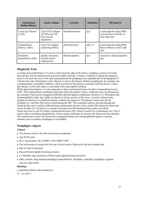
Autoimmune<br />
Bullous Disease<br />
L<strong>in</strong>ear IgA Disease<br />
(LAD)<br />
Epidermolysis<br />
Bullosa (EBA)<br />
Dermatitis<br />
herpetiformis (DH)<br />
Diagnostic Tests<br />
Target Antigen Location Antibodies DIF pattern<br />
Type XVII collagen<br />
(97-kDa <strong>and</strong> 120kDa<br />
cleaved<br />
fragments)<br />
Type VII collagen<br />
(anchor<strong>in</strong>g fibrils)<br />
gliad<strong>in</strong>, reticulum,<br />
smooth muscle<br />
endomysium<br />
Hemidesmosome IgA L<strong>in</strong>ear deposits along <strong>BMZ</strong>,<br />
roof <strong>and</strong> floor of blister <strong>in</strong><br />
NaCl split sk<strong>in</strong><br />
Sub-basal layer IgG, C3 L<strong>in</strong>ear deposits along <strong>BMZ</strong>,<br />
floor of blister <strong>in</strong> NaCl split<br />
sk<strong>in</strong><br />
Dermal papillae IgA Granular <strong>in</strong> dermal papillary<br />
tips<br />
A rout<strong>in</strong>e sk<strong>in</strong> punch biopsy (3-4 mm) is taken from the edge of the blister, <strong>in</strong>clud<strong>in</strong>g a portion of normal<br />
sk<strong>in</strong> <strong>and</strong> this sent for haematoxyl<strong>in</strong> <strong>and</strong> eos<strong>in</strong> (H&E) sta<strong>in</strong><strong>in</strong>g. A biopsy is helpful <strong>in</strong> mak<strong>in</strong>g the diagnosis<br />
s<strong>in</strong>ce it will show the level of the split (<strong>in</strong>traepidermal for pemphigus <strong>and</strong> subepidermal for pemphigoid). It<br />
will also show the <strong>in</strong>flammatory cells which are a clue to the disease. Bullous pemphigoid, for example, has<br />
a predom<strong>in</strong>antly eos<strong>in</strong>ophilic <strong>in</strong>filtrate, which accounts for the pruritus associated with the disease. L<strong>in</strong>ear<br />
IgA disease, on the other h<strong>and</strong>, is associated with a neutrophilic <strong>in</strong>filtrate.<br />
While do<strong>in</strong>g sk<strong>in</strong> biopsies, it is also important to take a perilesional biopsy for direct immunofluorescence<br />
(DIF). This method detects antibodies deposited with<strong>in</strong> the patient’s tissue. Antibodies have the fluorescent<br />
dye attached. Fluoresce<strong>in</strong>-conjugated antibodies directed aga<strong>in</strong>st complement fractions (C3, fibr<strong>in</strong>ogen) <strong>and</strong><br />
immunoglobul<strong>in</strong>s (IgG, IgA, IgM) are placed on frozen sections of the tissue. A positive fluorescence<br />
(graded on <strong>in</strong>tensity) <strong>in</strong> a particular pattern confirms the diagnosis. The patient’s tissue may also be<br />
<strong>in</strong>cubated <strong>in</strong> 1 mol/litre NaCl prior to perform<strong>in</strong>g the DIF. This technique <strong>in</strong>duces cleavage through the<br />
lam<strong>in</strong>a lucida, <strong>and</strong> is useful <strong>in</strong> differentiat<strong>in</strong>g autoimmune <strong>diseases</strong> with a similar DIF pattern by observ<strong>in</strong>g<br />
where the IgG or C3 localizes (i.e. dermal roof pattern for BP <strong>and</strong> dermal floor pattern for EBA).<br />
Serum may also be sent for <strong>in</strong>direct immunofluorescence (IIF) which is useful as a confirmatory test. This is<br />
used to detect circulat<strong>in</strong>g autoantibodies. In this sett<strong>in</strong>g, antibodies do not have the fluorescent dye attached.<br />
This method uses serum with fluoresce<strong>in</strong>-conjugated human anti-immunoglobul<strong>in</strong> aga<strong>in</strong>st a mucosal<br />
substrate such as monkey oesophagus or rat bladder.<br />
<strong>Pemphigus</strong> vulgaris<br />
Cl<strong>in</strong>ical<br />
• This disease <strong>in</strong>volves the sk<strong>in</strong> <strong>and</strong> mucous membranes<br />
• Age 40-60 years<br />
• HLA Associations: HLA DRB1*1454, DRB1*1401 3<br />
• The oral mucosa is usually the first site of <strong>in</strong>volvement, followed by the sk<strong>in</strong> months later<br />
• May be fatal if untreated<br />
• Flaccid blisters rapidly becom<strong>in</strong>g erosions<br />
• (+) Nikolsky sign (extension of blister upon apply<strong>in</strong>g lateral pressure)<br />
• Other variants: drug-<strong>in</strong>duced pemphigus (penicillam<strong>in</strong>e, nifedip<strong>in</strong>e, captopril), pemphigus vegetans<br />
(<strong>in</strong>volves sk<strong>in</strong> folds)<br />
Histology<br />
• Suprabasal blister with acantholysis<br />
IF - see table 1<br />
31