Pemphigus foliaceus: review of clinical signs & diagnosis in dogs ...
Pemphigus foliaceus: review of clinical signs & diagnosis in dogs ...
Pemphigus foliaceus: review of clinical signs & diagnosis in dogs ...

You also want an ePaper? Increase the reach of your titles
YUMPU automatically turns print PDFs into web optimized ePapers that Google loves.
<strong>Pemphigus</strong> <strong>foliaceus</strong>: <strong>review</strong> <strong>of</strong> <strong>cl<strong>in</strong>ical</strong> <strong>signs</strong> & <strong>diagnosis</strong> <strong>in</strong> <strong>dogs</strong> and cats<br />
Dr. Peter Hill BVSc PhD DVD DipACVD DipECVD MRCVS MACVSc<br />
Veter<strong>in</strong>ary Specialist Centre, North Ryde<br />
Classification and <strong>cl<strong>in</strong>ical</strong> <strong>signs</strong> <strong>of</strong> pemphigus <strong>in</strong> <strong>dogs</strong> and cats<br />
<strong>Pemphigus</strong> can broadly be classified <strong>in</strong>to superficial and deep forms<br />
<strong>Pemphigus</strong> <strong>foliaceus</strong><br />
The most common autoimmune sk<strong>in</strong> disease <strong>of</strong> <strong>dogs</strong> and cats.<br />
Lesions progress from erythematous macules to papules to pustules to crusts.<br />
N.B. Compared to pyoderma, PF pustules are usually more numerous, larger, may have irregular, highly<br />
erythematosus borders and may coalesce.<br />
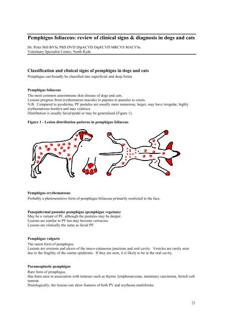
Distribution is usually facial/pedal or may be generalised (Figure 1).<br />
Figure 1 - Lesion distribution patterns <strong>in</strong> pemphigus <strong>foliaceus</strong><br />
<strong>Pemphigus</strong> erythematosus<br />
Probably a photosensitive form <strong>of</strong> pemphigus <strong>foliaceus</strong> primarily restricted to the face.<br />
Panepidermal pustular pemphigus (pemphigus vegetans)<br />
May be a variant <strong>of</strong> PF, although the pustules may be deeper.<br />
Lesions are similar to PF but may become verrucose.<br />
Lesions are <strong>cl<strong>in</strong>ical</strong>ly the same as facial PF.<br />
<strong>Pemphigus</strong> vulgaris<br />
The rarest form <strong>of</strong> pemphigus.<br />
Lesions are erosions and ulcers <strong>of</strong> the muco-cutaneous junctions and oral cavity. Vesicles are rarely seen<br />
due to the fragility <strong>of</strong> the can<strong>in</strong>e epidermis. If they are seen, it is likely to be <strong>in</strong> the oral cavity.<br />
Paraneoplastic pemphigus<br />
Rare form <strong>of</strong> pemphigus.<br />
Has been seen <strong>in</strong> association with tumours such as thymic lymphosarcoma, mammary carc<strong>in</strong>oma, Sertoli cell<br />
tumour.<br />
Histologically, the lesions can show features <strong>of</strong> both PV and erythema multiforme.<br />
21
Drug-<strong>in</strong>duced pemphigus<br />
Some cases <strong>of</strong> PF are thought to have been <strong>in</strong>duced by drugs.<br />
<strong>Pemphigus</strong> <strong>of</strong> chronic disease<br />
Some authors believe that <strong>dogs</strong> that have suffered from other sk<strong>in</strong> diseases for prolonged periods <strong>of</strong> time<br />
(allergy, chronic pyoderma) may develop pemphigus later <strong>in</strong> life. There is currently no evidence to support<br />
this anecdotal observation.<br />
Diagnosis <strong>of</strong> pemphigus<br />
As with other sk<strong>in</strong> diseases, the <strong>diagnosis</strong> <strong>of</strong> autoimmune sk<strong>in</strong> diseases relies on history, physical<br />
exam<strong>in</strong>ation and diagnostic tests (cytology and histopathology).<br />
History<br />
The history <strong>of</strong> <strong>dogs</strong> and cats with autoimmune sk<strong>in</strong> diseases is usually non-specific. Due to the severity <strong>of</strong><br />
many <strong>of</strong> these diseases, owners usually present their animals as soon as the disease appears. With some<br />
autoimmune sk<strong>in</strong> diseases such as pemphigus <strong>foliaceus</strong>, the lesions may appear <strong>in</strong> waves and go through<br />
periods <strong>of</strong> wax<strong>in</strong>g and wan<strong>in</strong>g. Systemic manifestations such as lethargy and anorexia are variable. Some<br />
people believe that <strong>dogs</strong> that have suffered from recurrent sk<strong>in</strong> disease (especially allergic disease and<br />
pyoderma) for a long time can ultimately develop a form <strong>of</strong> pemphigus <strong>foliaceus</strong>. A true cause and effect<br />
has yet to be proven.<br />
Physical exam<strong>in</strong>ation<br />
The ma<strong>in</strong> objectives <strong>of</strong> the physical exam<strong>in</strong>ation are to determ<strong>in</strong>e:<br />
1. The nature <strong>of</strong> the sk<strong>in</strong> lesions – are they superficial and pustular/crust<strong>in</strong>g or are they deep and<br />
erosive/ulcerative.<br />
2. The distribution <strong>of</strong> the sk<strong>in</strong> lesions – are they conf<strong>in</strong>ed to certa<strong>in</strong> body regions such as the face or mucocutaneous<br />
junctions or are they generalised.<br />
3. Are there any other abnormalities such as jo<strong>in</strong>t swell<strong>in</strong>g, anaemia etc.<br />
Based on the physical exam<strong>in</strong>ation alone, it is possible to draw up a list <strong>of</strong> differential diagnoses that<br />
<strong>in</strong>cludes or excludes some <strong>of</strong> the above auto-immune sk<strong>in</strong> diseases, as well as other, non immune-mediated<br />
diseases. However, without further diagnostic tests, it is not possible to make a specific <strong>diagnosis</strong>.<br />
Diagnostic tests<br />
The two most useful tests <strong>in</strong> the <strong>diagnosis</strong> <strong>of</strong> autoimmune sk<strong>in</strong> diseases are cytology and histopathology.<br />
Cytology<br />
Cytological exam<strong>in</strong>ation should be performed <strong>in</strong> all cases <strong>of</strong> suspected autoimmune sk<strong>in</strong> disease. In cases <strong>of</strong><br />
pemphigus <strong>foliaceus</strong> (and the <strong>cl<strong>in</strong>ical</strong>ly similar variants), cytology can be virtually diagnostic because <strong>of</strong> the<br />
presence <strong>of</strong> acantholytic kerat<strong>in</strong>ocytes (Figure 2). In all autoimmune sk<strong>in</strong> diseases, cytology can help to<br />
confirm the presence <strong>of</strong> secondary bacterial <strong>in</strong>fection or other differential diagnoses.<br />
22
Figure 2 – Microscopic field show<strong>in</strong>g the cytological appearance <strong>of</strong> PF<br />
Acantholysis refers to a process <strong>in</strong> which the<br />
kerat<strong>in</strong>ocytes from deeper layers with<strong>in</strong> the epidermis<br />
become separated from each other. The separated cells<br />
are known as acantholytic kerat<strong>in</strong>ocytes or<br />
acanthocytes. Acanthocytes are very large cells with a<br />
centrally placed nucleus that are about 3-5 times the<br />
diameter <strong>of</strong> a neutrophil. The presence <strong>of</strong> large<br />
numbers <strong>of</strong> acanthocytes amongst a background <strong>of</strong><br />
neutrophils is a characteristic f<strong>in</strong>d<strong>in</strong>g <strong>in</strong> pemphigus<br />
<strong>foliaceus</strong>. However, culture <strong>of</strong> pustules and biopsy is<br />
<strong>in</strong>dicated whenever this picture is seen because bacterial<br />
<strong>in</strong>fections can occasionally cause this pattern <strong>of</strong><br />
<strong>in</strong>flammation (X400).<br />
Biopsy and Histopathology<br />
Although cytology is a very useful test that can be performed <strong>in</strong> the cl<strong>in</strong>ic, it is essential to biopsy all cases <strong>of</strong><br />
suspected autoimmune sk<strong>in</strong> diseases. With the appropriate samples it may be possible to make specific<br />
diagnoses <strong>of</strong> pemphigus <strong>foliaceus</strong>, pemphigus erythematosus, panepidermal pustular pemphigus, pemphigus<br />
vulgaris, and cutaneous lupus. It is not possible to dist<strong>in</strong>guish between bullous pemphigoid, mucous<br />
membrane pemphigoid, l<strong>in</strong>ear IgA bullous dermatosis and epidermolysis bullosa acquisita on histopathology.<br />
When tak<strong>in</strong>g biopsies from autoimmune sk<strong>in</strong> diseases, the follow<strong>in</strong>g tips may help to get diagnostic results:<br />
• Biopsy punches are acceptable for the majority <strong>of</strong> lesions but ellipse biopsies may be preferable for large<br />
bullae or ulcers.<br />
• Always take at least five biopsies.<br />
• For pustular/crusted lesions, try and biopsy pustules. If none are present, biopsy papules or macules –<br />
they may conta<strong>in</strong> microscopic pustules. If you have to biopsy crusted lesions, make sure that the crusts<br />
are submitted along with the sk<strong>in</strong>.<br />
• If you suspect pemphigus <strong>foliaceus</strong> but there are no primary lesions, clip the fur <strong>of</strong>f an area <strong>of</strong> sk<strong>in</strong> and<br />
wait for 24-48 hours. New lesions may develop <strong>in</strong> the clipped area (or other areas).<br />
• If ulcers are biopsied, sample the junction between normal and ulcerated sk<strong>in</strong>. Make sure that the biopsy<br />
is orientated so that the pathologist sections the sk<strong>in</strong> the right way.<br />
• If there are ulcerative lesions, rub an unaffected area <strong>of</strong> sk<strong>in</strong> with a pencil eraser for about a m<strong>in</strong>ute. If<br />
there is dermo-epidermal weakness, this can artefactually create a new lesion to biopsy.<br />
• If the first set <strong>of</strong> biopsies are non-diagnostic but you still suspect an autoimmune disease, biopsy the<br />
animal aga<strong>in</strong>.<br />
Histopathological changes <strong>in</strong> the sk<strong>in</strong><br />
The key histopathological f<strong>in</strong>d<strong>in</strong>gs <strong>of</strong> the various diseases can be summarised as follows:<br />
• <strong>Pemphigus</strong> <strong>foliaceus</strong> – Intracorneal or subcorneal pustules conta<strong>in</strong><strong>in</strong>g neutrophils and acantholytic<br />
kerat<strong>in</strong>ocytes; surface neutrophilic crusts conta<strong>in</strong><strong>in</strong>g degenerated acanthocytes.<br />
• <strong>Pemphigus</strong> erythematosus – Epidermal changes as for pemphigus <strong>foliaceus</strong>, but with lichenoid or<br />
<strong>in</strong>terface changes at the dermo-epidermal junction. These may <strong>in</strong>clude hydropic degeneration <strong>of</strong> basal<br />
cells, basal cell apoptosis, abnormal appearance <strong>of</strong> the basement membrane and a sub-epidermal<br />
<strong>in</strong>filtrate <strong>of</strong> mononuclear cells, ma<strong>in</strong>ly lymphocytes.<br />
• <strong>Pemphigus</strong> vulgaris – Suprabasilar acantholysis leads to cleft formation above the basal cell layer <strong>of</strong> the<br />
epidermis. The basal cells may rema<strong>in</strong> attached to the basement membrane like a row <strong>of</strong> tombstones.<br />
• Paraneoplastic pemphigus – this disease has features <strong>of</strong> both PV and erythema multiforme i.e.<br />
suprabasilar cleft formation with apoptosis at multiple layers throughout the epidermis.<br />
Other tests that should be performed when a sk<strong>in</strong> disease is suspected to be auto-immune <strong>in</strong>clude fungal<br />
culture (<strong>in</strong> suspected cases <strong>of</strong> PF), bacterial culture and sensitivity (<strong>in</strong> suspected cases <strong>of</strong> PF and if rods are<br />
seen on cytology), sk<strong>in</strong> scrap<strong>in</strong>gs to rule out demodicosis, and a rout<strong>in</strong>e haematology and biochemistry panel<br />
to allow monitor<strong>in</strong>g <strong>of</strong> therapy and to look for evidence <strong>of</strong> systemic <strong>in</strong>volvement.<br />
23
Immun<strong>of</strong>luorescence and immunohistochemistry<br />
These techniques allow detection <strong>of</strong> the autoantibodies directed aga<strong>in</strong>st sk<strong>in</strong> targets. However, they are not<br />
widely available <strong>in</strong> commercial histopathology labs, although they have been used extensively <strong>in</strong> research.<br />
The pr<strong>in</strong>ciples underly<strong>in</strong>g these tests are illustrated below.<br />
Direct immun<strong>of</strong>luorescence<br />
Indirect immun<strong>of</strong>luorescence<br />
Immunohistochemistry<br />
1. Sk<strong>in</strong> section taken from a dog with autoimmune<br />
sk<strong>in</strong> disease<br />
5. Fluorescent marker visualised under UV light microscope<br />
4. Specific antibody labelled with fluorescent marker is added<br />
Specific antibody labelled with iod<strong>in</strong>e 125<br />
3. Autoantibody or complement components to be detected (already<br />
bound to diseased sk<strong>in</strong>)<br />
2. Tissue section<br />
1. Sk<strong>in</strong>, lip, oesophagus section taken from a normal dog<br />
5. Fluorescent marker visualised under UV light microscope<br />
4. Specific antibody labelled with fluorescent marker is added<br />
Specific antibody labelled with iod<strong>in</strong>e 125<br />
3. Serum from a dog with autoimmune sk<strong>in</strong> disease conta<strong>in</strong><strong>in</strong>g<br />
autoantibodies to sk<strong>in</strong> components is added<br />
2. Tissue section<br />
1. Tissue section taken from an animal with sk<strong>in</strong><br />
disease (autoimmune or tumour)<br />
5. Enzyme reaction with substrate allows visualisation <strong>of</strong> specific<br />
prote<strong>in</strong>s under microscope<br />
4. Specific antibody labelled with enzyme marker is added<br />
Specific antibody labelled with iod<strong>in</strong>e 125<br />
3. Autoantibody or tumour-specific prote<strong>in</strong> to be detected<br />
(already bound to, or part <strong>of</strong>, diseased sk<strong>in</strong>)<br />
2. Tissue section<br />
24