Pemphigus foliaceus: review of clinical signs & diagnosis in dogs ...
Pemphigus foliaceus: review of clinical signs & diagnosis in dogs ...
Pemphigus foliaceus: review of clinical signs & diagnosis in dogs ...

You also want an ePaper? Increase the reach of your titles
YUMPU automatically turns print PDFs into web optimized ePapers that Google loves.
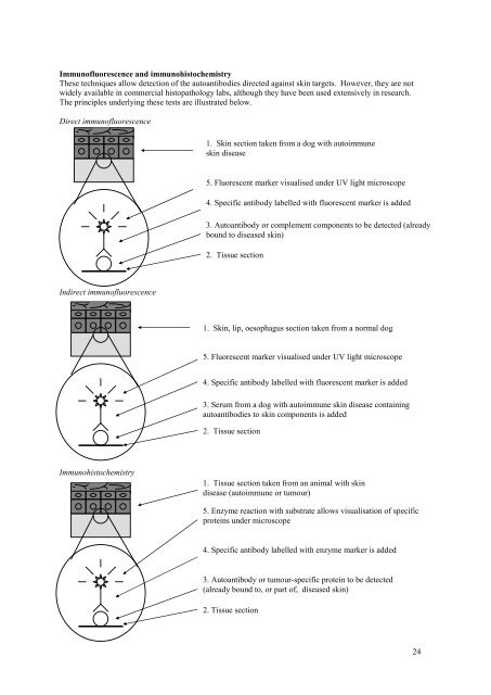
Immun<strong>of</strong>luorescence and immunohistochemistry<br />
These techniques allow detection <strong>of</strong> the autoantibodies directed aga<strong>in</strong>st sk<strong>in</strong> targets. However, they are not<br />
widely available <strong>in</strong> commercial histopathology labs, although they have been used extensively <strong>in</strong> research.<br />
The pr<strong>in</strong>ciples underly<strong>in</strong>g these tests are illustrated below.<br />
Direct immun<strong>of</strong>luorescence<br />
Indirect immun<strong>of</strong>luorescence<br />
Immunohistochemistry<br />
1. Sk<strong>in</strong> section taken from a dog with autoimmune<br />
sk<strong>in</strong> disease<br />
5. Fluorescent marker visualised under UV light microscope<br />
4. Specific antibody labelled with fluorescent marker is added<br />
Specific antibody labelled with iod<strong>in</strong>e 125<br />
3. Autoantibody or complement components to be detected (already<br />
bound to diseased sk<strong>in</strong>)<br />
2. Tissue section<br />
1. Sk<strong>in</strong>, lip, oesophagus section taken from a normal dog<br />
5. Fluorescent marker visualised under UV light microscope<br />
4. Specific antibody labelled with fluorescent marker is added<br />
Specific antibody labelled with iod<strong>in</strong>e 125<br />
3. Serum from a dog with autoimmune sk<strong>in</strong> disease conta<strong>in</strong><strong>in</strong>g<br />
autoantibodies to sk<strong>in</strong> components is added<br />
2. Tissue section<br />
1. Tissue section taken from an animal with sk<strong>in</strong><br />
disease (autoimmune or tumour)<br />
5. Enzyme reaction with substrate allows visualisation <strong>of</strong> specific<br />
prote<strong>in</strong>s under microscope<br />
4. Specific antibody labelled with enzyme marker is added<br />
Specific antibody labelled with iod<strong>in</strong>e 125<br />
3. Autoantibody or tumour-specific prote<strong>in</strong> to be detected<br />
(already bound to, or part <strong>of</strong>, diseased sk<strong>in</strong>)<br />
2. Tissue section<br />
24