Pemphigus foliaceus: review of clinical signs & diagnosis in dogs ...
Pemphigus foliaceus: review of clinical signs & diagnosis in dogs ...
Pemphigus foliaceus: review of clinical signs & diagnosis in dogs ...

Create successful ePaper yourself
Turn your PDF publications into a flip-book with our unique Google optimized e-Paper software.
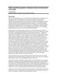
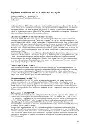
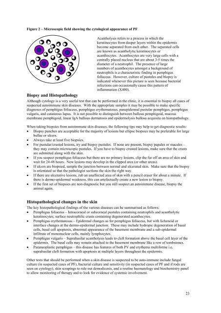
Figure 2 – Microscopic field show<strong>in</strong>g the cytological appearance <strong>of</strong> PF<br />
Acantholysis refers to a process <strong>in</strong> which the<br />
kerat<strong>in</strong>ocytes from deeper layers with<strong>in</strong> the epidermis<br />
become separated from each other. The separated cells<br />
are known as acantholytic kerat<strong>in</strong>ocytes or<br />
acanthocytes. Acanthocytes are very large cells with a<br />
centrally placed nucleus that are about 3-5 times the<br />
diameter <strong>of</strong> a neutrophil. The presence <strong>of</strong> large<br />
numbers <strong>of</strong> acanthocytes amongst a background <strong>of</strong><br />
neutrophils is a characteristic f<strong>in</strong>d<strong>in</strong>g <strong>in</strong> pemphigus<br />
<strong>foliaceus</strong>. However, culture <strong>of</strong> pustules and biopsy is<br />
<strong>in</strong>dicated whenever this picture is seen because bacterial<br />
<strong>in</strong>fections can occasionally cause this pattern <strong>of</strong><br />
<strong>in</strong>flammation (X400).<br />
Biopsy and Histopathology<br />
Although cytology is a very useful test that can be performed <strong>in</strong> the cl<strong>in</strong>ic, it is essential to biopsy all cases <strong>of</strong><br />
suspected autoimmune sk<strong>in</strong> diseases. With the appropriate samples it may be possible to make specific<br />
diagnoses <strong>of</strong> pemphigus <strong>foliaceus</strong>, pemphigus erythematosus, panepidermal pustular pemphigus, pemphigus<br />
vulgaris, and cutaneous lupus. It is not possible to dist<strong>in</strong>guish between bullous pemphigoid, mucous<br />
membrane pemphigoid, l<strong>in</strong>ear IgA bullous dermatosis and epidermolysis bullosa acquisita on histopathology.<br />
When tak<strong>in</strong>g biopsies from autoimmune sk<strong>in</strong> diseases, the follow<strong>in</strong>g tips may help to get diagnostic results:<br />
• Biopsy punches are acceptable for the majority <strong>of</strong> lesions but ellipse biopsies may be preferable for large<br />
bullae or ulcers.<br />
• Always take at least five biopsies.<br />
• For pustular/crusted lesions, try and biopsy pustules. If none are present, biopsy papules or macules –<br />
they may conta<strong>in</strong> microscopic pustules. If you have to biopsy crusted lesions, make sure that the crusts<br />
are submitted along with the sk<strong>in</strong>.<br />
• If you suspect pemphigus <strong>foliaceus</strong> but there are no primary lesions, clip the fur <strong>of</strong>f an area <strong>of</strong> sk<strong>in</strong> and<br />
wait for 24-48 hours. New lesions may develop <strong>in</strong> the clipped area (or other areas).<br />
• If ulcers are biopsied, sample the junction between normal and ulcerated sk<strong>in</strong>. Make sure that the biopsy<br />
is orientated so that the pathologist sections the sk<strong>in</strong> the right way.<br />
• If there are ulcerative lesions, rub an unaffected area <strong>of</strong> sk<strong>in</strong> with a pencil eraser for about a m<strong>in</strong>ute. If<br />
there is dermo-epidermal weakness, this can artefactually create a new lesion to biopsy.<br />
• If the first set <strong>of</strong> biopsies are non-diagnostic but you still suspect an autoimmune disease, biopsy the<br />
animal aga<strong>in</strong>.<br />
Histopathological changes <strong>in</strong> the sk<strong>in</strong><br />
The key histopathological f<strong>in</strong>d<strong>in</strong>gs <strong>of</strong> the various diseases can be summarised as follows:<br />
• <strong>Pemphigus</strong> <strong>foliaceus</strong> – Intracorneal or subcorneal pustules conta<strong>in</strong><strong>in</strong>g neutrophils and acantholytic<br />
kerat<strong>in</strong>ocytes; surface neutrophilic crusts conta<strong>in</strong><strong>in</strong>g degenerated acanthocytes.<br />
• <strong>Pemphigus</strong> erythematosus – Epidermal changes as for pemphigus <strong>foliaceus</strong>, but with lichenoid or<br />
<strong>in</strong>terface changes at the dermo-epidermal junction. These may <strong>in</strong>clude hydropic degeneration <strong>of</strong> basal<br />
cells, basal cell apoptosis, abnormal appearance <strong>of</strong> the basement membrane and a sub-epidermal<br />
<strong>in</strong>filtrate <strong>of</strong> mononuclear cells, ma<strong>in</strong>ly lymphocytes.<br />
• <strong>Pemphigus</strong> vulgaris – Suprabasilar acantholysis leads to cleft formation above the basal cell layer <strong>of</strong> the<br />
epidermis. The basal cells may rema<strong>in</strong> attached to the basement membrane like a row <strong>of</strong> tombstones.<br />
• Paraneoplastic pemphigus – this disease has features <strong>of</strong> both PV and erythema multiforme i.e.<br />
suprabasilar cleft formation with apoptosis at multiple layers throughout the epidermis.<br />
Other tests that should be performed when a sk<strong>in</strong> disease is suspected to be auto-immune <strong>in</strong>clude fungal<br />
culture (<strong>in</strong> suspected cases <strong>of</strong> PF), bacterial culture and sensitivity (<strong>in</strong> suspected cases <strong>of</strong> PF and if rods are<br />
seen on cytology), sk<strong>in</strong> scrap<strong>in</strong>gs to rule out demodicosis, and a rout<strong>in</strong>e haematology and biochemistry panel<br />
to allow monitor<strong>in</strong>g <strong>of</strong> therapy and to look for evidence <strong>of</strong> systemic <strong>in</strong>volvement.<br />
23