OCULAR TRAUMA - Michigan Optometric Association
OCULAR TRAUMA - Michigan Optometric Association
OCULAR TRAUMA - Michigan Optometric Association

You also want an ePaper? Increase the reach of your titles
YUMPU automatically turns print PDFs into web optimized ePapers that Google loves.
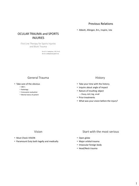
<strong>OCULAR</strong> <strong>TRAUMA</strong> and SPORTS<br />
INJURIES<br />
First Line Therapy for Sports Injuries<br />
and Blunt Trauma<br />
General Trauma<br />
• Take care of the obvious<br />
• ABC’s<br />
• Radiology<br />
• Concussion evaluation<br />
• Mental status of patient<br />
Vision<br />
Derek N. Cunningham, OD, FAAO<br />
derek.n.cunningham@gmail.com<br />
• Must Check VISION<br />
• Paramount Duty both legally and medically<br />
Previous Relations<br />
• Abbott, Allergan, B+L, Inspire, Ista<br />
History<br />
• Take your time with the history.<br />
• Inquire about angle of impact<br />
• Nature of insulting object<br />
– Sharp, dull, big, small<br />
• Prior treatments<br />
• What was your vision before the injury?<br />
Start with the most serious<br />
• Open globe<br />
• Major orbital trauma<br />
• Intaocular foreign body<br />
• Head/Neck trauma<br />
1
CT<br />
• If you suspect any of the previous, a CT scan is<br />
indicated<br />
• Axial and Coronal sections (1mm) needed for<br />
suspected blow out<br />
• No MRI for fear of metallic foreign body<br />
• Do not patch<br />
• Shield Eye<br />
• Send to ER<br />
11<br />
Open Globe<br />
• Disproportionate conjunctival edema could be<br />
a possible indication of scleral rupture<br />
Endophthalmitis – Strain Susceptibility<br />
Methicillin-Resistant S. aureus<br />
Gentamicin<br />
81%<br />
Trimethoprim<br />
74%<br />
Moxifloxacin<br />
45%<br />
Gatifloxacin<br />
32%<br />
Open Globe<br />
• Check VA - reduced<br />
• Seidel’s sign<br />
– Fl stain<br />
• Displaced pupil/expelled contents<br />
• Non-reactive pupil<br />
• Low IOP<br />
• Poor reflex<br />
• Hyphema<br />
10<br />
Endophthalmitis – Strain Susceptibility<br />
Coagulase Negative Staphlococci<br />
Gentamicin<br />
97%<br />
Trimethoprim<br />
66%<br />
Moxifloxacin<br />
53%<br />
Gatifloxacin<br />
50%<br />
Blunt Trauma<br />
• Check VA<br />
• Proptosis from retrobulabar hemorrhage<br />
• Contusion/sub-conj hemorrhage<br />
• Retinal detachment<br />
• Commotio Retinae<br />
• Traumatic uveitis or hyphema<br />
• Traumatic cataract<br />
• Blow out fracture<br />
2
Contusion<br />
• Need to get eye open<br />
– Will dictate urgency of consult<br />
• Asses lids and globe for debris or lacerations<br />
• Check pupil response (round pupil)<br />
• Red Reflex?<br />
• Severe<br />
• Palpate orbital rim<br />
• Check VA<br />
Black Eyes<br />
Blow out fracture<br />
• Base and medial walls of orbit are very thin<br />
• Does not need to be a major trauma<br />
• Look for trapped extra-ocular muscles<br />
(reduced versions) - strabismus<br />
Contusion<br />
• Do eyes move well together<br />
• Instill Fl to check for abrasions<br />
• Check IOP if all else is clear<br />
• Palpate bony orbital rim checking for tightness or<br />
crepitus (orbital emphysema)<br />
• Check for orbital step off<br />
MVA trauma case<br />
Blow out fracture<br />
• Sunken Eye - hypo-ophthalmos<br />
• Infraorbital hypoesthesia<br />
• Diplopia<br />
• Pain on eye movement or nausea<br />
3
Need coronal cuts<br />
Repair?<br />
• Within 2 weeks<br />
– Symptomatic diplopia within 30º of primary gaze<br />
– Muscle entrapment (prevent ischemia and necrosis)<br />
– Fracture greater than 50% of orbit floor<br />
– Displaced orbital rim fracture<br />
– > 3mm of enophthamos, significant hypo-ophthalmos<br />
• Monitor<br />
– Diplopia outside central 30º<br />
– Modest isolated fractures<br />
– Improvement over first 2 weeks<br />
Lid lacerations<br />
• Check VA<br />
• Difficult to suture because of tarsal plate and<br />
margin function<br />
• Refer to ophtho<br />
• Tetanus prophylaxis<br />
• Upper lid skin has no subcutaneous fat<br />
•Does it hurt to chew?<br />
Orbital Trauma in Children<br />
• Trap door orbital floor fractures are very<br />
common.<br />
– More elastic orbits<br />
– More common to get muscle entrapment<br />
• Evaluation for repair typically in 5-7 days vs 2<br />
weeks for adults<br />
• Diplopia with ct evidence entrapped muscle with<br />
nonresolved oculocardial reflex<br />
– Bradycardia, heart block, nausea, vomiting, syncope<br />
Upper Lip<br />
• Must consider levator/aponeurosis<br />
• NO subcutaneous fat<br />
4
Lower lid<br />
• Lacerated canthus<br />
• Lacrimal drainage system<br />
• Quality reconstruction necessary<br />
• Wound closure can be delayed for up to 3 days<br />
with satisfactory surgical outcomes in adults and<br />
12-36 hours in children<br />
– Can be beneficial to allow swelling to go down,<br />
leading to better visualization of tissue reapproximation<br />
Periocular Infection<br />
• Any antibiotic regimen should have adequate<br />
central nervous system penetration to<br />
minimize the risk of meningitis and cavernous<br />
sinus thrombosis<br />
• Systemic steroid use is controversial and<br />
should only be used after sufficient antibiotic<br />
loading and on immunocompetent patients<br />
• Treatment?<br />
Conjunctival Lacerations<br />
MRSA<br />
• Treat all athletes and healthcare workers as<br />
though they have MRSA<br />
Conjunctival Lacerations<br />
NEVER PATCH !!!<br />
• Patching creates a great anaerobic<br />
environment<br />
• Patient can not tell if things are getting worse<br />
• Oxygen speeds healing<br />
• If a patch is needed let an eye doc make the<br />
decision<br />
– Patch for pain until they get into your office?<br />
5
Corneal abrasions<br />
• Check VA<br />
• Important to know what abraded the cornea –<br />
Organic vs Inorganic<br />
• Did the patient put anything into their eye<br />
afterwards?<br />
• Grade the level of pain/light sensativity<br />
• NEVER PATCH unless lid immobilization is<br />
necessary<br />
Corneal Abrasion While Gardening<br />
Bandage Lens and 1 day p/0 Missed 3 day p/o, seen on day 7<br />
LASIK<br />
• Any corneal abrasion on a flap is serious.<br />
• Microkeratome flaps can easily come off years<br />
after surgery<br />
• Femtosecond flaps incredibly stable, but can<br />
still have issues<br />
Fluorescein<br />
• Always instill Fl for a suspected corneal<br />
abrasion<br />
• Need to use a cobalt blue light to excite the Fl<br />
• Be careful with the use of topical anesthetics<br />
6
Abrasion Treatment<br />
• Minor abrasion require only prophylactic<br />
antibiotic and ocular lubricants (topical<br />
NSAIDS?)<br />
• Moderate to severe – cycloplegic, oral<br />
analgesic, bandage contact lens, 4 th Gen<br />
Fluoroquinolone<br />
– Clean up margins?<br />
– Doxy?<br />
UV keratitis treatment<br />
• Artificial tears<br />
• Oral analgesics<br />
• Antibiotic is infection is suspected<br />
• No topical anesthetics<br />
• cyclo<br />
Chemical Burns<br />
• Check VA<br />
• Alkali Burn is way worse than Acid<br />
• Check pH if possible<br />
• Immediate irrigation<br />
– Do not wait until they are at your office<br />
• Absolute Emergency – 1 day consult at most<br />
for minor cases<br />
Photokeratitis/Snow blindness<br />
• Check VA<br />
• Caused by UVB(C) exposure to the cornea – 320-<br />
290nm<br />
• Painful !!!!!<br />
• Superficial punctate keratopathy about 6 hours<br />
after exposure (corneal sun burn)<br />
• Typically self limiting<br />
• Welders flash, tanning beds, skiing, desert, sailing<br />
Radiation and Chemicals<br />
• Ultraviolet/Infrared<br />
• Chemicals involved<br />
– Acids<br />
– Bases<br />
– Duration of chemical contact<br />
Corneal Foreign Body<br />
• Remove if visible and not completely<br />
penetrating<br />
• Always document depth of FB<br />
• Stain cornea with Fl to look for tract marks<br />
from upper lid – invert upper lid<br />
• Anesthetize eye for patient comfort and to<br />
allow a better view.<br />
7
Traumatic Hyphema<br />
• Sports Injuries account for 60%of hyphemas<br />
(usually Pediatric)<br />
• Complications<br />
• Elevated IOP<br />
• Posterior Synechiae<br />
• Peripheral anterior synechiae<br />
• Corneal blood staining<br />
• Optic atrophy<br />
• Angle recession glaucoma (usually >180º)<br />
Traumatic Hyphema<br />
Red blood cells in the TM<br />
Immediate Rise in IOP<br />
Ghost cell glaucoma<br />
IOP rise weeks post-trauma<br />
Angle recession glaucoma<br />
IOP rise years post-trauma<br />
Traumatic Hyphema Treatments<br />
• Steriods – immediate<br />
use is debatable<br />
• Use after 4-5 days likely<br />
helpful to reduce risk of<br />
scarring<br />
Traumatic Hyphema<br />
• Draw the level of the clot and record the level<br />
of free cells<br />
• Tear usually occurs at the anterior aspect of<br />
the ciliary body in the angle<br />
• Uncomplicated hyphemas usually last 5-6 days<br />
Traumatic Hyphema Treatments<br />
Elevate head and shield the eye<br />
• Pain – acetaminophen ( no Asprin)<br />
• Cycloplegics – decrease risk of posterior<br />
synechiae<br />
• Miotics – increase surface area for iris<br />
reabsorption<br />
• Steriods – immediate use is debatable<br />
• Use after 4-5 days likely helpful to reduce risk of<br />
scarring<br />
Traumatic Hyphema Treatments<br />
• Aminocaproic (antifibrinolytic) acid may be<br />
used for larger hyphemas or with increased<br />
risk of re-bleeds<br />
• Oral osmotic agents can be used to control<br />
IOP<br />
– Debatable whether any topical medications have a<br />
therapeutic advantage in the acute phase<br />
• Greater than 75% hyphema should be referred<br />
8
Traumatic Hyphema Treatments<br />
• Aminocaproic Acid<br />
– May require inpatient care due to side effects<br />
• Mild<br />
• Syncope<br />
• Nausea<br />
• Vomiting<br />
• Hypotension<br />
Black Eyes<br />
Purpose of Hyphema Treatments<br />
• Prevent IOP increase<br />
• Prevent Secondary Hemorrhage<br />
• Prevent Corneal blood Staining<br />
• Sickle Cell Anemia complicates things<br />
• Check VA<br />
• Light sensitive<br />
• Ciliary flush<br />
Traumatic Uveitis<br />
• Decreased VA<br />
• Decreased pupil response<br />
• Sub-conj hemorrhage<br />
Traumatic uveitis Retinal Detachment<br />
9
Commotio Retinae<br />
• Energy is transferred to the opposite side of<br />
the globe.<br />
• Inflammation will usually be on posterior<br />
nasal retina<br />
• Symptoms<br />
– Eye pain<br />
– Diplopia<br />
Valsalva Retinopathy<br />
Retrobulbar hemorrhage<br />
– Vision loss<br />
– Reduced ocular motility<br />
– Proptosis<br />
Nature.com<br />
Retinal Hemorrhage<br />
High Altitude Retinopathy<br />
Intraretinal hemorrhage, retinal venous dilation, optic disc edema<br />
Associated with HACE<br />
Retrobulbar Hemorrhage<br />
• Signs<br />
– Proptosis<br />
– Increases IOP<br />
– Ecchymosis<br />
– Ophthlmoplegia<br />
– APD<br />
– Papilledema<br />
– Central retinal artery pulsation<br />
10
Acute orbital compatment syndrome<br />
• Build up of volume is only held back by medial<br />
and lateral canthal tendons<br />
Fat<br />
Prolapse<br />
Proptosis<br />
CRA<br />
Ischemia<br />
Decreased<br />
Perfusion<br />
Lateral Canthotomy Cantholysis<br />
Increased<br />
IOP<br />
www.jcdp.com Neuro-ophthalmologic Trauma<br />
• Third, fourth and sixth nerve palsies can all<br />
happen<br />
– Third nerve palsies associated with worst outcome<br />
– Sixth nerve palsies associated with best outcomes<br />
11
Neuro-ophthalmologic Traum<br />
• Note<br />
– It has been shown that multiple cranial<br />
neuropathies were associated with less severe<br />
head injury and more significant extremity injury.<br />
» Dhaliwal et al.<br />
Traumatic Optic Neuropathy<br />
• Visual outcome is poor<br />
– Regardless of treatment (high dose<br />
corticosteroids, optic nerve sheath fenestration,<br />
or optic canal decompression), outcome is poor<br />
• RAPD presence is the most useful diagnostic<br />
test<br />
Thank you<br />
derek.n.cunningham@gmail.com<br />
Traumatic Optic Neuropathy<br />
• Can often be the only ophthalmic injury after<br />
significant head trauma<br />
• Believed to happen in the canalicular portion<br />
of the nerve<br />
The Ridiculous<br />
12