Wnt Signaling Polarizes an Early C. elegans ... - MCD Biology
Wnt Signaling Polarizes an Early C. elegans ... - MCD Biology
Wnt Signaling Polarizes an Early C. elegans ... - MCD Biology

Create successful ePaper yourself
Turn your PDF publications into a flip-book with our unique Google optimized e-Paper software.
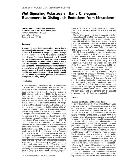
Cell, Vol. 90, 695–705, August 22, 1997, Copyright ©1997 by Cell Press<br />
<strong>Wnt</strong> <strong>Signaling</strong> <strong>Polarizes</strong> <strong>an</strong> <strong>Early</strong> C. eleg<strong>an</strong>s<br />
Blastomere to Distinguish Endoderm from Mesoderm<br />
Christopher J. Thorpe, Ann Schlesinger, might act solely by regulating cytoskeletal polarity in<br />
J. Clayton Carter <strong>an</strong>d Bruce Bowerm<strong>an</strong>*<br />
EMS, influencing gene expression in E <strong>an</strong>d MS only<br />
Institute of Molecular <strong>Biology</strong><br />
indirectly.<br />
University of Oregon<br />
The maternal gene pop-1 also is required to distin-<br />
Eugene, Oregon 97403<br />
guish the fates of E <strong>an</strong>d MS, in <strong>an</strong> apparently cell autonomous<br />
m<strong>an</strong>ner (Lin et al., 1995). In pop-1 mut<strong>an</strong>t embryos<br />
E <strong>an</strong>d MS both adopt E-like fates <strong>an</strong>d produce twice<br />
Summary<br />
the normal number of intestinal cells. pop-1 encodes a<br />
protein with a single high mobility group (HMG) DNA<br />
A polarizing signal induces endoderm production by binding domain similar to vertebrate T cell factor 1<br />
a 4-cell stage blastomere in C. eleg<strong>an</strong>s called EMS. We (TCF-1) <strong>an</strong>d mouse lymphoid enh<strong>an</strong>cer-binding factor<br />
identified 16 mutations in five genes, mom-1 through 1 (LEF-1). Recent work indicates that these proteins are<br />
mom-5, required for EMS to produce endoderm. targets for activation by the <strong>Wnt</strong> signal tr<strong>an</strong>sduction<br />
mom-1, mom-2, <strong>an</strong>d mom-3 are required in the signal- pathway (Behrens et al., 1996; Miller <strong>an</strong>d Moon, 1996;<br />
ing cell, P2, while mom-4 is required in EMS. P2 signal-<br />
ing downregulates <strong>an</strong> HMG domain protein, POP-1, in<br />
one EMS daughter. The sequence of mom-2 predicts<br />
that it encodes a member of the <strong>Wnt</strong> family of secreted<br />
glycoproteins, which in other systems activate HMG<br />
domain proteins. Defective mitotic spindle orientations<br />
in mom mut<strong>an</strong>t embryos indicate that <strong>Wnt</strong> signal-<br />
ing influences cytoskeletal polarity in blastomeres<br />
throughout the early embryo.<br />
Riese et al., 1996; Brunner et al., 1997; v<strong>an</strong> de Wetering<br />
et al., 1997 [but see Merriam et al., 1997]). POP-1 is<br />
present in the nuclei of all 4-cell stage blastomeres, but<br />
at the 8-cell stage POP-1 levels are higher in MS th<strong>an</strong><br />
in E, suggesting that down-regulation of POP-1 in E<br />
permits specification of endoderm (see Figure 1).<br />
We have identified 5 maternally expressed C. eleg<strong>an</strong>s<br />
genes required for endoderm induction. Mutations in<br />
these genes (called mom for more mesoderm) result in<br />
E <strong>an</strong>d MS both adopting MS-like fates. We show that<br />
Introduction<br />
mom-2 encodes a <strong>Wnt</strong> family member, <strong>an</strong>d we suggest<br />
that <strong>Wnt</strong> signaling regulates cytoskeletal polarity in<br />
To generate cellular asymmetry, extrinsic <strong>an</strong>d intrinsic<br />
processes c<strong>an</strong> polarize parent cells prior to division,<br />
localizing different developmental regulators to their<br />
daughters (for reviews, see Horvitz <strong>an</strong>d Herskowitz,<br />
1992; Gonczy <strong>an</strong>d Hym<strong>an</strong>, 1996). In the nematode<br />
responding cells. mom-1, mom-2, <strong>an</strong>d mom-3 are<br />
required in P2 for signaling, while mom-4 functions<br />
autonomously in EMS. POP-1 functions downstream of<br />
the mom genes, with <strong>Wnt</strong> signaling from P2 acting to<br />
downregulate this HMG domain protein in E.<br />
Caenorhabditis eleg<strong>an</strong>s, <strong>an</strong> inductive polarization regulates<br />
the development of a 4-cell stage embryonic<br />
Results<br />
cell, or blastomere, called EMS (Goldstein, 1992, 1993,<br />
1995a). In response to polarization, the two EMS daughters,<br />
E <strong>an</strong>d MS, adopt different fates. E makes all of the<br />
endoderm, or intestinal cells, in the embryo, while MS<br />
produces mesoderm, which in C. eleg<strong>an</strong>s includes phar-<br />
ynx <strong>an</strong>d body wall muscle (Sulston et al., 1983).<br />
A signal from the sister of EMS, a blastomere called<br />
P2, polarizes EMS to induce endoderm (Figure 1). This<br />
induction c<strong>an</strong> be <strong>an</strong>alyzed in vitro by culturing blasto-<br />
meres isolated from early embryos (Goldstein, 1992; Ed-<br />
gar, 1995; Shelton <strong>an</strong>d Bowerm<strong>an</strong>, 1996). If EMS is isolated<br />
<strong>an</strong>d left to develop alone, it fails to make endoderm<br />
<strong>an</strong>d instead producestwo MS-like daughters (Goldstein,<br />
1993). When P2 <strong>an</strong>d EMS are placed in contact suffi-<br />
ciently early, the EMS daughter born next to P2 makes<br />
endoderm, while the other adopts <strong>an</strong> MS-like fate. EMS<br />
polarization may involve reorg<strong>an</strong>ization of the cytoskele-<br />
ton: exposure of 4-cell stage embryos to chemicals that<br />
depolymerize either microfilaments or microtubules pre-<br />
vents endoderm development, while similar treatments<br />
at the 8-cell stage have no effect (Goldstein, 1995a).<br />
Signal(s) from P2 also orients the mitotic spindle in EMS,<br />
indicating that the microtubule cytoskeleton is one tar-<br />
get of a P2 signal (Goldstein, 1995b). Indeed, P2 signaling<br />
Genetic Identification of Five mom Loci<br />
To study the induction of cell polarity in EMS, we used<br />
a genetic screen to identify recessive, maternal-effect,<br />
embryonic-lethal mut<strong>an</strong>ts in which both EMS daughters<br />
adopt <strong>an</strong> MS-like fate <strong>an</strong>d produce excess mesoderm<br />
at the expense of endoderm. We identified 16 such mutations,<br />
defining 5 genes, mom-1 through mom-5. All<br />
the mut<strong>an</strong>t mom alleles are recessive <strong>an</strong>d fully penetr<strong>an</strong>t<br />
for embryonic lethality (Experimental Procedures). For<br />
the stronger alleles, most mut<strong>an</strong>t embryos entirely lack<br />
intestinal cells <strong>an</strong>d instead make large amounts of phar-<br />
ynx (Table 1, Figure 2A). In addition, mom mut<strong>an</strong>t em-<br />
bryos are severely defective in morphogenesis, producing<br />
amorphous clumps of differentiated tissues whether<br />
or not they make endoderm (Figure 2A). Thus, mutational<br />
inactivation of the mom genes affects not just the polarization<br />
of EMS but also elongation of the embryo into<br />
a long, thin worm. The morphogenesis defect c<strong>an</strong> be<br />
partially rescued by paternal contribution of a wild-type<br />
allele, but the mom-2 requirement for endoderm specifi-<br />
cation is strictly maternal (Figure 2A, Experimental Procedures).<br />
Although we detect no zygotic phenotypes<br />
associated with mut<strong>an</strong>t alleles of mom-2, mom-4, <strong>an</strong>d<br />
mom-5, homozygous mom-1 <strong>an</strong>d homozygous mom-3<br />
mut<strong>an</strong>t hermaphrodites withoutexception have protrud-<br />
*To whom correspondence should be addressed.<br />
ing, nonfunctional vulvae <strong>an</strong>d are mildly uncoordinated
Cell<br />
696<br />
E <strong>an</strong>d MS Both Adopt MS-like Fates in mom<br />
Mut<strong>an</strong>t Embryos<br />
Terminally differentiated mom mut<strong>an</strong>t embryos that lack<br />
intestinal cells also appear to make excess pharynx (Figure<br />
2A), suggesting that E might produce <strong>an</strong> MS-like<br />
pattern of cell fate. We tested this possibility in three<br />
ways. We used tissue-specific <strong>an</strong>tibodies to determine<br />
if E in mom-2 mut<strong>an</strong>t embryos produces mesodermal<br />
cell types normally made by MS, we tested the genetic<br />
requirements for ectopic pharyngeal cell production by<br />
E, <strong>an</strong>d we <strong>an</strong>alyzed the cell lineage of some E descend<strong>an</strong>ts<br />
to determine if they adopt cell cycle times <strong>an</strong>d<br />
cleavage patterns similar to those made by a wild-type<br />
MS blastomere. As described below, our results indicate<br />
that E adopts <strong>an</strong> MS-like fate in mom-2 mut<strong>an</strong>t embryos.<br />
To examine the fate of E, we first used a laser microbeam<br />
focused through a microscope to kill every cell<br />
in early mom-2 mut<strong>an</strong>t embryos except for E. These<br />
partial embryos produced differentiated descend<strong>an</strong>ts<br />
that were fixed <strong>an</strong>d stained with cell type–specific monoclonal<br />
<strong>an</strong>tibodies (Table 2, Figure 2B). Similar laser ablation<br />
data were obtained for mut<strong>an</strong>t embryos lacking<br />
mom-1, mom-3 <strong>an</strong>d mom-4 function (A. S., C. J. T.,<br />
Figure 1. EMS Development Requires Both Autonomously Acting M. Meneghini, <strong>an</strong>d B. B., unpublished data). In control<br />
POP-1 <strong>an</strong>d a Signal from P2 experiments using wild-type embryos, E always pro-<br />
POP-1 protein is found in all nuclei at the 4-cell stage (dark shading). duced intestinal cells but not two cell types made by<br />
A signal from P2 to EMS early in the 4-cell stage (curved arrow)<br />
MS: pharyngeal muscle <strong>an</strong>d body wall muscle. For two<br />
polarizes EMS such that the potential to produce endoderm is acstrong<br />
mom2 alleles, E in most operated embryos<br />
quired by only the E daughter of EMS. E contains relatively low levels<br />
of POP-1<strong>an</strong>d makes only intestinal cells, whileMS has relativelyhigh failed to produce <strong>an</strong>y intestinal cells. Instead, when E<br />
levels of POP-1 <strong>an</strong>d produces mesoderm. Sister cells are indicated failed to make gut, it produced four cell types normally<br />
by short connecting lines. made by MS: pharyngeal <strong>an</strong>d body wall muscle cells,<br />
pharyngeal gl<strong>an</strong>d cells, <strong>an</strong>d pharyngeal marginal cells<br />
(Table 2). MS in mom mut<strong>an</strong>t embryos also produces<br />
(C. J. T., A. S., <strong>an</strong>d B. B., unpublished data). Here we pharynx <strong>an</strong>d body wall muscle (Table 2). As no other<br />
focus on the maternal-effect phenotypes caused by mu- early blastomere besides MS in wild-type embryos pro-<br />
tations in the mom genes. We note that because some duces both pharynx <strong>an</strong>d body wall muscle (Sulston et al.,<br />
mom genes have zygotic functions, the maternal-effect 1983), E <strong>an</strong>d MS in mom mut<strong>an</strong>t embryos both appear to<br />
alleles we have isolated may not be null. Indeed, the adopt MS-like fates.<br />
different penetr<strong>an</strong>ce of the gut phenotypes associated<br />
If E in mom-2 embryos develops like MS, then it should<br />
with our different mom alleles suggests that most alleles<br />
require skn-1 but not glp-1 to produce pharyngeal mus-<br />
are not null (Table 1). However, double mut<strong>an</strong>t embryos<br />
cle cells (Priess et al., 1987; Bowerm<strong>an</strong> et al., 1992). MS<br />
from mothers homozygous for both mom-2 <strong>an</strong>d mom-4<br />
makes about half the pharyngeal cells produced during<br />
have a completely penetr<strong>an</strong>t gut defect (Table 1). Thus,<br />
embryogenesis, with the remainder made by two gr<strong>an</strong>d-<br />
daughters of the 4-cell stage blastomere ABa (Sulston<br />
the mom gene products, whether they function in a<br />
et al., 1983). The production of pharyngeal cells by ABa<br />
single pathway or in parallel, are essential for the specifidescend<strong>an</strong>ts<br />
requires <strong>an</strong> inductive signal from MS at<br />
cation of endoderm in early C. eleg<strong>an</strong>s embryos.<br />
about the 12-cell stage (Priess et al., 1987; Hutter <strong>an</strong>d<br />
In addition to their lack of endoderm, mom-1, mom-2,<br />
Schnabel, 1994; M<strong>an</strong>go et al., 1994a); glp-1 encodes<br />
mom-3, <strong>an</strong>d mom-5 mut<strong>an</strong>t embryos also are defective<br />
a putative receptor required for ABa descend<strong>an</strong>ts to<br />
in orienting the mitotic spindle of <strong>an</strong> 8-cell stage blastoreceive<br />
the MS signal (Priess et al., 1987; Austin <strong>an</strong>d<br />
mere called ABar (Table 1, Figure 3). In wild-type em-<br />
Kimble, 1989; Yochem <strong>an</strong>d Greenwald, 1989). Mutations<br />
bryos, ABar divides along a largely left–right (l/r) axis,<br />
in glp-1 result in <strong>an</strong> absence of ABa-derived pharyngeal<br />
roughly orthogonal to the three other AB descend<strong>an</strong>ts<br />
cells but do not affect the production of pharyngeal cells<br />
that all divide along the <strong>an</strong>terior–posterior (a/p) axis with<br />
by MS (Priess et al., 1987). Mutations in skn-1 result in<br />
a pronounced dorsal–ventral (d/v) tilt (Figure 3D). In all a loss of all pharyngeal cells, as skn-1 is required both<br />
but mom-4 mut<strong>an</strong>ts, ABar divides roughly parallel to the to specify MS fate <strong>an</strong>d to activate the MS signal that<br />
other AB descend<strong>an</strong>ts (Table 1, Figure 3H). Because induces ABa descend<strong>an</strong>ts to make pharyngeal cells<br />
the paternal rescue of morphogenesis described above (Bowerm<strong>an</strong> et al., 1992; Mello et al., 1992; Shelton <strong>an</strong>d<br />
does not correlate with rescue of the ABar spindle orien- Bowerm<strong>an</strong>, 1996). By staining fixed,terminally differentitation<br />
(Experimental Procedures), the abnormal axis of ated embryos from double mut<strong>an</strong>t mothers with cellthe<br />
mitotic spindle in ABar c<strong>an</strong>not fully account for the type specific <strong>an</strong>tibodies (Experimental Procedures), we<br />
highly penetr<strong>an</strong>t morphogenesis defect observed in found that skn-1; mom-2 embryos produce few or no<br />
mom mut<strong>an</strong>t embryos.<br />
pharyngeal cells: 17 of 46 double mut<strong>an</strong>t embryos made
Cell Polarity <strong>an</strong>d Endoderm Specification<br />
697<br />
Table 1. Penetr<strong>an</strong>ce of Endoderm Defect <strong>an</strong>d ABar Cleavage Abnormality in mom Mut<strong>an</strong>t Embryos<br />
Fraction of Embryos<br />
% Embryos with Aberr<strong>an</strong>t ABar<br />
Gene Allele Lacking Gut (n) a Cleavage b<br />
mom-1 or10 85 (215) 10/10<br />
or46 52 (160) 7/7<br />
or65 50 (275)<br />
or83 46 (153)<br />
or70 39 (321) 13/13<br />
mom-2 or85 88 (485)<br />
or9 77 (226)<br />
or48 74 (380)<br />
or42 72 (194) 28/31<br />
or33 50 (210)<br />
or77 8 (65) 15/15<br />
mom-3 or78 65 (361) 30/30<br />
mom-4 or39 40 (100) 0/12<br />
or49 24 (181) 0/12<br />
or11 1 (228) 2/17<br />
mom-5 or57 5 (167) 9/9<br />
skn-1; mom-2 zu67; or42 100 (241) —<br />
mom-4; mom-2 or39; or42 100 (124) —<br />
a To assay production of intestinal cells, embryos were collected from mom mut<strong>an</strong>t hermaphrodites <strong>an</strong>d allowed to develop at least 10 hr at<br />
20C. The presence of intestinal cells was scored using polarizing light microscopy to detect intestine-specific birefringent gut gr<strong>an</strong>ules.<br />
b The orientation of the mitotic spindle of the ABar blastomere was examined in lateral views of 8-cell stage embryos using Nomarski optics.<br />
In wild-type embryos, the <strong>an</strong>terior daughter of ABar contacts the MS blastomere (Figure 3D). ABar cleavage was scored as defective if its<br />
posterior daughter contacted MS (Figure 3H).<br />
between 1 <strong>an</strong>d 5 pharyngeal muscle cells while the re- genetic interval, we obtained rescue of the mut<strong>an</strong>t phemainder<br />
made none, compared to the 39 made in wild notype by germline tr<strong>an</strong>sformation with the cosmids<br />
type (Sulston et al., 1983). In contrast, both E <strong>an</strong>d MS F52E1 <strong>an</strong>d ZK427. Sequence data from the C. eleg<strong>an</strong>s<br />
from glp-1; mom-2 double mut<strong>an</strong>t embryos produce Genome Center predicts a gene, F38E1.7, on ZK427 that<br />
pharynx: 9 of 10 embryos in which E was isolated, <strong>an</strong>d would encode a member of the <strong>Wnt</strong> family of secreted<br />
6 of 6 in which MS was isolated, made large numbers signaling molecules (Experimental Procedures). To deof<br />
pharyngeal muscle cells. Because E <strong>an</strong>d MS in mom-2 termine if F38E1.7 is the mom-2 locus, we microinjected<br />
mut<strong>an</strong>t embryos require skn-1 but not glp-1 function to <strong>an</strong>tisense RNA from one exon of the predicted gene into<br />
produce pharyngeal cells, we conclude that both EMS<br />
daughters adopt <strong>an</strong> MS-like fate.<br />
To define their fates more precisely, we compared<br />
the cell lineages E <strong>an</strong>d MS produce in mom-2 mut<strong>an</strong>t<br />
embryos to the wild-type MS lineage (Figure 2C). C.<br />
eleg<strong>an</strong>s embryos produce a nearly invari<strong>an</strong>t cell lineage,<br />
as determined by scoring the relative positions of<br />
daughter cells following each division <strong>an</strong>d observing<br />
their eventual fates (Sulston et al., 1983). In wild-type<br />
embryos, E <strong>an</strong>d MS become different shortly after their<br />
birth: Ea <strong>an</strong>d Ep are the first cells to migrate inside the<br />
embryo during gastrulation, <strong>an</strong>d they divide about 15<br />
min later th<strong>an</strong> do MSa <strong>an</strong>d MSp. In mom-2 embryos, Ea<br />
<strong>an</strong>d Ep fail to gastrulate, <strong>an</strong>d they divide nearly synchronously<br />
with MSa <strong>an</strong>d MSp. Subsequent divisions of the<br />
E descend<strong>an</strong>ts also occur with approximately MS-like<br />
timing, <strong>an</strong>d several cell deaths that occur in a wild-type<br />
the syncitial gonad of wild-type <strong>an</strong>imals. All injected<br />
<strong>an</strong>imals produced dead embryos with a Mom mut<strong>an</strong>t<br />
phenotype (Experimental Procedures). To confirm fur-<br />
ther the gene identity, we sequenced six mut<strong>an</strong>t alleles<br />
of mom-2. Each allele contains a single lesion within<br />
the coding sequence, except for one mutation at a splice<br />
junction (Figure 4). We conclude that mom-2 is F38E1.7<br />
<strong>an</strong>d that it encodes a member of the <strong>Wnt</strong> family of secreted<br />
signaling glycoproteins. The predicted MOM-2/<strong>Wnt</strong> pro-<br />
tein is 363 amino acids in length <strong>an</strong>d includes 24 highly<br />
conserved cysteine residues. BLAST searches indicated<br />
that the predicted MOM-2 protein is most closely related<br />
to mammali<strong>an</strong> <strong>Wnt</strong>-2, sharing approximately 40% amino<br />
acid identity with mouse <strong>an</strong>d hum<strong>an</strong> <strong>Wnt</strong>-2. We con-<br />
clude that the polarization of gut potential in EMS requires<br />
the <strong>Wnt</strong> signal tr<strong>an</strong>sduction pathway.<br />
MS lineage also occur at the corresponding points in<br />
the lineage of E in some mom-2 mut<strong>an</strong>t embryos. How- mom-1, mom-2, <strong>an</strong>d mom-3 Are Required<br />
ever, we often do not observe characteristic cell deaths in P2 for Endoderm Induction, while<br />
in both the E <strong>an</strong>d MS lineages (Figure 2C). We conclude mom-4 Is Required in EMS<br />
that E <strong>an</strong>d MS in mom-2 mut<strong>an</strong>t embryos both adopt To determine which mom genes are required in P2 for<br />
fates similar but not identical to a wild-type MS.<br />
signaling <strong>an</strong>d which are required in EMS for responding,<br />
we assembled genetically mosaic partial embryos in<br />
mom-2 Encodes a <strong>Wnt</strong> Family Member<br />
vitro (Figure 5 <strong>an</strong>d Experimental Procedures). Using<br />
We used positional cloning to identify the wild-type blastomeres isolated from wild-type <strong>an</strong>d mom mut<strong>an</strong>t<br />
mom-2 gene (Figure 4). After mapping mom-2 to a small embryos, we found that mom-1, mom-2, <strong>an</strong>d mom-3
Cell<br />
698<br />
Figure 2. E Adopts <strong>an</strong> MS-like Identity in mom-2 Embryos<br />
(A) Immunofluorescence <strong>an</strong>d light micrographs of wild-type (left column), mom-2 (middle column), <strong>an</strong>d mom-2/ embryos (right column) from<br />
mom-2/mom-2 mothers. Embryos were allowed to develop 15 hr (a, b, e, <strong>an</strong>d f), or 8 hr (c <strong>an</strong>d d) at 20C. (a, b, <strong>an</strong>d c) Living embryos viewed<br />
with Nomarski optics. Pharyngeal tissue is surrounded by a prominent basement membr<strong>an</strong>e (arrowheads), <strong>an</strong>d contains a secreted cuticle<br />
(wide arrows). Visible in wild-type embryos, but not in mom-2 or mom-2/ embryos, are intestinal cells with characteristically large nuclei<br />
containing a single large nucleolus (thin arrows in [a]). While homozygous mom-2 embryos fail to undergo <strong>an</strong>y morphogenesis <strong>an</strong>d invariably<br />
arrest as unelongated clumps of differentiated tissue (b), mom-2/ embryos, even those lacking intestine, often elongate into short, stubby<br />
worms (c <strong>an</strong>d i). Paternal contribution of a wild-type copy of mom-2 does not rescue the intestine defect (Experimental Procedures). (d, e,<br />
<strong>an</strong>d f) Intact embryos stained with J126, a monoclonal <strong>an</strong>tibody (MAb) that recognizes intestinal cells. (g, h, <strong>an</strong>d i) Intact embryos stained<br />
with 9.2.1, a MAb that recognizes pharyngeal muscle cells.<br />
(B) Immunofluorescence micrographs of operated mom-2 embryos in which all blastomeres in the embryos except E were killed with a laser<br />
microbeam. Embryos were allowed to develop 8 hours (a <strong>an</strong>d c), or 16 hours (b) at 20C before fixing <strong>an</strong>d staining. MAb J126 (a) recognizes<br />
intestinal cells, MAb 9.2.1 (b) recognizes pharyngeal muscle cells, <strong>an</strong>d MAb 5.6 (c) recognizes body-wall muscle.<br />
(C) Cell deaths scored in E <strong>an</strong>d MS lineages from mom-2 mut<strong>an</strong>t embryos (below) compared to the wild-type MS lineage (above). Vertical<br />
bars represent time; horizontal bars represent cell division. Programmed cell deaths are indicated by Xs. The fraction of embryos in which<br />
the corresponding E <strong>an</strong>d MS lineages in mom-2 mut<strong>an</strong>t embryos produced cell deaths are indicated below each X.
Cell Polarity <strong>an</strong>d Endoderm Specification<br />
699<br />
Figure 3. Mitotic Spindle Axes Are Misoriented<br />
in mom-2 <strong>an</strong>d mom-2; mom-1 Mut<strong>an</strong>t<br />
Embryos<br />
All embryos are shown in lateral views with<br />
<strong>an</strong>terior to the left <strong>an</strong>d ventral up; maternal<br />
genotypes are indicated at the top of each<br />
column. (A, E, <strong>an</strong>d I ) Wild-type, mom-2, <strong>an</strong>d<br />
mom-2; mom-1 embryos at the 4-cell stage.<br />
(B, F, <strong>an</strong>d J) EMS cleavage. In wild-type <strong>an</strong>d<br />
mom-2 embryos, the EMS spindle aligns on<br />
the a/p axis, while in some mom-2; mom-1<br />
embryos, it is tilted along the d/v axis, as<br />
shown here. (C, G, <strong>an</strong>d K) 8-cell stage. E <strong>an</strong>d<br />
MS are positioned normally in the mom-2;<br />
mom-1 embryo following cytokinesis. (D, H,<br />
<strong>an</strong>d L) 12-cell stage. In wild-type embryos,<br />
ABar divides along a mostly l/r axis, tr<strong>an</strong>sverse<br />
to that of the other AB descend<strong>an</strong>ts; the<br />
division axes of ABal <strong>an</strong>d ABpr are marked for<br />
comparison. The <strong>an</strong>terior daughter of ABar,<br />
ABara, touches MS. In mom-2 mut<strong>an</strong>t embryos,<br />
ABar divides parallel to the other AB<br />
descend<strong>an</strong>ts <strong>an</strong>d ABarp instead of ABara<br />
contacts MS. In a small fraction of mom-2;<br />
mom-1 embryos (2 of 21 embryos scored, <strong>an</strong><br />
example of which is shown here), we observe<br />
dramatic rearr<strong>an</strong>gements of blastomeres, beginning<br />
at the 8-cell stage. In both cases, posterior<br />
blastomeres moved <strong>an</strong>teriorly, such<br />
that E <strong>an</strong>d MS became the <strong>an</strong>teriormost cells<br />
in the embryo. Other mom-2; mom-1 embryos<br />
showed the same ABar cleavage axis defect observed in mom-2 mut<strong>an</strong>t embryos. Surprisingly, double mut<strong>an</strong>t embryos made intestinal<br />
cells more often th<strong>an</strong> either single mut<strong>an</strong>t: 52% of mom-2; mom-1 embryos (n197) compared to 15% for or10 <strong>an</strong>d 28% for or42 embryos<br />
(Table 1). Laser ablation experiments with double mut<strong>an</strong>t embryos indicate that E but not MS produces endoderm: Of 14, 7 isolated E’s made<br />
intestinal cells, compared to 0 of 15 isolated MS blastomeres.<br />
are required in P2, while mom-4 is required in EMS. mom-4 <strong>an</strong>d the Interpretation of Cell<br />
Because of the low penetr<strong>an</strong>ce of the endoderm pheno- Polarity in EMS<br />
type in mom-5 mut<strong>an</strong>t embryos (Table 1), we have not Our blastomere mosaic <strong>an</strong>alysis indicates that mom-4<br />
been able to determine which blastomere requires is required in EMS to respond to the P2 signal (Table 3).<br />
mom-5 function. The development of P2 appears normal To determine how mom-4 functions with respect to the<br />
in mom-1, mom-2, <strong>an</strong>d mom-3 mut<strong>an</strong>t embryos (Experi- establishment or interpretation of cell polarity, we exammental<br />
Procedures), suggesting that the wild-type ined mom-4 function in pie-1 mut<strong>an</strong>t embryos. Muta-<br />
genes function specifically in P2 signaling. Finally, our tions in pie-1 cause P2 to develop like EMS, producing<br />
identification of mom-2 as a <strong>Wnt</strong> gene is consistent with excess pharynx <strong>an</strong>d intestine (Mello et al., 1992). Unlike<br />
its function being required in P2 for signaling.<br />
EMS, P2 in pie-1 embryos does not require a signal from<br />
Table 2. Cell Types Produced by E <strong>an</strong>d MS in mom-2 Mut<strong>an</strong>t Embryos<br />
Body Pharyngeal Pharyngeal<br />
Blastomere Wall Pharyngeal Gl<strong>an</strong>d Marginal<br />
Genotype Isolated Intestinal Cells Muscle Muscle Cells Cells<br />
Wild type E 13/13 0/7 0/10 — —<br />
mom-2(or42) E 9/170 17/17 15/15 10/10 16/17<br />
mom-2(or9) E 2/16 — 8/10 — —<br />
Wild type MS 0/8 11/11 12/12 8/8 9/9<br />
mom-2(or42) MS 0/64 6/6 8/10 13/14 12/13<br />
mom-2(or9) MS 0/9 — 6/6 — —<br />
All blastomeres in the embryo except MS or E were killed using a laser microbeam. ABa, ABp, <strong>an</strong>d P2 were killed at the 4-cell stage, then<br />
either MS or E was killed following division of EMS. The operated embryos were allowed to develop overnight <strong>an</strong>d then scored for the presence<br />
of intestinal cells using polarizing optics. Operated embryos not making gut were fixed <strong>an</strong>d stained with MAbs to detect the tissues indicated<br />
(see Experimental Procedures). When E was isolated by laser ablation at the 8-cell stage, 9 of 22 operated embryos made intestinal cells.<br />
The nine that made intestine were double stained with J126 <strong>an</strong>d 3NB12; one made both pharyngeal <strong>an</strong>d intestinal cells while the remainder<br />
made only intestine.
Cell<br />
700<br />
Table 3. P2 <strong>an</strong>d EMS Blastomere Mosaics<br />
may be sufficient to specify E fate in P3, suggesting that<br />
Blastomeres Recombined<br />
Fraction of Recombin<strong>an</strong>t<br />
the intrinsic polarity of P2 <strong>an</strong>d the induced polarity of<br />
EMS share common properties.<br />
P2 EMS Embryos Making Gut To position mom-4 relative to the establishment of<br />
WT WT 46/47<br />
cell polarity during endoderm induction, we constructed<br />
WT<br />
mom-1<br />
WT<br />
mom-2<br />
mom-1<br />
WT<br />
mom-2<br />
WT<br />
7/7<br />
2/7<br />
10/12<br />
1/8<br />
a mom-4; pie-1 strain <strong>an</strong>d asked if mom-4 is required<br />
for P2 to produce endoderm in pie-1 mut<strong>an</strong>t embryos<br />
(Table 4). By two criteria, P2 in mom-4; pie-1 double<br />
mut<strong>an</strong>t embryos remains polarized. P2 divides asymmet-<br />
rically (24 of 24 embryos observed by light microscopy),<br />
WT<br />
mom-3<br />
mom-3<br />
WT<br />
11/11<br />
4/12<br />
<strong>an</strong>d P gr<strong>an</strong>ules are segregated to P3 (11 of 11 embryos;<br />
see Experimental Procedures). While intact pie-1 em-<br />
WT mom-4 2/10 bryos make extra intestinal cells, mom-4; pie-1 double<br />
mom-4 WT 7/7<br />
mut<strong>an</strong>t embryos usually make no endoderm (C. J. T.<br />
P2 <strong>an</strong>d EMS blastomeres were isolated from wild-type <strong>an</strong>d mom <strong>an</strong>d B. B., unpublisheddata). Using laser ablation experimut<strong>an</strong>t<br />
embryos <strong>an</strong>d recombined to form genetically mosaic partial ments to isolate the daughters of P2 <strong>an</strong>d EMS in double<br />
embryos. The genotypes of the recombined blastomeres are shown<br />
in the left column.The mut<strong>an</strong>t alleles used were: mom-1(or10), mom-<br />
2(or42), mom-3(or78), <strong>an</strong>d mom-4(or39). The fraction of partial em-<br />
bryos making intestinal cells in each experiment is shown in the<br />
right column.<br />
mut<strong>an</strong>t embryos, we found that both E <strong>an</strong>d P3 usually<br />
failed to produce intestinal cells (Table 4). Because<br />
mom-4 is required for P3 to make intestinal cells in pie-1<br />
mut<strong>an</strong>t embryos, even though P2 remains polarized,<br />
mom-4 may be required to interpret, not to establish,<br />
polarity in a wild-type EMS blastomere.<br />
<strong>an</strong>y neighboring blastomere to produce intestinal cells;<br />
in isolation it still divides into one E-like <strong>an</strong>d one MSlike<br />
daughter (Goldstein, 1995a). The ability of a pie-1<br />
mut<strong>an</strong>t P2 to produce endoderm autonomously raises<br />
the possibility that <strong>an</strong> intrinsic polarity within P2 is sufficient<br />
to specify endoderm.<br />
Several lines of evidence indicate that P2 possesses<br />
<strong>an</strong> intrinsic polarity that might specify endoderm in pie-1<br />
mut<strong>an</strong>t embryos. First, P2 isolated from wild-type or<br />
pie-1 embryos always divides asymmetrically, producing<br />
one larger <strong>an</strong>d one smaller daughter, called C <strong>an</strong>d P3 An alternative expl<strong>an</strong>ation for the lack of gut in pie-1;<br />
mom-4 double mut<strong>an</strong>t embryos is that autocrine signal-<br />
ing might polarize P2 in a pie-1 mut<strong>an</strong>t. If so, mom-4<br />
might play a role in tr<strong>an</strong>sducing <strong>an</strong> autocrine signal. To<br />
determine if P2 uses autocrine, mom-dependent signal-<br />
ing to specify endoderm in pie-1 mut<strong>an</strong>t embryos, we<br />
constructed double mut<strong>an</strong>ts of pie-1 with mom-1,<br />
mom-2, <strong>an</strong>d mom-3. In all cases, EMS but not P2 fails<br />
to produce endoderm (Table 4). We conclude that the<br />
production of endoderm by P2 in pie-1 mut<strong>an</strong>t embryos<br />
is largely independent of <strong>Wnt</strong> signaling, <strong>an</strong>d that mom-4<br />
may be unique among the mom genes in operating<br />
in wild type. Moreover, the smaller P3 daughter inherits downstream of, or parallel to, the establishment of cell<br />
cytoplasmic particles called P gr<strong>an</strong>ules, which are seg- polarity during endoderm induction. However, because<br />
regated to P3 as P2 divides (Strome <strong>an</strong>d Wood, 1983;<br />
Hird et al., 1996). Finally, although it is not known to<br />
both P2 <strong>an</strong>d EMS fail to make gut in 5% of intact pie-1;<br />
mom-2 mut<strong>an</strong>t embryos (10 of 191) <strong>an</strong>d in 1% of pie-1;<br />
have <strong>an</strong>y function in P2, the HMG domain protein POP-1 mom-3 embryos (4 of 471), <strong>Wnt</strong> signaling might have a<br />
accumulates asymmetrically in the daughters of P2, with<br />
C having higher levels th<strong>an</strong> P3 (Lin et al., 1995; Figure<br />
minor role in polarizing P2.<br />
1). In pie-1 mut<strong>an</strong>t embryos, C nearly always becomes<br />
MS-like, while P3 always adopts <strong>an</strong> E-like fate, consistent<br />
<strong>Wnt</strong>-Dependent P2 <strong>Signaling</strong> Restricts<br />
POP-1 Function to MS<br />
with lower levels of POP-1 permitting the specification As a final approach to positioning the mom genes in a<br />
of endoderm in both E <strong>an</strong>d P3 in pie-1 mut<strong>an</strong>t embryos. pathway of endoderm specification, we examined inter-<br />
To summarize, in pie-1 embryos P2’s intrinsic polarity actions of the mom genes with pop-1. Whereas the two<br />
Figure 4. Molecular Cloning of mom-2<br />
(A) mom-2 maps near pos-1, on LGV. mom-2<br />
mut<strong>an</strong>ts are rescued by a mix of cosmids<br />
ZK427 <strong>an</strong>d F52E1, but not F52E1 alone. Injection<br />
of <strong>an</strong>tisense RNA to exon 5 of F38E1.7,<br />
a predicted gene on the cosmid F38E1, which<br />
overlaps extensively with ZK427, into the gonads<br />
of wild-type <strong>an</strong>imals results in a phenotype<br />
indistinguishable from that of mom-2<br />
mut<strong>an</strong>t embryos. All six alleles of mom-2 have<br />
lesions in this gene, as depicted above <strong>an</strong>d<br />
below the exon/intron diagram.
Cell Polarity <strong>an</strong>d Endoderm Specification<br />
701<br />
permits the production of endoderm (Lin et al., 1995).<br />
Because inactivation of the P2 signal in mom mut<strong>an</strong>ts<br />
causes E to become like MS, P 2 signaling might be required<br />
to downregulate POP-1 in E. Consistent with this<br />
model, pop-1; mom-2 <strong>an</strong>d pop-1 mom-4 double mut<strong>an</strong>t<br />
embryos phenotypically resemble pop-1 single mut<strong>an</strong>ts<br />
(Figure 6A). We also found that most mom-2 mut<strong>an</strong>t<br />
embryos show equal levels of POP-1 in MS <strong>an</strong>d E (Figure<br />
6B). Therefore, one function of mom-2/<strong>Wnt</strong> signaling is<br />
to reduce the nuclear levels of POP-1 in the posterior<br />
daughter of EMS. We conclude that pop-1 may function<br />
downstream of all mom genes <strong>an</strong>d that down-regulation<br />
of POP-1 in E results from polarization of EMS by a <strong>Wnt</strong><br />
signal from P2.<br />
mom-2; mom-1 Double Mut<strong>an</strong>t Embryos Exhibit<br />
Widespread Defects in Mitotic Spindle Orientation<br />
<strong>an</strong>d Blastomere Positioning<br />
The misorientation of the ABar mitotic spindle in mom<br />
mut<strong>an</strong>t embryos (Table 1) <strong>an</strong>d the observation that a<br />
signal from P2 orients the mitotic spindle in EMS (Goldstein,<br />
1995b) prompted us to consider if the mom genes<br />
Figure 5. mom-2 Is Required in P2 <strong>an</strong>d Not in EMS for the Induction might be required for proper orientation of mitotic spinof<br />
Endoderm in C. eleg<strong>an</strong>s dles in other blastomeres. Normally, the EMS spindle<br />
P2 <strong>an</strong>d EMS blastomeres were isolated from wild-type <strong>an</strong>d mom-2 sets up along the l/r axis <strong>an</strong>d then rotates to lie along<br />
embryos, then recombined in culture medium to form genetically the a/p axis, pointing toward P2 (Hym<strong>an</strong> <strong>an</strong>d White,<br />
mosaic partial embryos (see Experimental Procedures). Partial em- 1987). We examined the orientation of the EMS spindle<br />
bryos were allowed to develop for 20 hr <strong>an</strong>d intestinal cell production<br />
in mom mut<strong>an</strong>t embryos <strong>an</strong>d observed subtle misalignassayed<br />
using polarizing optics. (A <strong>an</strong>d D) Recombined blastomeres<br />
viewed using Nomarski optics. The partial embryo in (A) is composed ment of the mitotic spindle in some mom-1, mom-3,<br />
<strong>an</strong>d mom-5 mut<strong>an</strong>t embryos (A. S., C. J. T., <strong>an</strong>d B. B.,<br />
of a wild-type P2 blastomere (smaller cell) <strong>an</strong>d a mom-2 EMS. The<br />
partial embryo in (D) was made by recombining a mom-2 P2 with a unpublished data).<br />
wild-type EMS. (B <strong>an</strong>d E) EMS division in the same partial embryos. To test more stringently if the mom genes are required<br />
P2 has been previously shown to orient the EMS mitotic spindle generally for mitotic spindle orientations in the early<br />
such that it “points” at P2. In both partial embryos shown here, the<br />
EMS spindle axis is oriented correctly. (C <strong>an</strong>d F) The differentiated<br />
descend<strong>an</strong>ts of these partial embryos viewed using polarized light<br />
optics to detect gut gr<strong>an</strong>ules, present only in (C).<br />
embryo, we constructed a mom-2(or42); mom-1(or10)<br />
double mut<strong>an</strong>t strain, combining two of our strongest<br />
mom alleles (Experimental Procedures, Table 1). We<br />
observed partially penetr<strong>an</strong>t but widespread defects in<br />
mitotic spindle orientation <strong>an</strong>d blastomere positioning<br />
daughters of EMS adopt MS-like fates in mom mut<strong>an</strong>t in mom-2; mom-1 double mut<strong>an</strong>ts (Figure 3). For exam-<br />
embryos, mutational inactivation of pop-1 results in both ple, in m<strong>an</strong>y mom-2; mom-1 embryos, the mitotic spin-<br />
EMS daughters adopting E-like fates (Lin et al., 1995). dle in EMS is misaligned (10 of 22 EMS cleavages<br />
POP-1 is present at higher levels in the nucleus of MS scored). EMS often divided more along a l/r or a d/v<br />
th<strong>an</strong> in the nucleus of E in most wild-type embryos, axis (Figure 3I). In addition to the relatively frequent EMS<br />
suggesting that down-regulation of nuclear POP-1 in E spindle orientation defects, a small fraction of mom-2;<br />
Table 4. P3 <strong>an</strong>d E Fate in pie-1; mom Double Mut<strong>an</strong>t Embryos<br />
Fraction of Operated<br />
Genotype Blastomere Isolated Embryos Making Gut<br />
pie-1(zu127) P3 14/16<br />
E 13/13<br />
mom-1(or10); pie-1(zu127) P3 3/3<br />
E 0/2<br />
mom-2(or42); pie-1(zu127) P3 7/7<br />
E 6/42<br />
mom-3(or78); pie-1(zu127) P3 7/7<br />
E 3/18<br />
mom-4(or39); pie-1(zu127) P3 0/13<br />
E 2/10<br />
All blastomeres in the embryo except P3 or E were killed using a laser microbeam. The operated embryos were allowed to develop overnight,<br />
<strong>an</strong>d the production of intestinal cells was assayed using polarizing optics to detect gut gr<strong>an</strong>ules.
Cell<br />
702<br />
Discussion<br />
We isolated mutations in 5 maternally expressed genes,<br />
mom-1 through mom-5, each required for induction of<br />
endoderm during embryogenesis in C. eleg<strong>an</strong>s. One<br />
gene required for endoderm induction, mom-2, is predicted<br />
to encode a member of the widely conserved<br />
<strong>Wnt</strong> family of secreted glycoproteins. These signaling<br />
proteins polarize cell fates across tissues <strong>an</strong>d in some<br />
cases are known to polarize individual cells (Nusse <strong>an</strong>d<br />
Varmus, 1992; Parr <strong>an</strong>d McMahon, 1994; Herm<strong>an</strong> et al.,<br />
1995; Miller <strong>an</strong>d Moon, 1996). mom-2/<strong>Wnt</strong> signaling<br />
specifies endoderm by posttr<strong>an</strong>scriptionally down-regulating<br />
the HMG domain protein POP-1 in the posterior<br />
daughter of <strong>an</strong> early blastomere. Widespread defects in<br />
mitotic spindle orientation in mom-2; mom-1 double<br />
mut<strong>an</strong>t embryos suggest that <strong>Wnt</strong> signaling influences<br />
the cytoskeleton to regulate polarity in blastomeres<br />
throughout the early embryo.<br />
Figure 6. mom-2 Acts to Negatively Regulate pop-1 in E <strong>Wnt</strong> <strong>Signaling</strong> Specifies Endoderm<br />
(A) Immunofluorescence micrographs of pop-1, mom-2, <strong>an</strong>dpop-1; in C. eleg<strong>an</strong>s Embryos<br />
mom-2 embryos fixed <strong>an</strong>d stained with J126 to detect intestinal Our genetic <strong>an</strong>d molecular <strong>an</strong>alyses of mom-2 indicate<br />
cells. pop-1 embryos produce twicethe normal amount of endoderm<br />
(a), while most mom-2(or42) embryos fail to make <strong>an</strong>y (b). pop-1;<br />
mom-2 embryos make twice the normal amount (c); laser ablation<br />
experiments show that both MS <strong>an</strong>d E make gut in pop-1; mom-2<br />
double mut<strong>an</strong>t embryos, as in pop-1 alone (see text). When pop-1<br />
that <strong>Wnt</strong> signaling from P2 at the 4-cell stage polarizes<br />
the ventralmost blastomere EMS, thereby distinguishing<br />
endoderm from mesoderm. The <strong>Wnt</strong> signal tr<strong>an</strong>sduction<br />
pathway has been conserved throughout <strong>an</strong>imal evolu-<br />
function was eliminated in homozygous mom-4 mut<strong>an</strong>t mothers tion, being found in metazo<strong>an</strong> org<strong>an</strong>isms as diverse as<br />
by RNA-mediated gene silencing (see Experimental Procedures), hum<strong>an</strong>s, fish, frogs, insects, mice, <strong>an</strong>d nematodes<br />
mut<strong>an</strong>t embryos produced intestinal cells comparable in number to<br />
those made by pop-1 mut<strong>an</strong>t embryos (n 43).<br />
(B) Immunofluorescence micrographs of wild-type <strong>an</strong>d mom-2(or42)<br />
embryos, stained with POP-1 <strong>an</strong>tiserum. The embryos are oriented<br />
with the <strong>an</strong>terior end to the left, <strong>an</strong>d the ventral surface toward the<br />
(Nusse <strong>an</strong>d Varmus, 1992; Parr <strong>an</strong>d McMahon, 1994;<br />
Herm<strong>an</strong> et al., 1995). Intriguingly, we have shown that<br />
three genes, mom-1, mom-2, <strong>an</strong>d mom-3, are required<br />
in P2 for signaling; only two genes, porcupine <strong>an</strong>d wing-<br />
lower part of the photo. In the wild-type embryo, MS (indicated by less, are known to be required in signaling cells for <strong>Wnt</strong><br />
the arrow) contains higher levels of POP-1 th<strong>an</strong> does E (arrowhead). pathway function in Drosophila (Kadowaki et al., 1996).<br />
We observed this pattern in 19 of 24 wild-type embryos, similar to<br />
the frequency reported in Lin et al., 1995. In 39 of 53 mom-2(or42)<br />
embryos, <strong>an</strong> example of which is shown here, E <strong>an</strong>d MS accumulate<br />
equal levels of POP-1. In both of the 7-cell stage embryos shown<br />
here, no staining is visible in P2, which is in mitosis. Equal levels of<br />
staining are visible in the four AB descend<strong>an</strong>ts.<br />
Homozygous mom-1 <strong>an</strong>d mom-3 hermaphrodites, in ad-<br />
dition to producing mom mut<strong>an</strong>t embryos, have protrud-<br />
ing vulvae, exhibit a highly penetr<strong>an</strong>t egg-laying defect,<br />
are mildly uncoordinated <strong>an</strong>d often rupture at the vulva<br />
upon reaching adulthood (C. J. T., A. S., <strong>an</strong>d B. B.,<br />
unpublished data). As mom-2 <strong>an</strong>imals do not show these<br />
zygotic defects, mom-1 <strong>an</strong>d mom-3 may regulate the<br />
mom-1 embryos exhibited widespread abnormalities in function of other C. eleg<strong>an</strong>s <strong>Wnt</strong>(s) during larval devel-<br />
mitotic spindle orientation <strong>an</strong>d blastomere positioning.<br />
In some embryos, C, a daughter of P2, divided d/v in-<br />
opment.<br />
stead of a/p, <strong>an</strong>d 16-cell stage AB descend<strong>an</strong>ts divided mom-2/<strong>Wnt</strong> <strong>Signaling</strong> Down-Regulates<br />
with r<strong>an</strong>dom orientations (data not shown). In a few the HMG Domain Protein POP-1<br />
cases, posterior <strong>an</strong>d ventral blastomeres moved to the Our <strong>an</strong>alysis of endoderm induction demonstrates that<br />
<strong>an</strong>terior end of the embryo, displacing the cells normally <strong>Wnt</strong>-mediated signaling downregulates POP-1 in the E<br />
there (compare Figures 3D <strong>an</strong>d 3I). No defects were daughter of EMS. For example, the opposite phenotypes<br />
observed in the orientation of the mitotic spindles in the of pop-1 <strong>an</strong>d mom-2 mut<strong>an</strong>ts <strong>an</strong>d the finding that pop-1;<br />
germline precursors P0,P1,P2, <strong>an</strong>d P3 (Figure 3K, data mom-2 double mut<strong>an</strong>ts resemble pop-1 single mut<strong>an</strong>ts<br />
not shown), lending support to the notion that germline suggest that mom-2 acts upstream to regulate pop-1<br />
precursors possess <strong>an</strong> intrinsic polarity (Schierenberg, negatively. Furthermore, in wild-type embryos POP-1 is<br />
1987). Whether the abnormal positioning of blastomeres present at high levels in the nucleus of MS but not of<br />
in some mom-2; mom-1 embryos results indirectly from E, <strong>an</strong>d only E makes endoderm (Lin et al., 1995). In<br />
spindle orientation defects or is caused by ch<strong>an</strong>ges in mom-2 mut<strong>an</strong>ts, nuclear POP-1 levels are high in both<br />
cell adhesion or migration is not clear. These data indi- EMS daughters, <strong>an</strong>d both adopt MS-like fates. Thus,<br />
cate that <strong>Wnt</strong> signaling influences cell polarity in somatic <strong>Wnt</strong> signaling from P2 polarizes EMS such that nuclear<br />
blastomeres throughout the early embryo <strong>an</strong>d suggest levels of POP-1 are down-regulated in E, permitting en-<br />
that <strong>Wnt</strong> signaling regulates cytoskeletal polarity.<br />
doderm fate. As pop-1 mRNA is maternally provided,
Cell Polarity <strong>an</strong>d Endoderm Specification<br />
703<br />
this regulation must occur posttr<strong>an</strong>scriptionally. It is not In other systems, <strong>Wnt</strong> signaling is thought to function,<br />
known whether the asymmetric accumulation of POP-1 at least in part, by regulating gene tr<strong>an</strong>scription in reinvolves<br />
<strong>an</strong> active localization of preexisting POP-1 to sponding nuclei. In C. eleg<strong>an</strong>s, the mom genes may<br />
MS, a degradation of POP-1 in E, or preferential tr<strong>an</strong>sla- control both endoderm induction <strong>an</strong>d mitotic spindle<br />
tion of pop-1 mRNA in MS. We note that POP-1 levels orientation solely by influencing polarity of the cytoskelcould<br />
be regulated solely by polarizing the cytoskeleton eton. In such a model, <strong>Wnt</strong> signaling causes a differential<br />
of EMS to localize factors that act posttr<strong>an</strong>scriptionally regulation of POP-1 in E <strong>an</strong>d MS due to their inherit<strong>an</strong>ce<br />
to restrict high levels of nuclear POP-1 to MS. of qualitatively distinct cytoplasms. Additional studies<br />
The negative regulation of pop-1 is surprising because in C. eleg<strong>an</strong>s promise to shed new light on how <strong>Wnt</strong><br />
POP-1 is related to tr<strong>an</strong>scription factors such as LEF-1 signaling c<strong>an</strong> influence the cytoskeleton in addition to<br />
<strong>an</strong>d TCF-1 that are activated, not down-regulated, by<br />
<strong>Wnt</strong> signaling (Lin <strong>an</strong>d Priess, 1995; Miller <strong>an</strong>d Moon,<br />
1996; but see Merriam et al., 1997). In both Xenopus<br />
regulating gene expression.<br />
<strong>an</strong>d Drosophila, LEF-1/TCF-1-like proteins bind -catenin<br />
<strong>an</strong>d appear to tr<strong>an</strong>slocate with it to the nucleus (Behrens<br />
Experimental Procedures<br />
et al., 1996; Riese et al., 1997; v<strong>an</strong> de Wetering et al., Strains <strong>an</strong>d Alleles<br />
1997). Indeed, LEF-1’s ability to induce axis duplication N2 Bristol was used as the wild-type strain; the basic methods of<br />
<strong>an</strong>d to activate target genes requires -catenin, suggesting<br />
that a complex of the two proteins tr<strong>an</strong>scription-<br />
ally activates target genes. Furthermore, mutational<br />
<strong>an</strong>alysis of the Drosophila TCF homolog p<strong>an</strong>golin indi-<br />
C. eleg<strong>an</strong>s culture, mutagenesis, <strong>an</strong>d genetics were as described<br />
(Brenner, 1974). The mutations <strong>an</strong>d bal<strong>an</strong>cer chromosomes used<br />
are listed by chromosome as follows: LGI: dpy-5(e61), him-1(e879),<br />
mom-4(or11, or39, or49), mom-5(or57), pop-1(zu189), hT1(I;V),<br />
hT2(I;III); szT1(I;X). LGII: mom-3(or78), rol-6(e187), mnC1(inversion<br />
cates that wingless signaling activates p<strong>an</strong>golin (Brun- bal<strong>an</strong>cer: dpy-10; unc-52). LGIII: dpy-18(e364), glp-1(e2141ts), piener<br />
et al., 1997; Riese et al., 1997). We conclude that 1(zu127), qC1(inversion bal<strong>an</strong>cer: dpy-19, glp-1, mog-1). LGIV: skn-<br />
although <strong>Wnt</strong> signaling activates HMG domain tr<strong>an</strong>scription<br />
factors in other systems, it c<strong>an</strong> regulate <strong>an</strong><br />
HMG domain protein negatively during endoderm<br />
induction in C. eleg<strong>an</strong>s. POP-1 is similar to P<strong>an</strong>golin<br />
1(zu67), DnT1(IV;V), nT1(IV;V). LGV: dpy-11(e224), mom-2(or9, or33,<br />
or42, or48, or77, or85), pos-1(zu148), rol-3(e754), unc-23(e324), unc-<br />
42(e270), unc-76(e911). LGX: dpy-6(e14), lin-2(e1309), lon-2(e678),<br />
mom-1(or10, or46, or65, or70, or83), unc-6(n102). The double mu-<br />
t<strong>an</strong>t strains used in this study have the following genotypes: dpy-5<br />
<strong>an</strong>d LEF-1 in the region required for interacting with mom-4(or39)/hT1; mom-2(or42)/hT1; dpy-11 mom-2(or42)/;<br />
-catenin (Riese et al., 1997), raising the interesting pos- unc-6 mom-1(or10)/, pop-1 dpy-5/hT1; mom-2(or42) unc-42/<br />
sibility that such interactions might be capable of both hT1, dpy-18 pie-1/qC1; mom-2(or42) unc-42/Dnt1, skn-1/nT1;<br />
activating <strong>an</strong>d repressing the function of HMG-domain mom-2(or42) unc-42/Dnt1, <strong>an</strong>d glp-1(ts); mom-2(or42)/DnT1. St<strong>an</strong>-<br />
tr<strong>an</strong>scription factors.<br />
dard linkage group <strong>an</strong>d three-factor <strong>an</strong>alysis were used to map the<br />
mom genes: mom-1 maps near the center of LGX (more precise<br />
<strong>Wnt</strong> <strong>Signaling</strong> Regulates Cytoskeletal Polarity<br />
data available via Acedb), mom-2 is located on the right arm of LGV<br />
(see below), mom-3 maps on the extreme right arm of LGII, <strong>an</strong>d<br />
throughout the <strong>Early</strong> Embryo<br />
The partially penetr<strong>an</strong>t spindle orientation defects ob-<br />
mom-4 <strong>an</strong>d mom-5 are on the right arm of LGI.<br />
served in mom-2; mom-1 double mut<strong>an</strong>t embryos suggest<br />
that <strong>Wnt</strong> signaling regulates polarity in somatic<br />
blastomeres throughout the early embryo. Consistent<br />
with a role for the mom genes in polarizing the fates of<br />
blastomeres other th<strong>an</strong> EMS, defects were observed<br />
Isolation of mom Alleles<br />
The mom-1 allele or46 <strong>an</strong>d the mom-5 allele or57 were isolated<br />
in a screen for Tc1 tr<strong>an</strong>sposon-induced mutations, as described<br />
elsewhere (Mello et al., 1994). All other alleles of mom genes were<br />
isolated by a screen for maternal effect embryonic lethal mutations<br />
also in the cell fate patterns produced by the 4-cell stage after meth<strong>an</strong>esulfonic acid ethyl ester mutagenesis (Kemphues et<br />
blastomere ABp in mom-1, mom-2 <strong>an</strong>d mom-3 embryos al., 1988). mom-2 is required maternally, as no viable self-progeny<br />
(C. J. T., A. S., <strong>an</strong>d B. B., unpublished data). Moreover, were produced by hermaphrodites homozygous for mom-2(or42)<br />
misorientation of the mitotic spindle in ABar is the mostly<br />
highly penetr<strong>an</strong>t defect we have observed in mom-1,<br />
mom-2, mom-3, <strong>an</strong>d mom-5 single mut<strong>an</strong>t embryos. It<br />
(0%, n 5000). mom-2 is not required zygotically; all embryos<br />
produced by a mom-2(or42)/ hermaphrodite are viable (100%, n <br />
1086). However, paternal contribution of a wild-type mom-2 allele<br />
c<strong>an</strong> weakly rescue the morphogenesis defect. Of 638 mom-2/<br />
is possible that the MOM-2 secreted by P2 is a diffusible embryos obtained by mating wild-type males into purged momlig<strong>an</strong>d<br />
that polarizes blastomeres throughout the em- 2(or42) hermaphrodites, 8 hatched, <strong>an</strong>d 5 others elongated subst<strong>an</strong>-<br />
bryo. Alternatively, mom-2 signaling from P2 might be tially but did not hatch. All of the hatchers died as very young larvae.<br />
required indirectly for orienting the ABar mitotic spindle, Visual inspection of unhatched embryos indicated that morphogen-<br />
or MOM-2 might be expressed by cells other th<strong>an</strong> P2. Finally, mom-1; mom-2 mut<strong>an</strong>t embryos show extensive<br />
abnormalities in orienting mitotic spindles in somatic<br />
blastomeres throughout the early embryo, while mom-1<br />
esis of the pharynx was more normal in the mom-2/ embryos: 30<br />
of 48 embryos scored had a well-elongated pharynx with a buccal<br />
cavity, compared with 5 of 98 mom-2/mom-2 embryos. The pene-<br />
tr<strong>an</strong>ce of the gut defect in unhatched mom-2/ embryos was only<br />
slightly ch<strong>an</strong>ged: 35% made gut, compared to 28% for mom-2/<br />
<strong>an</strong>d mom-2 single mut<strong>an</strong>ts do not. One possible expla- mom-2 (Table 1), suggesting that only the morphogenesis defect<br />
nation for these observations is that mom-2 acts primarwas rescued. In 61 of 63 mom-2/ embryos scored, ABar divided<br />
ily to polarize gut potential, while a second <strong>Wnt</strong> functions along <strong>an</strong> aberr<strong>an</strong>t axis as it does in mom-2/mom-2 embryos. The<br />
primarily to orient mitotic spindles. Such functional dis-<br />
tinctions among <strong>Wnt</strong>s may exist in Xenopus, as ectopic<br />
two embryos in which ABar divided along its normal axis showed<br />
no signs of elongation, while <strong>an</strong>other embryo in which ABar divided<br />
abnormally showed subst<strong>an</strong>tial morphogenesis, elongating to nearly<br />
expression of Xwnt-5A causes defects in cell–cell adhesion,<br />
while ectopic X<strong>Wnt</strong>8 causes duplication of the d/v<br />
the two-fold stage. Thus, the rescue of the elongation defect ob-<br />
served in mom-2/ embryos does not appear to correlate with<br />
axis (Du et al., 1995).<br />
rescue of the ABar cleavage axis defect.
Cell<br />
704<br />
Genetic Analysis pharyngeal muscle cells, respectively. An intermediate filament <strong>an</strong>ti-<br />
mom-2(or42) was mapped to the right arm of LGV, near rol-3, using body, TIB-131 (Pruss et al., 1981), was used to detect pharyngeal<br />
st<strong>an</strong>dard two- <strong>an</strong>d three-factor genetic <strong>an</strong>alysis. The other alleles marginal cells. Intestinal cells were detected using either polarizing<br />
were shown to be alleles of mom-2 by linkage group mapping <strong>an</strong>d light microscopy in living embryos to score birefringent gut gr<strong>an</strong>ules<br />
complementation tests. Rol, nonUnc <strong>an</strong>d Unc, nonRol recombin<strong>an</strong>ts or using the monoclonal Ab J126 (M<strong>an</strong>go et al., 1994b). P gr<strong>an</strong>ules<br />
were picked from a rol-3 mom-2(or42)/dpy-11 unc-42 strain. Of 71 were stained with MAb OIC1D4 from S. Strome. All P-gr<strong>an</strong>ule stain-<br />
Rol, nonUnc recombin<strong>an</strong>ts, 6 were mom-2(or42), <strong>an</strong>d 8 of 64 Unc, ing was done on mixed stage populations of embryos, which were<br />
nonRol recombin<strong>an</strong>ts were mom-2(or42). This data places mom-2 costained with DAPI to facilitate identification of early embryos in<br />
approximately 0.08 map units to the right of rol-3. Unc, nonPos-1 which the blastomere(s) containing P gr<strong>an</strong>ules could be unambigu-<br />
<strong>an</strong>d Dpy, nonMom-2 recombin<strong>an</strong>ts were examined from a dpy-11 ously determined. Staining of embryos with POP-1 <strong>an</strong>tiserum <strong>an</strong>d<br />
mom-2(or42)/pos-1 unc-42 strain. Of Unc, nonPos-1, 50 picked up time-lapse lineage <strong>an</strong>alysis were performed as described (Lin et al.,<br />
mom-2(or42), <strong>an</strong>d 52 of 52 Dpy, nonMom-2 <strong>an</strong>imalspicked up pos-1, 1995; Draper et al. 1996). The lineage data presented in Figure 2 is<br />
placing mom-2 extremely close to pos-1. mom-2(or42) was out- based on <strong>an</strong>alysis of 3 mom-2(or42) embryos in which E did not<br />
crossed 10 times to N2, <strong>an</strong>d both chromosomal arms were crossed<br />
off to ensure that no other mutations were responsible for the pheno-<br />
types described.<br />
make intestine.<br />
To determine if P2 develops normally in mom mut<strong>an</strong>t embryos,<br />
we examined the localization of germline-associated P gr<strong>an</strong>ules in<br />
P2 <strong>an</strong>d its descend<strong>an</strong>ts. At the 4-cell stage, normally only P2 contains<br />
Molecular Analysis of mom-2 <strong>an</strong>d RNA-Mediated<br />
Gene Silencing<br />
Cosmid rescue of mom-2(or42) was done as described (Mello et<br />
al., 1991). Cosmids were injected singly or in groups with a marker<br />
rol-6 plasmid into a mom-2 unc-23/dpy-11 unc-42 strain. Heterozygous<br />
F1 tr<strong>an</strong>sform<strong>an</strong>ts were scored for tr<strong>an</strong>smission of the array.<br />
From such lines, rolling unc-23 <strong>an</strong>imals were scored for rescue of<br />
the mom-2 phenotype. A mixture of cosmids F52E1 <strong>an</strong>d ZK427<br />
(injected at 5 ng/l each) rescued the mom-2 phenotype. All Unc<br />
rollers showed similar levels of rescue. While 100% of embryos<br />
from mom-2(or42) mothers fail to hatch (n 5000 embryos), in a<br />
P gr<strong>an</strong>ules. When P2 divides, the P gr<strong>an</strong>ules are segregated to P3,<br />
the next germline precursor, <strong>an</strong>d then into the germline daughter<br />
of P3,P4.Inmom-1(or10), mom-2(or42), mom-3(or78), mom-4(or39),<br />
<strong>an</strong>d mom-5(or57) mut<strong>an</strong>t embryos, Pgr<strong>an</strong>ules are localized normally<br />
at all stages (data not shown). Further, terminally differentiated mom<br />
mut<strong>an</strong>t embryos produce two germ cells that are normal in appear-<br />
<strong>an</strong>ce (data not shown). To further examine the fate of P2, we isolated<br />
P2 descend<strong>an</strong>ts in mom-2(or42) embryos by laser ablation <strong>an</strong>d then<br />
counted the number of muscle cells made by each after fixing <strong>an</strong>d<br />
staining with MAb 5.6. In wild-type embryos, two gr<strong>an</strong>ddaughters<br />
of P2, Ca <strong>an</strong>d Cp, each make 16 body wall muscle cells, while a<br />
third gr<strong>an</strong>ddaughter, D, makes 20 (Sulston et al., 1983). We observed<br />
typical rescued brood a few embryos (2% of the brood) did hatch, similar numbers of muscle cells produced by these blastomeres in<br />
<strong>an</strong>d several of the unhatched embryos showed signific<strong>an</strong>t rescue mom-2 embryos (Ca <strong>an</strong>d Cp each made 14–20 muscle cells, n <br />
of the elongation defect. Some hatched<strong>an</strong>imals grew to adulthood—<br />
those that still had the array showed continued rescue, while those<br />
that did not have the array laid broods of inviable mom embryos.<br />
11; D made 18–20, n 8). By these criteria, P2 develops normally<br />
in mom mut<strong>an</strong>t embryos.<br />
Injection of cosmid F52E1 alone at 10 ng/l did not rescue mom-2. Blastomere Isolation <strong>an</strong>d Culture<br />
Cosmid F38E1, sequenced by the C. eleg<strong>an</strong>s Genome Project, Blastomeres were isolated from early embryos as described (Edgar,<br />
overlaps with cosmid ZK427. Using PCR, we cloned exon 5 from 1995; Shelton <strong>an</strong>d Bowerm<strong>an</strong>, 1996). Wild-type <strong>an</strong>d mom mut<strong>an</strong>t<br />
the predicted gene F38E1.7 into pBluescript SK() (Stratagene, La <strong>an</strong>imals were cut open in separate watch glasses; 1-cell stage em-<br />
Jolla, CA). Antisense RNA was synthesized from this construct fol- bryos were selected <strong>an</strong>d processed in parallel. The P1 blastomere<br />
lowing the method of Guo <strong>an</strong>d Kemphues (1995), then injected into was separated from AB early in the 2-cell stage. The two daughters<br />
the gonads of wild-type hermaphrodites. We injected ten <strong>an</strong>imals of P1, EMS <strong>an</strong>d P2, were separated as quickly as possible to prevent<br />
<strong>an</strong>d tr<strong>an</strong>sferred them to fresh plates daily. Unhatched embryos were <strong>an</strong>y signaling from occurring. At this time, P2 <strong>an</strong>d EMS blastomeres<br />
removed from plates roughly 24 hr after fertilization <strong>an</strong>d tr<strong>an</strong>sferred of different genotypes were placed in contact with each other using<br />
to agar pads for microscopy. For all ten injected worms, some a stream of culture medium. The partial embryos were cultured<br />
embryonic lethality was visible on day 1 following injection, with the overnight in a humidity chamber <strong>an</strong>d then scored for the production<br />
unhatched embryos showing a morphogenesis defect but no gut<br />
defect. By day 2, 95% of embryos failed to hatch <strong>an</strong>d showed a<br />
of intestinal cells using polarizing optics.<br />
strong mom phenotype indistinguishable from mom-2(or42) embryos.<br />
Similar procedures were used to microinject pop-1 RNA into<br />
Acknowledgments<br />
eight homozygous mom-4(or39) mut<strong>an</strong>t mothers to produce pop-1 The authors th<strong>an</strong>k Bruce Draper, Sus<strong>an</strong> M<strong>an</strong>go, members of the<br />
mom-4 double mut<strong>an</strong>t embryos. All born more th<strong>an</strong> 12 hr after Bowerm<strong>an</strong> laboratory, Vivi<strong>an</strong> Siegel, <strong>an</strong>d <strong>an</strong>onymous reviewers for<br />
injection, 5–10 embryos/mother were examined for gut production helpful comments on this m<strong>an</strong>uscript. We are grateful to Russell<br />
by scoring morphology <strong>an</strong>d the presence of birefringent gut gr<strong>an</strong>- Hill for providing the pos-1 strain; to Rueyling Lin for providing the<br />
ules. pop-1 RNA was made in vitro using <strong>an</strong> RT–PCR cDNA product pop-1 strain <strong>an</strong>d POP-1 <strong>an</strong>tiserum; to David Miller for providing<br />
corresponding to a full-length pop-1 message with a T7 promoter MAbs 9.2.1 <strong>an</strong>d 5.6; to Sus<strong>an</strong> Strome for MAbs to P gr<strong>an</strong>ules; <strong>an</strong>d<br />
site present in the amplifying primers.<br />
to Will Downs, Jim Priess, Christi<strong>an</strong> Rocheleau, <strong>an</strong>d Craig Mello<br />
We used PCR to clone genomic mom-2 DNA from wild type, lin-2<br />
for valuable discussions <strong>an</strong>d for communication of data prior to<br />
(the strain in which we did the mutagenesis), <strong>an</strong>d all six of our mom-2<br />
alleles. Separate PCR reactions were used for sequencing each<br />
str<strong>an</strong>d, starting 500 bp upstream of the initiation codon, <strong>an</strong>d ending<br />
100 bp downstream of the stop codon. All sequencing was done at<br />
the University of Oregon DNA Sequencing Facility, using <strong>an</strong> ABI 377<br />
Prism automated fluorescent sequencer. Using RT-PCR (according<br />
to m<strong>an</strong>ufacturer’s directions, GIBCO–BRL Life Technologies), we<br />
cloned a nearly full-length cDNA from wild-type embryonic RNA.<br />
Both str<strong>an</strong>ds were sequenced; comparison to thewild-type genomic<br />
sequence allowed us to determine the gene structure shown in<br />
publication. Several strains were obtained from the C. eleg<strong>an</strong>s Genetic<br />
Center, funded by the NIH National Center for Research Re-<br />
sources (NCRR). Special th<strong>an</strong>ks to Aaron Severson for adv<strong>an</strong>ces in<br />
worm microinjection technique; to Chris Shelton for patient guid<strong>an</strong>ce<br />
with blastomere m<strong>an</strong>ipulations; <strong>an</strong>d to Chuck Carey, Craig<br />
Miller, David Rivers, <strong>an</strong>d Ari Sainz for constructing double mut<strong>an</strong>t<br />
strains. C. J. T. <strong>an</strong>d A. S. were supported by training gr<strong>an</strong>ts from<br />
the National Institutes of Health (HD07348 <strong>an</strong>d GM07413); J. C. C.<br />
<strong>an</strong>d B. B. were supported by gr<strong>an</strong>ts from the Americ<strong>an</strong> C<strong>an</strong>cer<br />
Society (DB-61) <strong>an</strong>d the National Institutes of Health (GM49869).<br />
Figure 4.<br />
Received May 16, 1997; revised July 2, 1997.<br />
Microscopy <strong>an</strong>d Laser Ablations<br />
Laser ablations, embryo fixation, <strong>an</strong>d <strong>an</strong>tibody staining were per-<br />
References<br />
formed as previously described (Avery <strong>an</strong>d Horvitz, 1989; Bowerm<strong>an</strong> Austin, J., <strong>an</strong>d Kimble, J. (1989). Tr<strong>an</strong>script <strong>an</strong>alysis of glp-1 <strong>an</strong>d<br />
et al., 1992). The monoclonal <strong>an</strong>tibodies 5.6 <strong>an</strong>d 9.2.1 (Miller et al., lin-12, homologous genes required for cell interactions during devel-<br />
1983) were used to detect the presence of body wall muscle <strong>an</strong>d<br />
opment of C. eleg<strong>an</strong>s. Cell 58, 565–571.
Cell Polarity <strong>an</strong>d Endoderm Specification<br />
705<br />
Avery, L., <strong>an</strong>d Horvitz, H.R. (1989). Pharyngeal pumping continues required to distinguish the fates of equivalent blastomeres in early<br />
after laser killing of the pharyngeal nervous system of C. eleg<strong>an</strong>s. C. eleg<strong>an</strong>s embryos. Development 120, 2305–2315.<br />
Neuron 3, 473–485. Mello, C.C., Kramer, J.M., Stinchcomb, D., <strong>an</strong>d Ambros, V. (1991).<br />
Behrens, J., von Kries, J.P., Kuhl, M., Bruhn, L., Wedlich, D., Efficient gene tr<strong>an</strong>sfer in C. eleg<strong>an</strong>s: extrachromosomal mainte-<br />
Grosschedl, R., <strong>an</strong>d Birchmeier, W. (1996). Functional interaction of n<strong>an</strong>ce <strong>an</strong>d integration of tr<strong>an</strong>sforming sequences. EMBO J. 10,<br />
-catenin with the tr<strong>an</strong>scription factor LEF-1. Nature 382, 638–642. 3959–3970.<br />
Bowerm<strong>an</strong>, B., Eaton, B.A., <strong>an</strong>d Priess, J.R. (1992). skn-1, a mater- Mello, C.C., Draper, B.W., Krause, M., Weintraub, H., <strong>an</strong>d Priess,<br />
nally expressed gene required to specify the fate of ventral blasto- J.R. (1992). The pie-1 <strong>an</strong>d mex-1 genes <strong>an</strong>d maternal control of<br />
meres in the early C. eleg<strong>an</strong>s embryo. Cell 68, 1061–1075. blastomere identity in early C. eleg<strong>an</strong>s embryos. Cell 70, 163–176.<br />
Brenner, S. (1974). The genetics of Caenorhabditis eleg<strong>an</strong>s. Genet- Mello, C.C., Draper, B.W., <strong>an</strong>d Priess, J.R. (1994). The maternal<br />
ics 77, 71–94. genes apx-1 <strong>an</strong>d glp-1 <strong>an</strong>d establishment of dorsal–ventral polarity<br />
Brunner, E., Peter, O., Schweizer, L., <strong>an</strong>d Basler, K. (1997). p<strong>an</strong>golin in the early C. eleg<strong>an</strong>s embryo. Cell 77, 95–106.<br />
encodes a Lef-1 homologue that acts downstream of armadillo to Merriam, J.M., Rubinstein, A.B., <strong>an</strong>d Klymkowsky, M.W. (1997). Cytr<strong>an</strong>sduce<br />
the wingless signal in Drosophila. Nature 385, 829–833. toplasmically <strong>an</strong>chored plakoglobin induces a WNT-like phenotype<br />
Draper, B.W., Mello, C.C., Bowerm<strong>an</strong>, B., Hardin, J., <strong>an</strong>d Priess, J.R. in Xenopus embryos. Dev. Biol. 185, 67–81.<br />
(1996). MEX-3 is a KH domain protein that regulates blastomere Miller, J.M., <strong>an</strong>d Moon, R.T. (1996). Signal tr<strong>an</strong>sduction through<br />
identity in early C. eleg<strong>an</strong>s embryos. Cell 87, 205–216. -catenin <strong>an</strong>d specification of cell fate during embryogenesis.<br />
Du, S.J., Purcell, S.M., Christi<strong>an</strong>, J.L., McGrew, L.L., <strong>an</strong>d Moon, R.T. Genes Dev. 10, 2527–2539.<br />
(1995). Identification of distinct classes <strong>an</strong>d functional domains of Miller, D.M.I., Ortiz, I., Berliner, G.C., <strong>an</strong>d Epstein, H.F. (1983). Differ-<br />
<strong>Wnt</strong>s through expression of wild-type <strong>an</strong>d chimeric proteins in Xeno- ential localization of two myosins within nematode thick filaments.<br />
pus embryos. Mol. Cell. Biol. 15, 2625–2634. Cell 34, 477–790.<br />
Edgar, L. (1995). Blastomere culture <strong>an</strong>d <strong>an</strong>alysis. In Caenorhabditis Nusse, R., <strong>an</strong>d Varmus, H.E. (1992). <strong>Wnt</strong> genes. Cell 69, 1073–1087.<br />
eleg<strong>an</strong>s: Modern Biological Analysis of <strong>an</strong> Org<strong>an</strong>ism. (S<strong>an</strong> Diego,<br />
CA: Academic Press), pp. 303–321.<br />
Goldstein, B. (1992). Induction of gut in Caenorhabditis eleg<strong>an</strong>s<br />
embryos. Nature 357, 255–257.<br />
Goldstein, B. (1993). Establishment of gut fate in the E lineage of<br />
C. eleg<strong>an</strong>s: the roles of lineage-dependent mech<strong>an</strong>isms <strong>an</strong>d cell<br />
interactions. Development 118, 1267–1277.<br />
Goldstein, B. (1995a). An <strong>an</strong>alysis of the response to gut induction<br />
in the C. eleg<strong>an</strong>s embryo. Development 121, 1227–1236.<br />
Parr, B.A., <strong>an</strong>d McMahon, A.P. (1994). <strong>Wnt</strong> genes <strong>an</strong>d vertebrate<br />
development. Curr. Opin. Genet. Dev. 4, 523–528.<br />
Priess, J.R., Schnabel, H. <strong>an</strong>d Schnabel, R. (1987). The glp-1 locus<br />
<strong>an</strong>d cellular interactions in early C. eleg<strong>an</strong>s embryos. Cell 51,<br />
601–611.<br />
Pruss, R.M., Mirsky, R., Raff, M.C., Thorpe, R., Dowding, A.J., <strong>an</strong>d<br />
Anderton, B.H. (1981). All classes of intermediate filaments share a<br />
common <strong>an</strong>tigenic determin<strong>an</strong>t defined by a monoclonal <strong>an</strong>tibody.<br />
Cell 27, 419–428.<br />
Goldstein, B. (1995b). Cell contacts orient some cell division axes<br />
in the Caenorhabditis eleg<strong>an</strong>s embryo. J. Cell Biol. 129, 1071–1080.<br />
Riese, J., Yu, X., Munnerlyn, A., Eresh, S., Hsu, S.C., Grosschedl, R.,<br />
<strong>an</strong>d Bienz, M. (1997). LEF-1, a nuclear factor coordinating signaling<br />
Gonczy, P., <strong>an</strong>d Hym<strong>an</strong>, A.A. (1996). Cortical domains <strong>an</strong>d the mech- inputs from wingless <strong>an</strong>d decapentaplegic. Cell 88, 777–787.<br />
<strong>an</strong>isms of asymmetric cell division. Trends Cell Biol. 6, 382–387.<br />
Schierenberg, E. (1987). Reversal of cellular polarity <strong>an</strong>d early cell–<br />
Herm<strong>an</strong>, M.A., <strong>an</strong>d Horvitz, H.R. (1994). The Caenorhabditis eleg<strong>an</strong>s cell interactions in the embryo of Caenorhabiditis eleg<strong>an</strong>s. Dev. Biol.<br />
gene lin-44 controls the polarity of asymmetric cell divisions. Devel- 122, 452–463.<br />
opment 120, 1035–1047.<br />
Shelton, C.A., <strong>an</strong>d Bowerm<strong>an</strong>, B. (1996). Time-dependent responses<br />
Herm<strong>an</strong>, M.A., Vassilieva, L.L., Horvitz, H.R., Shaw, J.E., <strong>an</strong>d Her- to glp-1-mediated inductions in early C. eleg<strong>an</strong>s embryos. Developm<strong>an</strong>,<br />
R.K. (1995). The C. eleg<strong>an</strong>s gene lin-44, which controls the ment 122, 2043–2050.<br />
polarity of certain asymmetric divisions, encodes a <strong>Wnt</strong> protein <strong>an</strong>d<br />
acts cell nonautonomously. Cell 83, 101–110.<br />
Hird, S.N., Paulsen, J.E., <strong>an</strong>d Strome, S. (1996). Segregation of germ<br />
gr<strong>an</strong>ules in living Caenorhabditis eleg<strong>an</strong>s embryos: cell-type-specific<br />
mech<strong>an</strong>isms for cytoplasmic localization. Development 122,<br />
1303–1312.<br />
Horvitz, H.R., <strong>an</strong>d Herskowitz, I. (1992). Mech<strong>an</strong>isms of asymmetric<br />
cell division: two Bs or not two Bs, that is the question. Cell 68,<br />
237–255.<br />
Hutter, H., <strong>an</strong>d Schnabel, R. (1994). glp-1<strong>an</strong>d inductions establishing<br />
embryonic axes in C. eleg<strong>an</strong>s. Development 120, 2051–2064.<br />
Hym<strong>an</strong>, A.A., <strong>an</strong>d White, J.G. (1987). Determination of cell division<br />
axes in the early embryogenesis of Caenorhabditis eleg<strong>an</strong>s. J. Cell<br />
Biol. 105, 2123–2135.<br />
Strome, S., <strong>an</strong>d Wood, W.B. (1983). Generation of asymmetry <strong>an</strong>d<br />
segregation of germ-line gr<strong>an</strong>ules in early C. eleg<strong>an</strong>s embryos. Cell<br />
35, 15–25.<br />
Sulston, J.E., Schierenberg, E., White, J.G., <strong>an</strong>d Thomson, J.N.<br />
(1983). The embryonic cell lineage of the nematode Caenorhabditis<br />
eleg<strong>an</strong>s. Developmental <strong>Biology</strong> 100, 64–119.<br />
v<strong>an</strong> den Heuvel, M., Harrym<strong>an</strong>-Samos, C., Klingensmith, J., Perri-<br />
mon, N., <strong>an</strong>d Nusse, R. (1993). Mutations in the segment polarity<br />
genes wingless <strong>an</strong>d porcupine impair secretion of the wingless protein.<br />
EMBO J. 12, 5293–5302.<br />
v<strong>an</strong> de Wetering, M., Cavallo, R., Dooijes, D., v<strong>an</strong> Beest, M., v<strong>an</strong> Es,<br />
J., Loureiro, J., Ypma, A., Hursh, D., Jones, T., Bejsovec, A., et al.<br />
(1997). Armadillo coactivates tr<strong>an</strong>scription driven by the product of<br />
the Drosophila segment polarity gene dTCF. Cell 88, 789–799.<br />
Kadowaki, T., Wilder, E., Klingensmith, J., Zachary, K.,<strong>an</strong>d Perrimon,<br />
N. (1996). The segment polarity gene porcupine encodes a putative<br />
multitr<strong>an</strong>smembr<strong>an</strong>e protein involved in Wingless processing.<br />
Genes Dev. 10, 3116–3128.<br />
Kemphues, K.J., Priess, J.R., Morton, D.G., <strong>an</strong>d Cheng, N.S. (1988).<br />
Identification of genes required for cytoplasmic localization in early<br />
C. eleg<strong>an</strong>s embryos. Cell 52, 311–320.<br />
Lin, R., Thompson, S., <strong>an</strong>d Priess, J.R. (1995). pop-1 encodes <strong>an</strong><br />
HMG box protein required for the specification of a mesoderm precursor<br />
in early C. eleg<strong>an</strong>s embryos. Cell 83, 599–609.<br />
M<strong>an</strong>go, S.E., Lambie, E.J., <strong>an</strong>d Kimble, J. (1994a). The pha-4 gene<br />
is required to generate thepharyngeal primordium of Caenorhabditis<br />
eleg<strong>an</strong>s. Development 120, 3019–3031.<br />
M<strong>an</strong>go, S.E., Thorpe, C.J., Martin, P.R., Chamberlain, S.H., <strong>an</strong>d<br />
Bowerm<strong>an</strong>, B. (1994b). Two maternal genes, apx-1 <strong>an</strong>d pie-1, are<br />
Yochem, J., <strong>an</strong>d Greenwald, I. (1989). glp-1 <strong>an</strong>d lin-12, genes implicated<br />
in distinct cell–cell interactions in C. eleg<strong>an</strong>s, encode similar<br />
tr<strong>an</strong>smembr<strong>an</strong>e proteins. Cell 58, 553–563.