Preliminary Evidence for White Matter Tract Abnormalities in Young ...
Preliminary Evidence for White Matter Tract Abnormalities in Young ...
Preliminary Evidence for White Matter Tract Abnormalities in Young ...

You also want an ePaper? Increase the reach of your titles
YUMPU automatically turns print PDFs into web optimized ePapers that Google loves.
230 BIOL PSYCHIATRY 2009;65:227–234 J. Choi et al.<br />
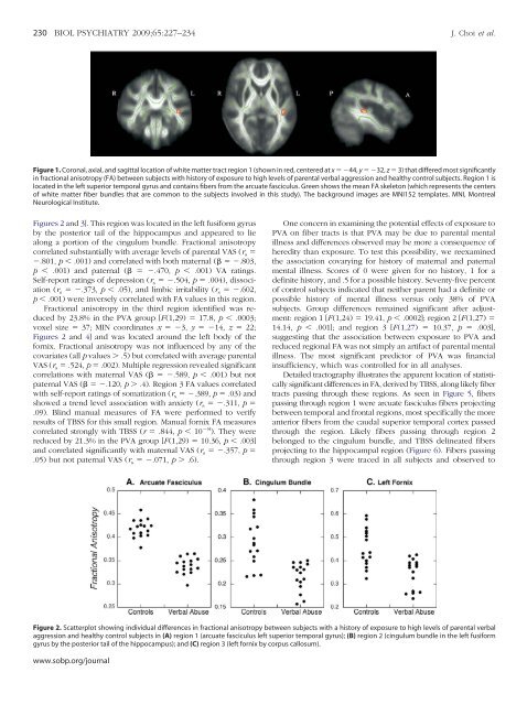
Figure 1. Coronal, axial, and sagittal location of white matter tract region 1 (shown <strong>in</strong> red, centered at x 44, y 32, z 3) that differed most significantly<br />
<strong>in</strong> fractional anisotropy (FA) between subjects with history of exposure to high levels of parental verbal aggression and healthy control subjects. Region 1 is<br />
located <strong>in</strong> the left superior temporal gyrus and conta<strong>in</strong>s fibers from the arcuate fasciculus. Green shows the mean FA skeleton (which represents the centers<br />
of white matter fiber bundles that are common to the subjects <strong>in</strong>volved <strong>in</strong> this study). The background images are MNI152 templates. MNI, Montreal<br />
Neurological Institute.<br />
Figures 2 and 3]. This region was located <strong>in</strong> the left fusi<strong>for</strong>m gyrus<br />
by the posterior tail of the hippocampus and appeared to lie<br />
along a portion of the c<strong>in</strong>gulum bundle. Fractional anisotropy<br />
correlated substantially with average levels of parental VAS (r s <br />
.801, p .001) and correlated with both maternal ( .803,<br />
p .001) and paternal ( .470, p .001) VA rat<strong>in</strong>gs.<br />
Self-report rat<strong>in</strong>gs of depression (r s .504, p .004), dissociation<br />
(r s .373, p .05), and limbic irritability (r s .602,<br />
p .001) were <strong>in</strong>versely correlated with FA values <strong>in</strong> this region.<br />
Fractional anisotropy <strong>in</strong> the third region identified was reduced<br />
by 23.8% <strong>in</strong> the PVA group [F(1,29) 17.8, p .0003;<br />
voxel size 37; MIN coord<strong>in</strong>ates x 3, y 14, z 22;<br />
Figures 2 and 4] and was located around the left body of the<br />
<strong>for</strong>nix. Fractional anisotropy was not <strong>in</strong>fluenced by any of the<br />
covariates (all p values .5) but correlated with average parental<br />
VAS (r s .524, p .002). Multiple regression revealed significant<br />
correlations with maternal VAS ( .589, p .001) but not<br />
paternal VAS ( .120, p .4). Region 3 FA values correlated<br />
with self-report rat<strong>in</strong>gs of somatization (r s .389, p .03) and<br />
showed a trend level association with anxiety (r s .311, p <br />
.09). Bl<strong>in</strong>d manual measures of FA were per<strong>for</strong>med to verify<br />
results of TBSS <strong>for</strong> this small region. Manual <strong>for</strong>nix FA measures<br />
correlated strongly with TBSS (r .844, p 10 8 ). They were<br />
reduced by 21.3% <strong>in</strong> the PVA group [F(1,29) 10.36, p .003]<br />
and correlated significantly with maternal VAS (r s .357, p <br />
.05) but not paternal VAS (r s .071, p .6).<br />
One concern <strong>in</strong> exam<strong>in</strong><strong>in</strong>g the potential effects of exposure to<br />
PVA on fiber tracts is that PVA may be due to parental mental<br />
illness and differences observed may be more a consequence of<br />
heredity than exposure. To test this possibility, we reexam<strong>in</strong>ed<br />
the association covary<strong>in</strong>g <strong>for</strong> history of maternal and paternal<br />
mental illness. Scores of 0 were given <strong>for</strong> no history, 1 <strong>for</strong> a<br />
def<strong>in</strong>ite history, and .5 <strong>for</strong> a possible history. Seventy-five percent<br />
of control subjects <strong>in</strong>dicated that neither parent had a def<strong>in</strong>ite or<br />
possible history of mental illness versus only 38% of PVA<br />
subjects. Group differences rema<strong>in</strong>ed significant after adjustment:<br />
region 1 [F(1,24) 19.41, p .0002]; region 2 [F(1,27) <br />
14.14, p .001]; and region 3 [F(1,27) 10.37, p .003],<br />
suggest<strong>in</strong>g that the association between exposure to PVA and<br />
reduced regional FA was not simply an artifact of parental mental<br />
illness. The most significant predictor of PVA was f<strong>in</strong>ancial<br />
<strong>in</strong>sufficiency, which was controlled <strong>for</strong> <strong>in</strong> all analyses.<br />
Detailed tractography illustrates the apparent location of statistically<br />
significant differences <strong>in</strong> FA, derived by TBSS, along likely fiber<br />
tracts pass<strong>in</strong>g through these regions. As seen <strong>in</strong> Figure 5, fibers<br />
pass<strong>in</strong>g through region 1 were arcuate fasciculus fibers project<strong>in</strong>g<br />
between temporal and frontal regions, most specifically the more<br />
anterior fibers from the caudal superior temporal cortex passed<br />
through the region. Likely fibers pass<strong>in</strong>g through region 2<br />
belonged to the c<strong>in</strong>gulum bundle, and TBSS del<strong>in</strong>eated fibers<br />
project<strong>in</strong>g to the hippocampal region (Figure 6). Fibers pass<strong>in</strong>g<br />
through region 3 were traced <strong>in</strong> all subjects and observed to<br />
Figure 2. Scatterplot show<strong>in</strong>g <strong>in</strong>dividual differences <strong>in</strong> fractional anisotropy between subjects with a history of exposure to high levels of parental verbal<br />
aggression and healthy control subjects <strong>in</strong> (A) region 1 (arcuate fasciculus left superior temporal gyrus); (B) region 2 (c<strong>in</strong>gulum bundle <strong>in</strong> the left fusi<strong>for</strong>m<br />
gyrus by the posterior tail of the hippocampus); and (C) region 3 (left <strong>for</strong>nix by corpus callosum).<br />
www.sobp.org/journal