Preliminary Evidence for White Matter Tract Abnormalities in Young ...
Preliminary Evidence for White Matter Tract Abnormalities in Young ...
Preliminary Evidence for White Matter Tract Abnormalities in Young ...

Create successful ePaper yourself
Turn your PDF publications into a flip-book with our unique Google optimized e-Paper software.
J. Choi et al. BIOL PSYCHIATRY 2009;65:227–234 231<br />
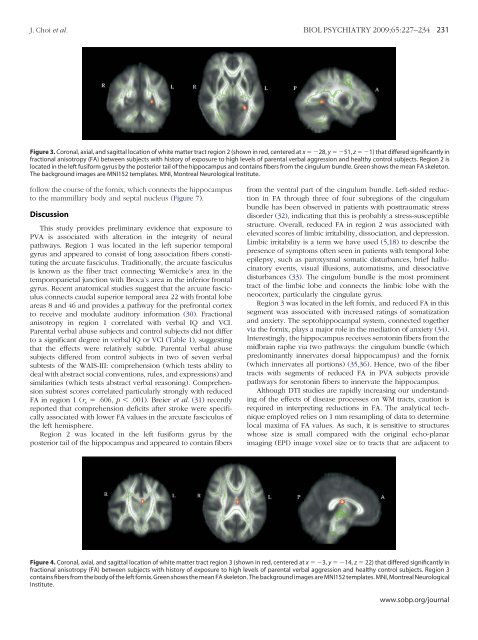
Figure 3. Coronal, axial, and sagittal location of white matter tract region 2 (shown <strong>in</strong> red, centered at x 28, y 51, z 1) that differed significantly <strong>in</strong><br />
fractional anisotropy (FA) between subjects with history of exposure to high levels of parental verbal aggression and healthy control subjects. Region 2 is<br />
located <strong>in</strong> the left fusi<strong>for</strong>m gyrus by the posterior tail of the hippocampus and conta<strong>in</strong>s fibers from the c<strong>in</strong>gulum bundle. Green shows the mean FA skeleton.<br />
The background images are MNI152 templates. MNI, Montreal Neurological Institute.<br />
follow the course of the <strong>for</strong>nix, which connects the hippocampus<br />
to the mammillary body and septal nucleus (Figure 7).<br />
Discussion<br />
This study provides prelim<strong>in</strong>ary evidence that exposure to<br />
PVA is associated with alteration <strong>in</strong> the <strong>in</strong>tegrity of neural<br />
pathways. Region 1 was located <strong>in</strong> the left superior temporal<br />
gyrus and appeared to consist of long association fibers constitut<strong>in</strong>g<br />
the arcuate fasciculus. Traditionally, the arcuate fasciculus<br />
is known as the fiber tract connect<strong>in</strong>g Wernicke’s area <strong>in</strong> the<br />
temporoparietal junction with Broca’s area <strong>in</strong> the <strong>in</strong>ferior frontal<br />
gyrus. Recent anatomical studies suggest that the arcuate fasciculus<br />
connects caudal superior temporal area 22 with frontal lobe<br />
areas 8 and 46 and provides a pathway <strong>for</strong> the prefrontal cortex<br />
to receive and modulate auditory <strong>in</strong><strong>for</strong>mation (30). Fractional<br />
anisotropy <strong>in</strong> region 1 correlated with verbal IQ and VCI.<br />
Parental verbal abuse subjects and control subjects did not differ<br />
to a significant degree <strong>in</strong> verbal IQ or VCI (Table 1), suggest<strong>in</strong>g<br />
that the effects were relatively subtle. Parental verbal abuse<br />
subjects differed from control subjects <strong>in</strong> two of seven verbal<br />
subtests of the WAIS-III: comprehension (which tests ability to<br />
deal with abstract social conventions, rules, and expressions) and<br />
similarities (which tests abstract verbal reason<strong>in</strong>g). Comprehension<br />
subtest scores correlated particularly strongly with reduced<br />
FA <strong>in</strong> region 1 (r s .606, p .001). Breier et al. (31) recently<br />
reported that comprehension deficits after stroke were specifically<br />
associated with lower FA values <strong>in</strong> the arcuate fasciculus of<br />
the left hemisphere.<br />
Region 2 was located <strong>in</strong> the left fusi<strong>for</strong>m gyrus by the<br />
posterior tail of the hippocampus and appeared to conta<strong>in</strong> fibers<br />
from the ventral part of the c<strong>in</strong>gulum bundle. Left-sided reduction<br />
<strong>in</strong> FA through three of four subregions of the c<strong>in</strong>gulum<br />
bundle has been observed <strong>in</strong> patients with posttraumatic stress<br />
disorder (32), <strong>in</strong>dicat<strong>in</strong>g that this is probably a stress-susceptible<br />
structure. Overall, reduced FA <strong>in</strong> region 2 was associated with<br />
elevated scores of limbic irritability, dissociation, and depression.<br />
Limbic irritability is a term we have used (5,18) to describe the<br />
presence of symptoms often seen <strong>in</strong> patients with temporal lobe<br />
epilepsy, such as paroxysmal somatic disturbances, brief halluc<strong>in</strong>atory<br />
events, visual illusions, automatisms, and dissociative<br />
disturbances (33). The c<strong>in</strong>gulum bundle is the most prom<strong>in</strong>ent<br />
tract of the limbic lobe and connects the limbic lobe with the<br />
neocortex, particularly the c<strong>in</strong>gulate gyrus.<br />
Region 3 was located <strong>in</strong> the left <strong>for</strong>nix, and reduced FA <strong>in</strong> this<br />
segment was associated with <strong>in</strong>creased rat<strong>in</strong>gs of somatization<br />
and anxiety. The septohippocampal system, connected together<br />
via the <strong>for</strong>nix, plays a major role <strong>in</strong> the mediation of anxiety (34).<br />
Interest<strong>in</strong>gly, the hippocampus receives seroton<strong>in</strong> fibers from the<br />
midbra<strong>in</strong> raphe via two pathways: the c<strong>in</strong>gulum bundle (which<br />
predom<strong>in</strong>antly <strong>in</strong>nervates dorsal hippocampus) and the <strong>for</strong>nix<br />
(which <strong>in</strong>nervates all portions) (35,36). Hence, two of the fiber<br />
tracts with segments of reduced FA <strong>in</strong> PVA subjects provide<br />
pathways <strong>for</strong> seroton<strong>in</strong> fibers to <strong>in</strong>nervate the hippocampus.<br />
Although DTI studies are rapidly <strong>in</strong>creas<strong>in</strong>g our understand<strong>in</strong>g<br />
of the effects of disease processes on WM tracts, caution is<br />
required <strong>in</strong> <strong>in</strong>terpret<strong>in</strong>g reductions <strong>in</strong> FA. The analytical technique<br />
employed relies on 1 mm resampl<strong>in</strong>g of data to determ<strong>in</strong>e<br />
local maxima of FA values. As such, it is sensitive to structures<br />
whose size is small compared with the orig<strong>in</strong>al echo-planar<br />
imag<strong>in</strong>g (EPI) image voxel size or to tracts that are adjacent to<br />
Figure 4. Coronal, axial, and sagittal location of white matter tract region 3 (shown <strong>in</strong> red, centered at x 3, y 14, z 22) that differed significantly <strong>in</strong><br />
fractional anisotropy (FA) between subjects with history of exposure to high levels of parental verbal aggression and healthy control subjects. Region 3<br />
conta<strong>in</strong>s fibers from the body of the left <strong>for</strong>nix. Green shows the mean FA skeleton. The background images are MNI152 templates. MNI, Montreal Neurological<br />
Institute.<br />
www.sobp.org/journal