Left to Right Shunts and their Calculation Ghada ... - CardioEgypt.com
Left to Right Shunts and their Calculation Ghada ... - CardioEgypt.com
Left to Right Shunts and their Calculation Ghada ... - CardioEgypt.com

You also want an ePaper? Increase the reach of your titles
YUMPU automatically turns print PDFs into web optimized ePapers that Google loves.
3/18/2013<br />
<strong>Left</strong> <strong>to</strong> <strong>Right</strong> <strong>Shunts</strong> <strong>and</strong><br />
<strong>their</strong> <strong>Calculation</strong><br />
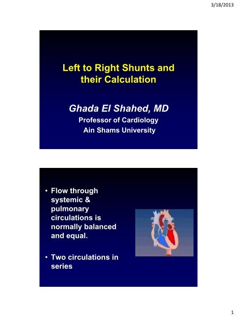
<strong>Ghada</strong> El Shahed, MD<br />
Professor of Cardiology<br />
Ain Shams University<br />
• Flow through<br />
systemic &<br />
pulmonary<br />
circulations is<br />
normally balanced<br />
<strong>and</strong> equal.<br />
• Two circulations in<br />
series<br />
1
3/18/2013<br />
• Flow through<br />
systemic &<br />
pulmonary<br />
circulations is<br />
normally balanced<br />
<strong>and</strong> equal.<br />
• Two circulations in<br />
series<br />
Oxygen Saturation<br />
Systemic vein<br />
Pulmonary vein<br />
RA<br />
65%<br />
LA<br />
100%<br />
RV<br />
65%<br />
LV<br />
100%<br />
Pulmonary artery<br />
Systemic artery<br />
2
3/18/2013<br />
<strong>Left</strong> <strong>to</strong> right shunts<br />
• “leak" of<br />
blood from<br />
the systemic<br />
<strong>to</strong> the<br />
pulmonary<br />
circulation<br />
<strong>Left</strong> <strong>to</strong> right shunts<br />
• pulmonary circulation<br />
receives :<br />
* blood that entered RA<br />
& RV from vena cavae<br />
plus<br />
* blood entering<br />
through ASD, VSD, or<br />
a PDA<br />
3
3/18/2013<br />
Causes<br />
Lesions resulting in left <strong>to</strong> right shunts<br />
include:<br />
• Ventricular septal defect (VSD)<br />
• Patent ductus arteriosus (PDA)<br />
• Atrial septal defect (ASD)<br />
• Atrioventricular defect (AVSD)<br />
Determinants of shunt size<br />
• In VSD <strong>and</strong> PDA:<br />
* size of defect<br />
And<br />
* relative resistance<br />
in the pulmonary<br />
<strong>and</strong> systemic<br />
circuits<br />
4
3/18/2013<br />
Determinants of shunt size<br />
• In ASD <strong>and</strong> AVSD:<br />
magnitude of the shunt<br />
depends mainly on<br />
relative ventricular<br />
<strong>com</strong>pliance.<br />
<strong>Left</strong> <strong>to</strong> right shunts:<br />
DETECTION AND<br />
QUANTIFICATION<br />
5
3/18/2013<br />
Clinical data: VSD<br />
• The intensity of the murmur<br />
is inversely proportional <strong>to</strong> the<br />
magnitude of the shunt<br />
• An apical mid-dias<strong>to</strong>lic murmur<br />
(increased flow across MV) indicates<br />
a large shunt<br />
Clinical data: PDA<br />
• Continuous or "machinery" murmur<br />
• An apical mid-dias<strong>to</strong>lic murmur<br />
denotes large shunt<br />
• Wide pulse pressure indicates a<br />
large left <strong>to</strong> right shunt<br />
6
3/18/2013<br />
Clinical data: ASD<br />
• Flow murmur in pulmonary area :<br />
>> large shunt<br />
• Mid-dias<strong>to</strong>lic murmur due <strong>to</strong> increased<br />
flow across the tricuspid valve<br />
• Wide splitting of S2 due <strong>to</strong> increased<br />
flow across the pulmonary valve<br />
7
3/18/2013<br />
“with a few limitations, the Doppler index RSV/LSV<br />
is clinically useful in the estimation of the<br />
magnitude of the shunt flow in patients with ASD”<br />
8
3/18/2013<br />
Shunt Detection :<br />
Indocyanine Green Method<br />
• Indocyanine green (1 cc) injected as a bolus in<strong>to</strong><br />
right side of circulation (pulmonary artery)<br />
• Concentration<br />
measured from<br />
peripheral artery<br />
Baim DS: Grossman’s Cardiac Catheterization, Angiography, <strong>and</strong> Intervention. 7 th Edition. Lippincot Williams<br />
<strong>and</strong> Wilkins, 2007.<br />
9
3/18/2013<br />
<strong>Left</strong>-<strong>to</strong>-<strong>Right</strong> Shunt<br />
Appearance <strong>and</strong> washout of dye produces initial 1 st<br />
pass curve followed by recirculation in normal adults<br />
Baim DS: Grossman’s Cardiac Catheterization, Angiography, <strong>and</strong> Intervention. 7 th Edition. Lippincot Williams<br />
<strong>and</strong> Wilkins, 2007.<br />
Shunt Detection:<br />
Indocyanine Green Method<br />
Bashore, TM. Congenital Heart Disease in Adults. The Measurement of Intracardiac <strong>Shunts</strong>. In: CATHSAP II.<br />
Bethesda: American College of Cardiology, 2001.<br />
10
3/18/2013<br />
Indica<strong>to</strong>r Dilution Method<br />
• More Sensitive for smaller<br />
shunts<br />
• Cannot localize the level of<br />
left <strong>to</strong> right shunt<br />
Oximetric Methods<br />
• Obtain O2 saturations<br />
in sequential<br />
chambers,<br />
identifying oxygen<br />
step-up<br />
• Insensitive for small<br />
shunts (< 1.3:1)<br />
Baim DS: Grossman’s Cardiac Catheterization, Angiography, <strong>and</strong> Intervention. 7 th Edition. Lippincot Williams<br />
<strong>and</strong> Wilkins, 2007.<br />
11
3/18/2013<br />
Shunt Detection & Measurement<br />
Oximetry Run<br />
• IVC, L4-5 level<br />
• IVC, above diaphragm<br />
• SVC, innominate<br />
• SVC, at RA<br />
• RA, high<br />
• RA, mid<br />
• RA, low<br />
• RV, mid<br />
• RV, apex<br />
• RV, outflow tract<br />
• PA, main<br />
• PA, right or left<br />
• <strong>Left</strong> ventricle<br />
• Aorta, distal <strong>to</strong> ductus<br />
x<br />
x<br />
x<br />
x<br />
x<br />
x<br />
x<br />
x<br />
x<br />
x<br />
x<br />
x<br />
x<br />
x<br />
x<br />
Baim DS: Grossman’s Cardiac Catheterization, Angiography, <strong>and</strong> Intervention. 7 th Edition. Lippincot Williams<br />
<strong>and</strong> Wilkins, 2007.<br />
Oximetry<br />
ASD VSD PDA<br />
Systemic arterial O2 Sat No change No change No change<br />
RA O2 Sat No change No change<br />
RV O2 Sat<br />
No change<br />
PA O2 Sat<br />
Pulmonary blood flow<br />
12
3/18/2013<br />
ASD<br />
Detection of <strong>Left</strong>-<strong>to</strong>-<strong>Right</strong> Shunt<br />
Level of shunt O 2 % Sat Differential diagnosis<br />
Atrial<br />
(SVC/IVC RA)<br />
Ventricular<br />
(RA RV)<br />
Great vessel<br />
(RV PA)<br />
7<br />
5<br />
5<br />
ASD, PAPVR, VSD with TR,<br />
Ruptured sinus of Valsalva,<br />
Coronary fistula <strong>to</strong> RA<br />
VSD, PDA with PR,<br />
Coronary fistula <strong>to</strong> RV<br />
Aor<strong>to</strong>-pulmonary window,<br />
Aberrant coronary origin,<br />
PDA<br />
13
3/18/2013<br />
VSD<br />
SHUNT QUANTIFICATION<br />
14
3/18/2013<br />
• <strong>Calculation</strong> of systemic <strong>and</strong> pulmonary<br />
flows (<strong>and</strong> <strong>their</strong> ratio) are used for shunt<br />
quantification<br />
• The most <strong>com</strong>mon methods:<br />
-Fick’s method<br />
-Indica<strong>to</strong>r dilution<br />
(Both have <strong>their</strong> assumptions & limitations<br />
that are important <strong>to</strong> remember)<br />
The Fick Method<br />
• Described by Adolph Fick in 1870<br />
• Is probably the most <strong>com</strong>mon<br />
method used <strong>to</strong> calculate cardiac<br />
flows in the pediatric cath. lab<br />
15
3/18/2013<br />
Concept<br />
• The <strong>to</strong>tal uptake (or release) of a substance<br />
by an organ is the product of blood flow <strong>to</strong><br />
that organ <strong>and</strong> the concentration difference<br />
of the substance in the arteries <strong>and</strong> veins<br />
leading in<strong>to</strong> <strong>and</strong> out of that organ<br />
Fick Oxygen Method<br />
• As applied <strong>to</strong> lungs, the substance<br />
released <strong>to</strong> the blood is oxygen<br />
>> oxygen consumption is the product of<br />
arteriovenous difference of oxygen across<br />
the lungs <strong>and</strong> pulmonary blood flow<br />
Q p =<br />
Oxygen consumption<br />
Arteriovenous O 2 difference<br />
Baim DS: Grossman’s Cardiac Catheterization, Angiography, <strong>and</strong> Intervention. 7 th Edition. Lippincot Williams<br />
<strong>and</strong> Wilkins, 2007.<br />
16
3/18/2013<br />
Concept<br />
• So, using arterial <strong>and</strong> venous oxygen<br />
content <strong>and</strong> oxygen consumption,<br />
one can easily calculate flow.<br />
• So the important equation is:<br />
(Multiply denomina<strong>to</strong>r by 10 <strong>to</strong> convert dL <strong>to</strong> L, as<br />
Hb is measured in g/dL)<br />
17
3/18/2013<br />
Calcluation of oxygen content<br />
of blood<br />
Oxygen Content (mL O2/dL plasma)<br />
= O2 bound <strong>to</strong> Hb + dissolved O2<br />
= [1.36 Hgb] X saturation] + [0.003 X PaO2]<br />
18
3/18/2013<br />
O2 content<br />
= 1.36 Hgb X saturation + 0.003 X PaO2<br />
• 1.36mL is the oxygen carrying capacity<br />
of1 gm of hemoglobin<br />
• If the saturation is 98%, then use 0.98<br />
in the formula, not 98<br />
• The relatively small amount of<br />
dissolved oxygen in plasma is 0.003mL<br />
O2/dL plasma/mmHg PaO2<br />
19
3/18/2013<br />
Oxygen consumption (VO2)<br />
• Oxygen consumption is often assumed,<br />
<strong>and</strong> is readily available in tables<br />
• There is variation based on sex, age, <strong>and</strong><br />
heart rate.<br />
• no data published for the very small infant.<br />
(Despite the limitations of assumed VO2, this<br />
method remains widely used)<br />
Measurement of VO2<br />
Baim DS: Grossman’s Cardiac Catheterization, Angiography, <strong>and</strong> Intervention. 7 th<br />
Edition. Lippincot Williams <strong>and</strong> Wilkins, 2007.<br />
20
3/18/2013<br />
• Using this principle, systemic <strong>and</strong><br />
pulmonary flows can be calculated<br />
Pulmonary blood flow<br />
QP=<br />
21
3/18/2013<br />
Shunt Measurement:<br />
Oximetric Methods<br />
• Fick Principle: The <strong>to</strong>tal uptake or<br />
release of any substance by an organ is<br />
the product of blood flow <strong>to</strong> the organ<br />
<strong>and</strong> the arteriovenous concentration<br />
difference of the substance.<br />
– Pulmonary circulation (Qp) utilizes PA<br />
<strong>and</strong> PV saturations<br />
RA (MV)<br />
RV<br />
Lungs<br />
LA (PV)<br />
LV<br />
PBF =<br />
O2 consumption<br />
(PvO 2 – PaO 2 ) x 10<br />
(O 2 content = 1.36 x Hgb x O 2 saturation)<br />
PA<br />
Ao<br />
(Alan Keith Berger, MD<br />
University of Minnesota,<br />
2003)<br />
Shunt Detection & Measurement<br />
Oximetric Methods<br />
• Fick Principle: The <strong>to</strong>tal uptake or<br />
release of any substance by an organ is<br />
the product of blood flow <strong>to</strong> the organ<br />
<strong>and</strong> the arteriovenous concentration<br />
difference of the substance.<br />
– Systemic circulation (Qs) utilizes MV <strong>and</strong><br />
Ao saturations<br />
O2 consumption<br />
SBF = (AoO 2 – MVO 2 ) x 10<br />
(O 2 content = 1.36 x Hgb x O 2 saturation)<br />
RA (MV)<br />
RV<br />
PA<br />
Body<br />
LA (PV)<br />
Ao<br />
LV<br />
(Alan Keith Berger, MD<br />
University of Minnesota,<br />
2003)<br />
22
3/18/2013<br />
Shunt Detection & Measurement<br />
Oximetric Methods<br />
• Fick Principle: The <strong>to</strong>tal uptake or<br />
release of any substance by an organ is<br />
the product of blood flow <strong>to</strong> the organ<br />
<strong>and</strong> the arteriovenous concentration<br />
difference of the substance.<br />
– Pulmonary circulation (Qp) utilizes PA<br />
<strong>and</strong> PV saturations<br />
– Systemic circulation (Qs) utilizes MV <strong>and</strong><br />
Ao saturations<br />
RA (MV)<br />
RV<br />
PA<br />
LA (PV)<br />
Ao<br />
LV<br />
PBF =<br />
O 2 consumption<br />
(PvO 2 – PaO 2 ) x 10<br />
SBF =<br />
O 2 consumption<br />
(AoO 2 – MVO 2 ) x 10<br />
Mixed venous O2 saturation:<br />
RA receives blood from several sources<br />
– SVC: Saturation most closely approximates<br />
true systemic venous saturation<br />
– IVC: Highly saturated because kidneys receive<br />
25% of CO <strong>and</strong> extract minimal oxygen<br />
– Coronary sinus: Markedly desaturated because<br />
heart maximizes O2 extraction<br />
Baim DS: Grossman’s Cardiac Catheterization, Angiography, <strong>and</strong> Intervention. 7 th Edition. Lippincot Williams<br />
<strong>and</strong> Wilkins, 2007.<br />
23
3/18/2013<br />
Mixed venous O2 saturation:<br />
• Phlamm Equation: Mixed venous saturation<br />
used <strong>to</strong> normalize for differences in blood<br />
saturations that enter RA<br />
Mixed venous saturation =<br />
3 (SVC) + IVC<br />
4<br />
Baim DS: Grossman’s Cardiac Catheterization, Angiography, <strong>and</strong> Intervention. 7 th Edition. Lippincot Williams<br />
<strong>and</strong> Wilkins, 2007.<br />
• For pediatric catheterization, we<br />
most often express the indexed flow<br />
(<strong>to</strong> BSA), distinguishing between<br />
cardiac output (CO; L/min) <strong>and</strong><br />
cardiac index (Qs; L/min/m2).<br />
Because the VO2 tables are usually<br />
indexed.<br />
24
3/18/2013<br />
• For pediatric catheterization, we<br />
most often express the indexed flow<br />
(<strong>to</strong> BSA), distinguishing between<br />
cardiac output (CO; L/min) <strong>and</strong><br />
cardiac index (Qs; L/min/m2).<br />
Because the VO2 tables are usually<br />
indexed.<br />
25
3/18/2013<br />
Example<br />
• Mixed venous saturation - 75%, PA<br />
saturations = 84%<br />
• pulmonary vein <strong>and</strong> aortic<br />
saturations = 99%.<br />
>>>>The net Qp:Qs in this patient is<br />
(99 – 75)/(99 – 84)=1.6<br />
Estimation of shunt size<br />
Flow Ratio (QP/QS):<br />
• < 1.5 = Small shunt<br />
• ≥ 2.0 = Large <strong>Left</strong> <strong>to</strong> <strong>Right</strong> Shunt<br />
• < 1.0 = Net <strong>Right</strong> <strong>to</strong> left Shunt<br />
No need for Oxygen consumption<br />
(Since this number will cancel out of<br />
the equation)<br />
26
3/18/2013<br />
Shunt Measurement:<br />
Effective Pulmonary Blood Flow<br />
• Effective Pulmonary Blood<br />
Flow: flow that would be<br />
present if no shunt were<br />
present<br />
• Requires<br />
– MV = PA saturation<br />
– PV – PA = PV - MV<br />
PBF<br />
Effective Pulmonary<br />
Blood Flow<br />
=<br />
O 2 consumption<br />
(Pv – MV O 2 ) x 10<br />
=<br />
O 2 consumption<br />
(Pv – Pa O 2 ) x 10<br />
Bashore, TM. Congenital Heart Disease in Adults. The Measurement of Intracardiac <strong>Shunts</strong>. In: CATHSAP II.<br />
Bethesda: American College of Cardiology, 2001.<br />
Shunt Detection & Measurement<br />
<strong>Left</strong>-<strong>to</strong>-<strong>Right</strong> Shunt<br />
• <strong>Left</strong> <strong>to</strong> right shunt: >> in step-up<br />
in O 2 between MV <strong>and</strong> PA<br />
• Shunt is the difference between<br />
pulmonary flow measured <strong>and</strong><br />
what it would be in the absence of<br />
shunt (EPBF)<br />
• Systemic Blood Flow = EPBF<br />
<strong>Left</strong>-<strong>Right</strong> Shunt = Pulmonary Blood Flow – Effective Blood Flow<br />
O 2 consumption<br />
=<br />
(PvO 2 – Pa O 2 ) x 10<br />
–<br />
O 2 consumption<br />
(PvO 2 – MVO 2 ) x 10<br />
Bashore, TM. Congenital Heart Disease in Adults. The Measurement of Intracardiac <strong>Shunts</strong>. In: CATHSAP II.<br />
Bethesda: American College of Cardiology, 2001.<br />
27
3/18/2013<br />
Shunt Measurement<br />
Limitations of Oximetric Method<br />
• Requires steady state with rapid collection of O 2<br />
samples<br />
• Insensitive <strong>to</strong> small shunts<br />
• Flow dependent<br />
– Normal variability of blood oxygen saturation in the right<br />
heart chambers is influenced by magnitude of SBF<br />
– High flow state may simulate a left-<strong>to</strong>-right shunt<br />
• When O 2 content is utilized (as opposed <strong>to</strong> O 2 sat),<br />
the step-up is dependent on hemoglobin.<br />
Baim DS: Grossman’s Cardiac Catheterization, Angiography, <strong>and</strong> Intervention. 7 th Edition. Lippincot Williams<br />
<strong>and</strong> Wilkins, 2007.<br />
SOME TIPS FOR QP, QS<br />
CALCULATIONS<br />
28
3/18/2013<br />
• If you do not directly obtain a<br />
pulmonary vein or LA saturation,<br />
assume something appropriate <strong>to</strong> the<br />
clinical status of the patient<br />
( ~95–100% in the absence of lung<br />
disease)<br />
• Always double check <strong>and</strong> confirm<br />
your math.<br />
• Always document all assumptions in<br />
your catheterization report.<br />
29
3/18/2013<br />
• Make sure your<br />
numbers make sense.<br />
[e.g. The SVC saturation should never be<br />
higher than the PA saturation<br />
(unless there is anomalous PV return or an<br />
oxygen-consuming tumor in the RV!)<br />
Fick Oxygen Method<br />
• Fick oxygen method <strong>to</strong>tal error 10%<br />
– Error in O2 consumption 6%<br />
– Error in AV O2 difference 5%. Narrow<br />
AV O2 differences more subject <strong>to</strong> error,<br />
<strong>and</strong> therefore Fick method is most<br />
accurate in low cardiac output states<br />
Baim DS: Grossman’s Cardiac Catheterization, Angiography, <strong>and</strong> Intervention. 7 th Edition. Lippincot Williams<br />
<strong>and</strong> Wilkins, 2007.<br />
30
3/18/2013<br />
Cardiac Output Measurement<br />
Fick Oxygen Method<br />
• Sources of Error<br />
– Spec<strong>to</strong>pho<strong>to</strong>metric determination of blood<br />
oxygen saturation<br />
– Changes in mean pulmonary volume<br />
• Patient may progressively increase or decrease<br />
pulmonary volume during sample collection. If<br />
patient relaxes <strong>and</strong> breathes smaller volumes, CO is<br />
underestimated<br />
– Improper collection of mixed venous blood<br />
sample<br />
• Contamination with PCW blood<br />
• Sampling from more proximal site<br />
Baim DS: Grossman’s Cardiac Catheterization, Angiography, <strong>and</strong> Intervention. 7 th Edition. Lippincot Williams<br />
<strong>and</strong> Wilkins, 2007.<br />
Indica<strong>to</strong>r Dilution Methods<br />
• Requirements<br />
– Bolus of indica<strong>to</strong>r substance which mixes<br />
<strong>com</strong>pletely with blood <strong>and</strong> whose concentration<br />
can be measured<br />
– Indica<strong>to</strong>r must go through a portion of circulation<br />
where all the blood of the body be<strong>com</strong>es mixed<br />
Baim DS: Grossman’s Cardiac Catheterization, Angiography, <strong>and</strong> Intervention. 7 th Edition. Lippincot Williams<br />
<strong>and</strong> Wilkins, 2007.<br />
31
Concentration<br />
3/18/2013<br />
Cardiac Output Measurement<br />
Indica<strong>to</strong>r Dilution Methods<br />
Stewart-Hamil<strong>to</strong>n Equation<br />
CO = <br />
0<br />
Indica<strong>to</strong>r amount<br />
C (t) dt<br />
C = concentration<br />
of indica<strong>to</strong>r<br />
CO =<br />
Indica<strong>to</strong>r amount (mg) x 60 sec/min<br />
mean indica<strong>to</strong>r concentration (mg/mL) x curve duration<br />
• Indica<strong>to</strong>rs<br />
– Indocyanine Green<br />
– Thermodilution (Indica<strong>to</strong>r = Cold)<br />
Baim DS: Grossman’s Cardiac Catheterization, Angiography, <strong>and</strong> Intervention. 7 th Edition. Lippincot Williams<br />
<strong>and</strong> Wilkins, 2007.<br />
Cardiac Output Measurement<br />
Indocyanine Green Method<br />
• Indocyanine green (volume <strong>and</strong> concentration<br />
fixed) injected as a bolus in<strong>to</strong> right side of<br />
circulation (pulmonary artery)<br />
• Samples taken from peripheral artery,<br />
withdrawing continuously at a fixed rate<br />
• Indocyanine green concentration measured by<br />
densi<strong>to</strong>metry<br />
CO =<br />
I<br />
(C x t )<br />
(C x t)<br />
Recirculation<br />
Extrapolation<br />
of plot<br />
Baim DS: Grossman’s Cardiac Catheterization, Angiography, <strong>and</strong> Intervention. 7 th Edition. Lippincot Williams<br />
<strong>and</strong> Wilkins, 2007.<br />
time<br />
CO inversely<br />
proportional<br />
<strong>to</strong> area under<br />
curve<br />
32
3/18/2013<br />
Cardiac Output Measurement<br />
Indocyanine Green Method<br />
• Sources of Error<br />
– Indocyanine green unstable over time <strong>and</strong> with<br />
exposure <strong>to</strong> light<br />
– Sample must be introduced rapidly as single bolus<br />
– Bolus size must be exact<br />
– Indica<strong>to</strong>r must mix thoroughly with blood, <strong>and</strong> should<br />
be injected just proximal or in<strong>to</strong> cardiac chamber<br />
– Dilution curve must have exponential downslope of<br />
sufficient length <strong>to</strong> extrapolate curve. Invalid in Low<br />
cardiac output states <strong>and</strong> shunts that lead <strong>to</strong> early<br />
recirculation<br />
– Withdrawal rate of arterial sample must be constant<br />
Baim DS: Grossman’s Cardiac Catheterization, Angiography, <strong>and</strong> Intervention. 7 th Edition. Lippincot Williams<br />
<strong>and</strong> Wilkins, 2007.<br />
THERMODILUTION<br />
33
3/18/2013<br />
• First introduced in the 1950s, this<br />
technique involves determining the<br />
extent <strong>and</strong> rate of thermal changes in the<br />
blood stream after injection of a fixed<br />
volume of fluid at a set temperature<br />
upstream.<br />
• From this temperature– time curve, the<br />
volume rate of flow can be calculated in a<br />
manner analogous <strong>to</strong> that used for dye<br />
<strong>and</strong> other indica<strong>to</strong>r–dilution methods.<br />
Common Problems<br />
• High cardiac output >> only a small<br />
temperature drop, increasing the risk for<br />
error.<br />
• If the proximal port is not in free flowing<br />
blood, there will be loss of temperature<br />
before reaching the thermis<strong>to</strong>r.<br />
• Slow injection of cooled solution will<br />
produce a smaller magnitude temperature<br />
change, falsely elevating the calculated<br />
cardiac output.<br />
34
3/18/2013<br />
Cardiac Output Measurement<br />
Thermodilution Method<br />
CO =<br />
V I (T B -T I ) (S I x C I / S B x C B ) x 60<br />
<br />
<br />
0<br />
T B dt<br />
V I = volume of injectate<br />
S I , S B = specific gravity of injectate <strong>and</strong> blood<br />
C I , C B = specific heat of injectate <strong>and</strong> blood<br />
T I = temperature of injectate<br />
T B = change in temperature measured downstream<br />
Baim DS: Grossman’s Cardiac Catheterization, Angiography, <strong>and</strong> Intervention. 7 th Edition. Lippincot Williams<br />
<strong>and</strong> Wilkins, 2007.<br />
Cardiac Output Measurement<br />
Thermodilution Method<br />
• Advantages<br />
– Withdrawal of blood not necessary<br />
– Arterial puncture not required<br />
– Indica<strong>to</strong>r (saline or D5W)<br />
– Virtually no recirculation, simplifying <strong>com</strong>puter<br />
analysis of primary curve sample<br />
Baim DS: Grossman’s Cardiac Catheterization, Angiography, <strong>and</strong> Intervention. 7 th Edition. Lippincot Williams<br />
<strong>and</strong> Wilkins, 2007.<br />
35
3/18/2013<br />
Summary of<br />
Fick Oxygen Method: AV O 2 Difference<br />
Step 1: Theoretical oxygen carrying capacity<br />
O 2 carrying capacity (mL O 2 / L blood) =<br />
1.36 mL O 2 / gm Hgb x 10 mL/dL x Hgb (gm/dL)<br />
Step 2: Determine arterial oxygen content<br />
Arterial O 2 content = Arterial saturation x O 2 carrying capacity<br />
Step 3: Determine venous oxygen content<br />
Mixed venous O 2 content = venous saturation x O 2 carrying capacity<br />
Step 3: Determine A-V O 2 oxygen difference<br />
AV O 2 difference = Arterial O2 content - venous O 2 content<br />
Baim DS: Grossman’s Cardiac Catheterization, Angiography, <strong>and</strong> Intervention. 7 th Edition. Lippincot Williams<br />
<strong>and</strong> Wilkins, 2007.<br />
Importance of accurate<br />
calculation & avoiding pitfalls<br />
• Shunt size >>>> surgical decision<br />
• Flow is used <strong>to</strong> calculate resistance<br />
36
3/18/2013<br />
Accurate measurements &<br />
calculations = correct decision<br />
• ‘‘A catheterization is<br />
like a puzzle:<br />
everything must fit<br />
with everything else.’’<br />
(If things don’t fit, your<br />
case is probably not<br />
<strong>com</strong>plete)<br />
37