A scanning and transmission electron microscopic study of ... - Digitum
A scanning and transmission electron microscopic study of ... - Digitum
A scanning and transmission electron microscopic study of ... - Digitum

You also want an ePaper? Increase the reach of your titles
YUMPU automatically turns print PDFs into web optimized ePapers that Google loves.
Chicken egg membrane<br />
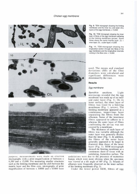
longitudinal section through lwo fibres <strong>of</strong> the<br />
e(i(i membrane <strong>and</strong> the lonaitudinal channels<br />
rÜñning in the fibre core. x 14,000<br />
used. The means <strong>and</strong> st<strong>and</strong>ard<br />
deviations (SD) <strong>of</strong> the fibre<br />
diameters were calculated <strong>and</strong><br />
significant differences were<br />
determined by the t-test.<br />
Results<br />
Egg membrane<br />
Semithin sections. Light<br />
microscopy revealed that the egg<br />
membrane was made up <strong>of</strong> an inner<br />
<strong>and</strong> outer layer ( ~ i 1). ~ : On its<br />
fibres appeared to adhere to it<br />
whereas the outer layer <strong>of</strong> fibres<br />
confronting the air-sac was not<br />
covered by a membrane.<br />
The thickness <strong>of</strong> each layer <strong>of</strong><br />
fibres was variable although the<br />
outer layer was generally thicker<br />
than the inner (Fig. 1). In addition,<br />
the fibres <strong>of</strong> the outer layer<br />
generally appeared to be large; in<br />
1 diameter than those <strong>of</strong> the inner<br />
purpose, measurements were made on <strong>electron</strong> undulations were due to the presence <strong>of</strong> numerous<br />
micrographs with a print magnification <strong>of</strong> between x humps which were more obvious when the specimen<br />
6,300 <strong>and</strong> x 15,000. For measuring smaller stmctures was viewed at a tilt angle <strong>of</strong> 60"Fig. 2). Str<strong>and</strong>s <strong>of</strong><br />
such as the limiting membrane <strong>and</strong> the cleft between the albumen were frequently observed on the inner surface<br />
mantle layer <strong>and</strong> the fibre core, micrographs <strong>of</strong> print <strong>of</strong> the limiting membrane (Fig. 3).<br />
magnification <strong>of</strong> between x 30000 <strong>and</strong> x 63000 were Under the lower power SEM, fibres <strong>of</strong> egg