Plant & Animal Cell LAB
Plant & Animal Cell LAB
Plant & Animal Cell LAB

Create successful ePaper yourself
Turn your PDF publications into a flip-book with our unique Google optimized e-Paper software.
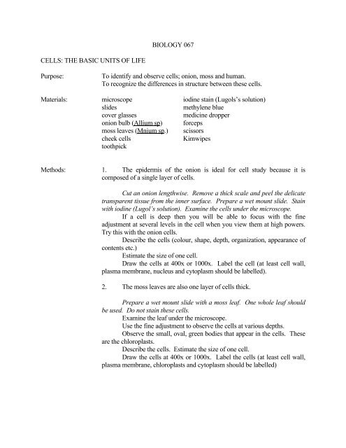
BIOLOGY 067<br />
CELLS: THE BASIC UNITS OF LIFE<br />
Purpose:<br />
To identify and observe cells; onion, moss and human.<br />
To recognize the differences in structure between these cells.<br />
Materials: microscope iodine stain (Lugols’s solution)<br />
slides<br />
methylene blue<br />
cover glasses<br />
medicine dropper<br />
onion bulb (Allium sp) forceps<br />
moss leaves (Mnium sp.) scissors<br />
cheek cells<br />
Kimwipes<br />
toothpick<br />
Methods: 1. The epidermis of the onion is ideal for cell study because it is<br />
composed of a single layer of cells.<br />
Cut an onion lengthwise. Remove a thick scale and peel the delicate<br />
transparent tissue from the inner surface. Prepare a wet mount slide. Stain<br />
with iodine (Lugol’s solution). Examine the cells under the microscope.<br />
If a cell is deep then you will be able to focus with the fine<br />
adjustment at several levels in the cell when you view them at high powers.<br />
Try this with the onion cells.<br />
Describe the cells (colour, shape, depth, organization, appearance of<br />
contents etc.)<br />
Estimate the size of one cell.<br />
Draw the cells at 400x or 1000x. Label the cell (at least cell wall,<br />
plasma membrane, nucleus and cytoplasm should be labelled).<br />
2. The moss leaves are also one layer of cells thick.<br />
Prepare a wet mount slide with a moss leaf. One whole leaf should<br />
be used. Do not stain these cells.<br />
Examine the leaf under the microscope.<br />
Use the fine adjustment to observe the cells at various depths.<br />
Observe the small, oval, green bodies that appear in the cells. These<br />
are the chloroplasts.<br />
Describe the cells. Estimate the size of one cell.<br />
Draw the cells at 400x or 1000x. Label the cells (at least cell wall,<br />
plasma membrane, chloroplasts and cytoplasm should be labelled)
3. By gently scraping your cheek, you will release several cheek cells.<br />
Gently scrape the inside of your cheek with a clean toothpick.<br />
Prepare a wet mount slide with the material that you have scraped. Add<br />
methylene blue stain.<br />
Examine the cells with a microscope.<br />
Describe the cells. Estimate the size of one cell.<br />
Draw the cells at 400x or 1000x. Label the cell (at least plasma<br />
membrane, nucleus and cytoplasm should be labelled).<br />
Results:<br />
Conclusions:<br />
Your drawings and observations are your results.<br />
Compare the cells you observed: discuss similarities and differences among<br />
the three cells using at least four characteristics.<br />
What differences might be related to plant vs. animal cell structure?